1.2: Exercise
- Page ID
- 103123
\( \newcommand{\vecs}[1]{\overset { \scriptstyle \rightharpoonup} {\mathbf{#1}} } \)
\( \newcommand{\vecd}[1]{\overset{-\!-\!\rightharpoonup}{\vphantom{a}\smash {#1}}} \)
\( \newcommand{\id}{\mathrm{id}}\) \( \newcommand{\Span}{\mathrm{span}}\)
( \newcommand{\kernel}{\mathrm{null}\,}\) \( \newcommand{\range}{\mathrm{range}\,}\)
\( \newcommand{\RealPart}{\mathrm{Re}}\) \( \newcommand{\ImaginaryPart}{\mathrm{Im}}\)
\( \newcommand{\Argument}{\mathrm{Arg}}\) \( \newcommand{\norm}[1]{\| #1 \|}\)
\( \newcommand{\inner}[2]{\langle #1, #2 \rangle}\)
\( \newcommand{\Span}{\mathrm{span}}\)
\( \newcommand{\id}{\mathrm{id}}\)
\( \newcommand{\Span}{\mathrm{span}}\)
\( \newcommand{\kernel}{\mathrm{null}\,}\)
\( \newcommand{\range}{\mathrm{range}\,}\)
\( \newcommand{\RealPart}{\mathrm{Re}}\)
\( \newcommand{\ImaginaryPart}{\mathrm{Im}}\)
\( \newcommand{\Argument}{\mathrm{Arg}}\)
\( \newcommand{\norm}[1]{\| #1 \|}\)
\( \newcommand{\inner}[2]{\langle #1, #2 \rangle}\)
\( \newcommand{\Span}{\mathrm{span}}\) \( \newcommand{\AA}{\unicode[.8,0]{x212B}}\)
\( \newcommand{\vectorA}[1]{\vec{#1}} % arrow\)
\( \newcommand{\vectorAt}[1]{\vec{\text{#1}}} % arrow\)
\( \newcommand{\vectorB}[1]{\overset { \scriptstyle \rightharpoonup} {\mathbf{#1}} } \)
\( \newcommand{\vectorC}[1]{\textbf{#1}} \)
\( \newcommand{\vectorD}[1]{\overrightarrow{#1}} \)
\( \newcommand{\vectorDt}[1]{\overrightarrow{\text{#1}}} \)
\( \newcommand{\vectE}[1]{\overset{-\!-\!\rightharpoonup}{\vphantom{a}\smash{\mathbf {#1}}}} \)
\( \newcommand{\vecs}[1]{\overset { \scriptstyle \rightharpoonup} {\mathbf{#1}} } \)
\( \newcommand{\vecd}[1]{\overset{-\!-\!\rightharpoonup}{\vphantom{a}\smash {#1}}} \)
Exercise 1: Identifying the parts of the microscope
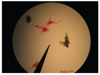
Figure \(\PageIndex{1}\): Side and front view of Olympus CX43 microscope, from user manual. Instructions: Identify & label the following parts of your microscope onto the image above, and fill-in-the blanks below. · Binocular head, Oculars: _______x · Arm · Revolving nosepiece · Objective lenses: ______x, _______x, _______x, _______x · Mechanical stage and X/Y knobs · Stage/slide clips · Condenser Turret __________, ___________, __________, ____________ · Iris diaphragm and how to adjust it · Coarse adjustment knob & fine adjustment knob · Light source; illuminator · Base · Dimmer; light intensity · Power switch & power cord · Total magnification calculation = Exercise 2: Using your MicroscopeThe genus Amoeba (sometimes referred to as Amoebozoa) are a group of eukaryotic single-cell organisms in the kingdom Protista. Some species can cause serious human diseases, such as Entamoeba histolytica which causes amoebic dysentery (bloody diarrhea), and Naegleria fowleri, commonly referred to as the "brain-eating" amoeba. Pseudopodia are foot-like extensions of the cell membrane and cytoplasm via actin microfilaments and are used for motility and ingestion of food (bacteria and other protists) via phagocytosis. Amoeba are fairly large multinucleated cells, meaning they contain more than one nucleus per cell.
Figure \(\PageIndex{2}\): Amoeba proteus. Amoebae with tubular and lobe-shaped pseudopodia are seen under a microscope. These isolates would be morphologically classified as amoebozoans. OpenStax Figure 23.13 used under the Creative Commons Attribution 4.0 International License. The genus Paramecium are a group of eukaryotic single-cell organisms in the kingdom Protista. They are typically found in freshwater or brackish (slightly salty) environments. Paramecium are called ciliates because they have specialized hair-like structures called cilia. Their cilia line the outside of the cell and are used for motility. Cilia also line the oral groove (primitive mouth) and are used to ingest of food such as bacteria. Contractile vacuoles allow the organism to excrete excess water. Paramecium have another distinguishing trait - two nuclei; a macronucleus that is used for transcription of genes, and a micronucleus that is used for sexual reproduction.
Figure \(\PageIndex{3}\): Paramecium. (credit “paramecium micrograph”: modification of work by NIH; scale-bar data from Matt Russell) OpenStax Figure 23.24 used under the Creative Commons Attribution 4.0 International License. Instructions: View both Paramecium and Amoeba slides under the 4x, 10x, and 40x objectives, and sketch and label each organism in the space below. Do NOT use the oil immersion lens today.
|
|---|