
8.8: Pancreas
- Page ID
- 103228

Giving yourself an injection can be difficult, but for someone with diabetes, it may be a matter of life or death. The person in the photo has diabetes and is injecting themself with insulin, the hormone that helps control the level of glucose in the blood. Insulin is produced by the pancreas.

Introduction to the Pancreas
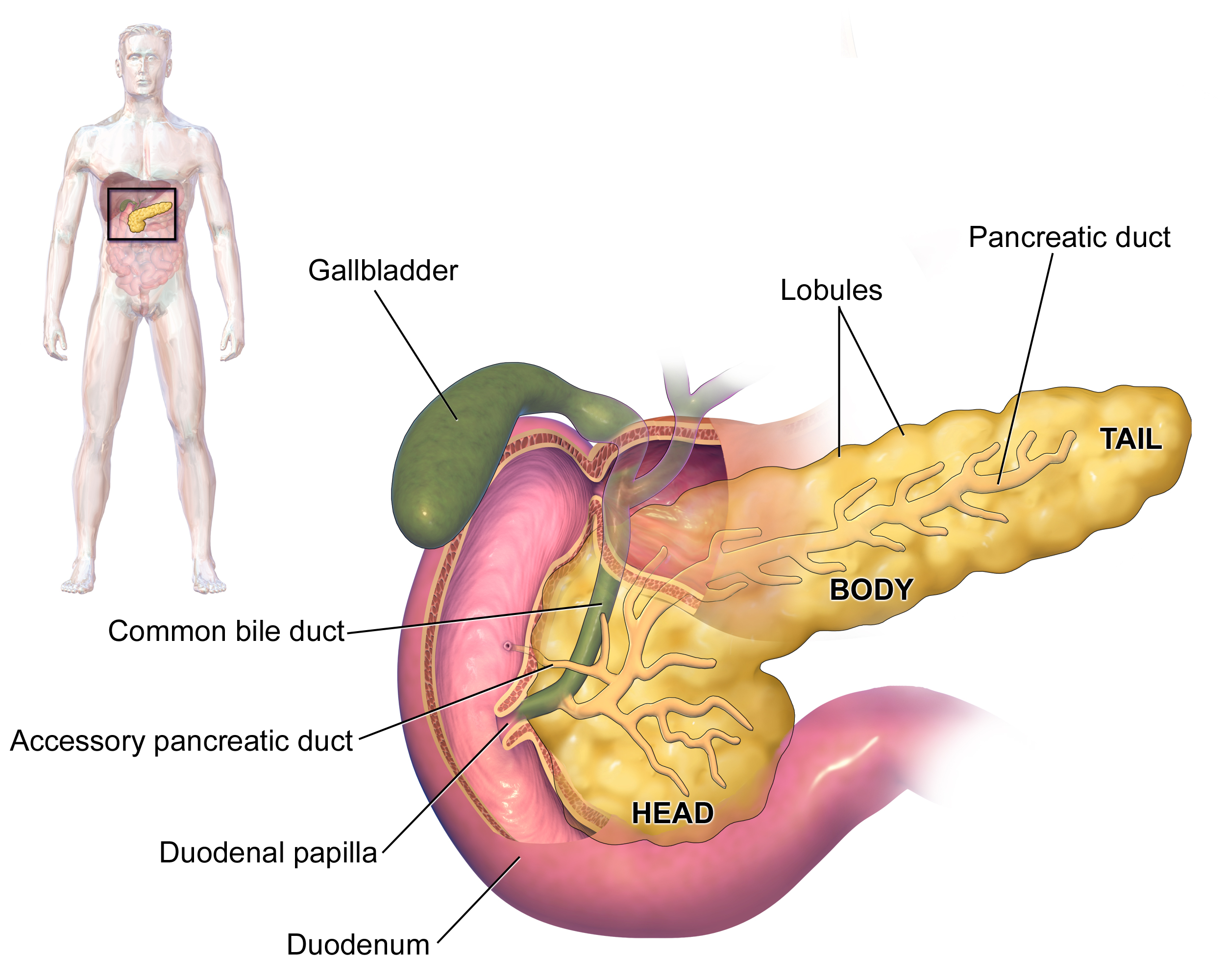
The pancreas is a long, slender organ, most of which is located in the upper left abdomen posterior to the bottom half of the stomach, as shown in the figure below. The pancreas is about 15 centimeters (6 in.) long; and it has a flat, oblong shape. Structurally, the pancreas is divided into a head, body, and tail. Functionally, the pancreas serves as both an endocrine gland and an exocrine gland.
- As an endocrine gland, the pancreas is part of the endocrine system. As such, it releases hormones, such as insulin, directly into the bloodstream for transport to cells throughout the body.
- As an exocrine gland, the pancreas is part of the digestive system. As such, it releases digestive enzymes into ducts that carry the enzymes to the gastrointestinal tract where they assist with digestion. In this concept, the focus is on the pancreas as an endocrine gland. You can read about the pancreas as an exocrine gland in the chapter Digestive System.
The Pancreas as an Endocrine Gland
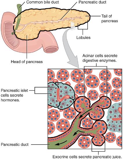
The tissues within the pancreas that have an endocrine role exist as clusters of cells called pancreatic islets. They are also called by the older term the islets of Langerhans. In Figure \(\PageIndex{3}\), you can see pancreatic tissue, including islets. There are approximately 3 million pancreatic islets, and they are crisscrossed by a dense network of capillaries. The capillaries are lined by layers of islet cells that have direct contact with the blood vessels, into which they secrete their endocrine hormones.
The pancreatic islets consist of four main types of cells, each of which secretes a different endocrine hormone. However, all of the hormones produced by the pancreatic islets play crucial roles in glucose metabolism and the regulation of blood glucose levels, among other functions.
- Islet cells called alpha (α) cells secrete the hormone glucagon; low blood glucose levels stimulate its release. The function of glucagon is to increase the level of glucose in the blood. It does this by stimulating the liver to convert stored glycogen into glucose, which is released into the bloodstream.
- Islet cells called beta (β) cells, which make up approximately 75 percent of each islet, secrete the hormone insulin. Elevated blood glucose levels stimulate the release of insulin. The function of insulin is to decrease the level of glucose in the blood. It does this by promoting the absorption of glucose from the blood into fat, liver, and skeletal muscle cells. In these tissues, the absorbed glucose is converted into glycogen, fats (triglycerides), or both.
- Islet cells called delta (δ) cells secrete the hormone somatostatin. This hormone is also called the growth hormone inhibiting hormone because it inhibits the anterior lobe of the pituitary gland from producing growth hormone. Somatostatin also inhibits the secretion of the pancreatic endocrine hormones glucagon and insulin and pancreatic exocrine enzymes.
- Islet cells called gamma (γ) cells secrete the hormone pancreatic polypeptide. The function of pancreatic polypeptide is to help regulate the secretion of both endocrine and exocrine substances by the pancreas. It is thought to play a role in appetite, as well as in the regulation of pancreatic exocrine and endocrine secretions. Pancreatic polypeptide released following a meal may reduce further food consumption; however, it is also released in response to fasting.
Regulation of Blood Glucose Levels by Insulin and Glucagon
Glucose is required for cellular respiration and is the preferred fuel for all body cells. The body derives glucose from the breakdown of the carbohydrate-containing foods and drinks we consume. Glucose not immediately taken up by cells for fuel can be stored by the liver and muscles as glycogen, or converted to triglycerides and stored in the adipose tissue. Hormones regulate both the storage and the utilization of glucose as required. Receptors located in the pancreas sense blood glucose levels, and subsequently the pancreatic cells secrete glucagon or insulin to maintain normal levels.
Glucagon
Receptors in the pancreas can sense the decline in blood glucose levels, such as during periods of fasting or during prolonged labor or exercise (Figure \(\PageIndex{4}\)). In response, the alpha cells of the pancreas secrete the hormone glucagon, which has several effects:
- It stimulates the liver to convert its stores of glycogen back into glucose. This response is known as glycogenolysis. The glucose is then released into the circulation for use by body cells.
- It stimulates the liver to take up amino acids from the blood and convert them into glucose. This response is known as gluconeogenesis.
- Taken together, these actions increase blood glucose levels. The activity of glucagon is regulated through a negative feedback mechanism; rising blood glucose levels inhibit further glucagon production and secretion.
Figure \(\PageIndex{4}\): Homeostatic Regulation of Blood Glucose Levels Blood glucose concentration is tightly maintained between 70 mg/dL and 110 mg/dL. If blood glucose concentration rises above this range, insulin is released, which stimulates body cells to remove glucose from the blood. If blood glucose concentration drops below this range, glucagon is released, which stimulates body cells to release glucose into the blood.
Insulin
The primary function of insulin is to facilitate the uptake of glucose into body cells. Red blood cells, as well as cells of the brain, liver, kidneys, and the lining of the small intestine, do not have insulin receptors on their cell membranes and do not require insulin for glucose uptake. Although all other body cells do require insulin if they are to take glucose from the bloodstream, skeletal muscle cells and adipose cells are the primary targets of insulin.
The presence of food in the intestine triggers the release of gastrointestinal tract hormones such as glucose-dependent insulinotropic peptide (previously known as gastric inhibitory peptide). This is in turn the initial trigger for insulin production and secretion by the beta cells of the pancreas. Once nutrient absorption occurs, the resulting surge in blood glucose levels further stimulates insulin secretion.
Precisely how insulin facilitates glucose uptake is not entirely clear. However, insulin appears to activate a tyrosine kinase receptor, triggering the phosphorylation of many substrates within the cell. These multiple biochemical reactions converge to support the movement of intracellular vesicles containing facilitative glucose transporters to the cell membrane. In the absence of insulin, these transport proteins are normally recycled slowly between the cell membrane and cell interior. Insulin triggers the rapid movement of a pool of glucose transporter vesicles to the cell membrane, where they fuse and expose the glucose transporters to the extracellular fluid. The transporters then move glucose by facilitated diffusion into the cell interior.
Insulin also reduces blood glucose levels by stimulating glycolysis, the metabolism of glucose for generation of ATP. Moreover, it stimulates the liver to convert excess glucose into glycogen for storage, and it inhibits enzymes involved in glycogenolysis and gluconeogenesis. Finally, insulin promotes triglyceride and protein synthesis. The secretion of insulin is regulated through a negative feedback mechanism. As blood glucose levels decrease, further insulin release is inhibited.
Hormones of the Pancreas
Associated hormones Chemical class Effect Insulin (beta cells) Protein Reduces blood glucose levels Glucagon (alpha cells) Protein Increases blood glucose levels Somatostatin (delta cells) Protein Inhibits insulin and glucagon release Pancreatic polypeptide (PP cells) Protein Role in appetite Table \(\PageIndex{1}\) The pancreatic hormones are summarized in Table \(\PageIndex{1}\).
Disorders of the Pancreas
There are a variety of disorders that affect the pancreas, including diabetes mellitus, pancreatitis, and pancreatic cancer.
Diabetes Mellitus
By far the most common type of pancreatic disorder is diabetes mellitus, more commonly called simply diabetes. It is caused by dysfunction of insulin production and secretion, as well as the target cells’ responsiveness to insulin. An increasingly common disease, diabetes mellitus has been diagnosed in more than 18 million adults in the United States, and more than 200,000 children. It is estimated that up to 7 million more adults have the condition but have not been diagnosed. In addition, approximately 79 million people in the US are estimated to have pre-diabetes, a condition in which blood glucose levels are abnormally high, but not yet high enough to be classified as diabetes.
There are several types of diabetes mellitus; the two major types are called type 1 diabetes and type 2 diabetes. The two types have different causes and may also have different treatments, but they generally produce the same initial symptoms, which include excessive urination and thirst. These symptoms occur because the kidneys excrete more urine in an attempt to rid the blood of excess glucose, and loss of water in urine stimulates greater thirst. Other signs and symptoms of diabetes are listed in Figure \(\PageIndex{5}\).

When diabetes is not well controlled, it is likely to have several serious long-term consequences. Most of these consequences are due to damage to small blood vessels because of high blood levels of glucose. Damage to blood vessels, in turn, may lead to an increased risk of coronary artery disease and stroke. Damage to blood vessels in the retina of the eye can result in gradual vision loss and blindness. Damage to blood vessels in the kidneys can lead to chronic kidney disease, sometimes requiring dialysis or a kidney transplant. Long-term consequences of diabetes may also include damage to the nerves of the body, known as diabetic neuropathy. In fact, this is the most common complication of diabetes. Symptoms of diabetic neuropathy may include numbness, tingling, and pain in the extremities.
Type 1 Diabetes
Type 1 diabetes is a chronic autoimmune disorder in which the immune system attacks the insulin-secreting beta cells of the pancreas. As a result, people with type 1 diabetes lack the insulin needed to keep blood glucose levels within the normal range. Type 1 diabetes may develop in people of any age but is most often diagnosed before adulthood. For type 1 diabetics, insulin injections are critical for survival. This form of diabetes accounts for less than five percent of all diabetes cases.
Type 2 Diabetes
Type 2 diabetes is the single most common form of diabetes, accounting for approximately 95 percent of all cases. It is acquired, and lifestyle factors such as poor diet, inactivity, and the presence of pre-diabetes greatly increase a person’s risk. About 80 to 90 percent of people with type 2 diabetes are overweight or obese. The cause of high blood glucose in this form of diabetes usually includes a combination of insulin resistance and impaired insulin secretion. In type 2 diabetes, cells become resistant to the effects of insulin. In response, the pancreas increases its insulin secretion, but over time, the beta cells become exhausted. In many cases, type 2 diabetes can be reversed by moderate weight loss, regular physical activity, and consumption of a healthy diet; however, if blood glucose levels cannot be controlled, the person with diabetes will eventually require insulin. Both genetic and environmental factors play roles in the development of type 2 diabetes. Management of type 2 diabetes includes changes in diet and physical activity, which may increase insulin sensitivity and help reduce blood glucose levels to normal ranges. Medications may also be used as part of the treatment, as may insulin injections.
Diagnosis, Symptoms, and Effects
Two of the early manifestations of diabetes are excessive urination and excessive thirst. They demonstrate how the out-of-control levels of glucose in the blood affect kidney function. The kidneys are responsible for filtering glucose from the blood. Excessive blood glucose draws water into the urine, and as a result the person eliminates an abnormally large quantity of sweet urine. The use of body water to dilute the urine leaves the body dehydrated, and so the person is unusually and continually thirsty. The person may also experience persistent hunger because the body cells are unable to access the glucose in the bloodstream.
Over time, persistently high levels of glucose in the blood injure tissues throughout the body, especially those of the blood vessels and nerves. Inflammation and injury of the lining of arteries lead to atherosclerosis and an increased risk of heart attack and stroke. Damage to the microscopic blood vessels of the kidney impairs kidney function and can lead to kidney failure. Damage to blood vessels that serve the eyes can lead to blindness. Blood vessel damage also reduces circulation to the limbs, whereas nerve damage leads to a loss of sensation, called neuropathy, particularly in the hands and feet. Together, these changes increase the risk of injury, infection, and tissue death (necrosis), contributing to a high rate of toe, foot, and lower leg amputations in people with diabetes. Uncontrolled diabetes can also lead to a dangerous form of metabolic acidosis called ketoacidosis. Deprived of glucose, cells increasingly rely on fat stores for fuel. However, in a glucose-deficient state, the liver is forced to use an alternative lipid metabolism pathway that results in the increased production of ketone bodies (or ketones), which are acidic. The build-up of ketones in the blood causes ketoacidosis, which—if left untreated—may lead to a life-threatening “diabetic coma.” Together, these complications make diabetes the seventh leading cause of death in the United States.
Diabetes is diagnosed when lab tests reveal that blood glucose levels are higher than normal, a condition called hyperglycemia. The treatment of diabetes depends on the type, the severity of the condition, and the ability of the patient to make lifestyle changes. As noted earlier, moderate weight loss, regular physical activity, and consumption of a healthful diet can reduce blood glucose levels. Some patients with type 2 diabetes may be unable to control their disease with these lifestyle changes, and will require medication. Historically, the first-line treatment of type 2 diabetes was insulin. Research advances have resulted in alternative options, including medications that enhance pancreatic function.
Some patients with type 1 diabetes have been given pancreatic islet cells transplants from other human donors. If the transplanted cells are not rejected by the recipient’s immune system, they can cure the patient of diabetes. However, only about 1,000 such surgeries have been performed over the past 10 years because of a shortage of appropriate human donors.
In June of 2016, a research team led by Dr. David K.C. Cooper at the Thomas E. Starzl Transplantation Institute in Pittsburgh, Pennsylvania, reported on their work developing pig islet cells for transplant into human diabetes patients. The researchers genetically engineered the pig islet cells to be protected from the human immune response. As a result, patients receiving transplanted cells would require only minimal suppression of their immune system after the surgery. The pig islet cells would also be less likely to transmit pathogenic agents because the animals could be raised in a controlled environment.
The researchers have successfully transplanted the pig islet cells into monkey models of type 1 diabetes. As of June 2016, the scientists were looking for funding to undertake clinical trials in humans with type 1 diabetes. Dr. Cooper predicted then that if the human trials go as well as expected, the pig islet cells could be available for curing patients in as little as two years.
Pancreatitis
Pancreatitis is inflammation of the pancreas. It has a variety of possible causes including gallstones, chronic alcohol use, infections such as measles or mumps, genetic causes, and certain medications. Pancreatitis occurs when digestive enzymes produced by the pancreas damage the gland’s tissues, which causes problems with fat digestion. The disorder is usually associated with intense pain in the central abdomen, and the pain may radiate to the back. Yellowing of the skin and whites of the eyes (Figure \(\PageIndex{6}\)), which is called jaundice, is a common sign of pancreatitis. People with pancreatitis may also have pale stools and dark urine. Treatment of pancreatitis includes administering drugs to manage pain and addressing the underlying cause of the disease, for example, by removing gallstones.

Pancreatic Cancer
There are several different types of pancreatic cancer that may affect either the endocrine or the exocrine tissues of the gland. Cancers affecting the endocrine tissues are all relatively rare. However, their incidence has been rising sharply. It is unclear to what extent this reflects increased detection, especially through medical imaging techniques. Unfortunately, pancreatic cancer is usually diagnosed at a relatively late stage when it is too late for surgery, which is the only way to cure the disorder. In the United States, pancreatic cancer is the fourth most common cause of death due to cancer.
Pancreatic cancer is rare before the age of 40 and occurs most often after the age of 60. Factors that increase the risk of developing pancreatic cancer include smoking, chronic pancreatitis, and diabetes. About one in four cases of pancreatic cancer are attributable to smoking. Certain rare genetic conditions are also risk factors for pancreatic cancer.
Review
- Describe the structure and location of the pancreas.
- Distinguish between the endocrine and exocrine functions of the pancreas.
- Identify the four types of pancreatic islet cells and the endocrine hormone each type of cell produces.
- What is pancreatitis? What are the possible causes and effects of pancreatitis?
- Describe the incidence, prognosis, and risk factors of cancer of the endocrine tissues of the pancreas.
- Compare and contrast type 1 and type 2 diabetes.
- If the alpha islet cells of the pancreas were damaged to the point that they no longer functioned, how would this affect blood glucose levels? Would the administration of insulin be more likely to help or hurt the condition? Explain your answer.
- Explain how the pancreas is able to regulate the production of its own endocrine hormones, to some extent.
- True or False. The pancreas is part of both the digestive system and the endocrine system.
- Give an example of how the pancreas can regulate the production of hormones from the pituitary gland.
- Which is the most common form of diabetes mellitus?
- Explain why diabetes causes excessive thirst.
- Damage to __________ is the underlying cause of many of the long-term consequences of diabetes.
- the adrenal gland
- gamma islet cells
- blood vessels
- the pituitary gland
Explore More
Attributions:
- Insulin Application by Mr. Hyde, public domain, via Czech Wikipedia
- Pancreas anatomy by Blausen.com staff (2014). "Medical gallery of Blausen Medical 2014". WikiJournal of Medicine 1 (2). DOI:10.15347/wjm/2014.010. ISSN 2002-4436. licensed CC BY 3.0 via Wikimedia Commons
- Exocrine and Endocrine Pancreas by OpenStax College, CC BY 3.0, via Wikimedia Commons
- Homeostatic Regulation of Blood Glucose by OpenStax licensed CC BY 4.0
- Symptoms of diabetes; licensed CC-0 via Wikimedia Commons
- Jaundice eye by CDC, public domain via Wikimedia Commons
- Text adapted from Human Biology by CK-12 licensed CC BY-NC 3.0
- Text adapted from Anatomy and Physiology e2 by OpenStax licensed CC BY 4.0.