
11.3: Neurons and Glial Cells
- Page ID
- 30694

\( \newcommand{\vecs}[1]{\overset { \scriptstyle \rightharpoonup} {\mathbf{#1}} } \)
\( \newcommand{\vecd}[1]{\overset{-\!-\!\rightharpoonup}{\vphantom{a}\smash {#1}}} \)
\( \newcommand{\id}{\mathrm{id}}\) \( \newcommand{\Span}{\mathrm{span}}\)
( \newcommand{\kernel}{\mathrm{null}\,}\) \( \newcommand{\range}{\mathrm{range}\,}\)
\( \newcommand{\RealPart}{\mathrm{Re}}\) \( \newcommand{\ImaginaryPart}{\mathrm{Im}}\)
\( \newcommand{\Argument}{\mathrm{Arg}}\) \( \newcommand{\norm}[1]{\| #1 \|}\)
\( \newcommand{\inner}[2]{\langle #1, #2 \rangle}\)
\( \newcommand{\Span}{\mathrm{span}}\)
\( \newcommand{\id}{\mathrm{id}}\)
\( \newcommand{\Span}{\mathrm{span}}\)
\( \newcommand{\kernel}{\mathrm{null}\,}\)
\( \newcommand{\range}{\mathrm{range}\,}\)
\( \newcommand{\RealPart}{\mathrm{Re}}\)
\( \newcommand{\ImaginaryPart}{\mathrm{Im}}\)
\( \newcommand{\Argument}{\mathrm{Arg}}\)
\( \newcommand{\norm}[1]{\| #1 \|}\)
\( \newcommand{\inner}[2]{\langle #1, #2 \rangle}\)
\( \newcommand{\Span}{\mathrm{span}}\) \( \newcommand{\AA}{\unicode[.8,0]{x212B}}\)
\( \newcommand{\vectorA}[1]{\vec{#1}} % arrow\)
\( \newcommand{\vectorAt}[1]{\vec{\text{#1}}} % arrow\)
\( \newcommand{\vectorB}[1]{\overset { \scriptstyle \rightharpoonup} {\mathbf{#1}} } \)
\( \newcommand{\vectorC}[1]{\textbf{#1}} \)
\( \newcommand{\vectorD}[1]{\overrightarrow{#1}} \)
\( \newcommand{\vectorDt}[1]{\overrightarrow{\text{#1}}} \)
\( \newcommand{\vectE}[1]{\overset{-\!-\!\rightharpoonup}{\vphantom{a}\smash{\mathbf {#1}}}} \)
\( \newcommand{\vecs}[1]{\overset { \scriptstyle \rightharpoonup} {\mathbf{#1}} } \)
\( \newcommand{\vecd}[1]{\overset{-\!-\!\rightharpoonup}{\vphantom{a}\smash {#1}}} \)
\(\newcommand{\avec}{\mathbf a}\) \(\newcommand{\bvec}{\mathbf b}\) \(\newcommand{\cvec}{\mathbf c}\) \(\newcommand{\dvec}{\mathbf d}\) \(\newcommand{\dtil}{\widetilde{\mathbf d}}\) \(\newcommand{\evec}{\mathbf e}\) \(\newcommand{\fvec}{\mathbf f}\) \(\newcommand{\nvec}{\mathbf n}\) \(\newcommand{\pvec}{\mathbf p}\) \(\newcommand{\qvec}{\mathbf q}\) \(\newcommand{\svec}{\mathbf s}\) \(\newcommand{\tvec}{\mathbf t}\) \(\newcommand{\uvec}{\mathbf u}\) \(\newcommand{\vvec}{\mathbf v}\) \(\newcommand{\wvec}{\mathbf w}\) \(\newcommand{\xvec}{\mathbf x}\) \(\newcommand{\yvec}{\mathbf y}\) \(\newcommand{\zvec}{\mathbf z}\) \(\newcommand{\rvec}{\mathbf r}\) \(\newcommand{\mvec}{\mathbf m}\) \(\newcommand{\zerovec}{\mathbf 0}\) \(\newcommand{\onevec}{\mathbf 1}\) \(\newcommand{\real}{\mathbb R}\) \(\newcommand{\twovec}[2]{\left[\begin{array}{r}#1 \\ #2 \end{array}\right]}\) \(\newcommand{\ctwovec}[2]{\left[\begin{array}{c}#1 \\ #2 \end{array}\right]}\) \(\newcommand{\threevec}[3]{\left[\begin{array}{r}#1 \\ #2 \\ #3 \end{array}\right]}\) \(\newcommand{\cthreevec}[3]{\left[\begin{array}{c}#1 \\ #2 \\ #3 \end{array}\right]}\) \(\newcommand{\fourvec}[4]{\left[\begin{array}{r}#1 \\ #2 \\ #3 \\ #4 \end{array}\right]}\) \(\newcommand{\cfourvec}[4]{\left[\begin{array}{c}#1 \\ #2 \\ #3 \\ #4 \end{array}\right]}\) \(\newcommand{\fivevec}[5]{\left[\begin{array}{r}#1 \\ #2 \\ #3 \\ #4 \\ #5 \\ \end{array}\right]}\) \(\newcommand{\cfivevec}[5]{\left[\begin{array}{c}#1 \\ #2 \\ #3 \\ #4 \\ #5 \\ \end{array}\right]}\) \(\newcommand{\mattwo}[4]{\left[\begin{array}{rr}#1 \amp #2 \\ #3 \amp #4 \\ \end{array}\right]}\) \(\newcommand{\laspan}[1]{\text{Span}\{#1\}}\) \(\newcommand{\bcal}{\cal B}\) \(\newcommand{\ccal}{\cal C}\) \(\newcommand{\scal}{\cal S}\) \(\newcommand{\wcal}{\cal W}\) \(\newcommand{\ecal}{\cal E}\) \(\newcommand{\coords}[2]{\left\{#1\right\}_{#2}}\) \(\newcommand{\gray}[1]{\color{gray}{#1}}\) \(\newcommand{\lgray}[1]{\color{lightgray}{#1}}\) \(\newcommand{\rank}{\operatorname{rank}}\) \(\newcommand{\row}{\text{Row}}\) \(\newcommand{\col}{\text{Col}}\) \(\renewcommand{\row}{\text{Row}}\) \(\newcommand{\nul}{\text{Nul}}\) \(\newcommand{\var}{\text{Var}}\) \(\newcommand{\corr}{\text{corr}}\) \(\newcommand{\len}[1]{\left|#1\right|}\) \(\newcommand{\bbar}{\overline{\bvec}}\) \(\newcommand{\bhat}{\widehat{\bvec}}\) \(\newcommand{\bperp}{\bvec^\perp}\) \(\newcommand{\xhat}{\widehat{\xvec}}\) \(\newcommand{\vhat}{\widehat{\vvec}}\) \(\newcommand{\uhat}{\widehat{\uvec}}\) \(\newcommand{\what}{\widehat{\wvec}}\) \(\newcommand{\Sighat}{\widehat{\Sigma}}\) \(\newcommand{\lt}{<}\) \(\newcommand{\gt}{>}\) \(\newcommand{\amp}{&}\) \(\definecolor{fillinmathshade}{gray}{0.9}\)This colorful picture could be an abstract work of modern art. You might imagine it hanging in an art museum or art gallery. In fact, the picture illustrates real life rather than an artistic creation. It is a micrograph of human nervous tissue. The neon green structures in the picture are neurons. The neuron is one of two basic types of cells in the nervous system, the other type being the glial cell.
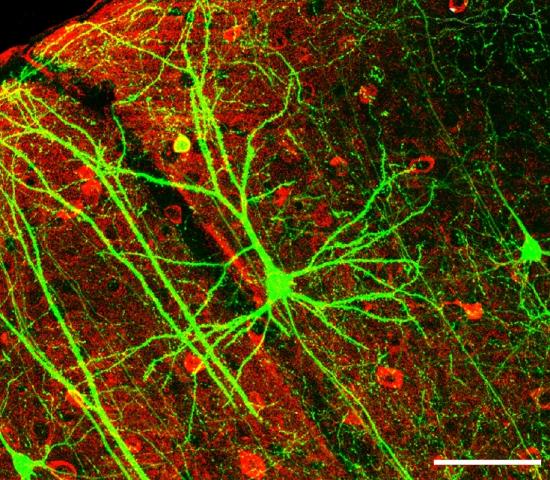
Neurons, also called nerve cells, are electrically excitable cells that are the main functional units of the nervous system. Their function is to transmit nerve impulses. They are the only type of human cells that can carry out this function.
Neuron Structure
Figure \(\PageIndex{2}\) shows the structure of a typical neuron. The main parts of a neuron are labeled in the figure and described below.
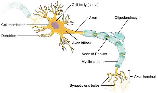
Figure \(\PageIndex{2}\): Somatic Motor Neuron with cell body, axon, axon, myelin sheath, nodes of Ranvier, axon terminal, dendrites, synaptic end of the bulbs, and other associated structures.
- The cell body is the part of a neuron that contains the cell nucleus and other cell organelles. It is usually quite compact, and may not be much wider than the nucleus.
- Dendrites are thin structures that are extensions of the cell body. Their function is to receive nerve impulses from other cells and carry them to the cell body. A neuron may have many dendrites, and each dendrite may branch repeatedly to form a dendrite “tree” with more than 1,000 “branches.” The end of each branch can receive nerve impulses from another cell, allowing a given neuron to communicate with tens of thousands of other cells.
- The axon is a long, thin extension of the cell body. It transmits nerve impulses away from the cell body and toward other cells. The axon branches at the end, forming multiple axon terminals. These are the points where nerve impulses are transmitted to other cells, often to the dendrites of other neurons. An area called a synapse occurs at each axon terminal. Synapses are complex membrane junctions that transmit signals to other cells. An axon may branch hundreds of times, but there is never more than one axon per neuron.
- Spread out along axons, especially the long axons of nerves, are many sections of the myelin sheath. These are lipid layers that cover sections of the axon. The myelin sheath is a very good electrical insulator, similar to the plastic or rubber that encases an electrical cord.
- Regularly spaced gaps between sections of myelin sheath occur along the axon. These gaps are called nodes of Ranvier, and they allow the transmission of nerve impulses along the axon. Nerve impulses skip from node to node, allowing nerve impulses to travel along the axon very rapidly.
- A Schwann cell (also on an axon) is a type of glial cell. Its function is to produce the myelin sheath that insulates axons in the peripheral nervous system. In the central nervous system, a different type of glial cell, called an oligodendrocyte, produces the myelin sheath.
Neurogenesis
Fully differentiated neurons, with all their special structures, cannot divide and form new daughter neurons. Until recently, scientists thought that new neurons could no longer be formed after the brain developed prenatally. In other words, they thought that people were born with all the brain neurons they would ever have, and as neurons died, they would not be replaced. However, new evidence shows that additional neurons can form in the brain, even in adults, from the division of undifferentiated neural stem cells that are found throughout the brain. The production of new neurons is called neurogenesis. The extent to which it can occur is not known, but it is not likely to be very great in humans.
Neurons in Nervous Tissues
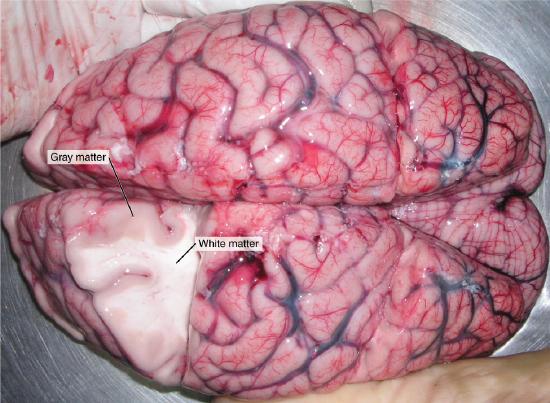
The nervous tissue in the brain and spinal cord consists of gray matter and white matter. Gray matter contains mainly the cell bodies of neurons. It is gray only in cadavers; living gray matter is actually more pink than gray (see image below). White matter consists mainly of axons covered with myelin sheath, which gives them their white color. White matter also makes up nerves of the peripheral nervous system. Nerves consist of long bundles of myelinated axons that extend to muscles, organs, or glands throughout the body. The axons in each nerve are bundled together like wires in a cable. Axons in nerves may be more than a meter long in an adult. The longest nerve runs from the base of the spine to the toes.
Types of Neurons
There are hundreds of different types of neurons in the human nervous system. These types exhibit a variety of structures and functions. Nonetheless, many neurons can be classified functionally based on the direction in which they carry nerve impulses.

- Sensory (also called afferent) neurons carry nerve impulses from sensory receptors in tissues and organs to the central nervous system. They change physical stimuli such as touch, light, and sound into nerve impulses.
- Motor (also called efferent) neurons, like the one in figure \(\PageIndex{2}\), carry nerve impulses from the central nervous system to muscles and glands. They change nerve signals into the activation of these structures.
- Interneurons carry nerve impulses back and forth often between sensory and motor neurons within the spinal cord or brain.
Glial Cells
Besides neurons, nervous tissues also consist of glial cells (also called neuroglia). They are now known to play many vital roles in the nervous system. There are several different types of glial cells, each with a different function. Schwann cells and Oligodendrocytes are glial cells that produce myelin sheath.
Would you like your brain to make new neurons that could help you become a better learner? What college student wouldn’t want a little more brainpower when it comes to learning new things? If research on rats applies to humans, then sustained aerobic exercise such as running can increase neurogenesis in the adult brain, and specifically in the hippocampus, a brain structure important for learning temporally and/or spatially complex tasks as well as memory. Although the research is still at the beginning stages, it suggests that exercise may actually lead to a “smarter” brain. However, even if the research results are not confirmed in the future for humans, it can’t hurt to get more aerobic exercise, because it is certainly beneficial for your body if not for your brain.
Review
- Identify the three main parts of a neuron and their functions.
- Describe the myelin sheath and nodes of Ranvier. How does their arrangement allow nerve impulses to travel very rapidly along axons?
- What is a synapse?
- Define neurogenesis. What is the potential for neurogenesis in the human brain?
- Relate neurons to different types of nervous tissues.
- Compare and contrast sensory and motor neurons.
- Identify the role of interneurons.
- For each type of neuron below, identify whether it is a sensory neuron, motor neuron, or interneuron.
- A neuron in the spinal cord receives touch information and then transmits that information to another spinal cord neuron that controls the movement of an arm muscle.
- A neuron that takes taste information from your tongue and sends it to your brain.
- A spinal cord neuron stimulates a muscle to contract.
- The myelin sheath is made by:
- Sensory neurons
- White neurons
- Peripheral nervous system neurons
- Glial cells
- True or False. Synapses often exist where a dendrite and an axon terminal meet.
- True or False. There is only one axon terminal per neuron.
Explore More
Multiple sclerosis (MS) is a progressive degenerative disease that is caused by the demyelination of axons in the central nervous system. When myelin degrades, the conduction of nerve impulses along the nerve can be impaired or lost, and the nerve eventually withers. Watch this inspirational TED talk in which the speaker shares how being diagnosed with MS changed her life and led her to become an MS nurse.
After his death in 1955, Albert Einstein's brain was studied by scientists worldwide—all wanting to gain insight into the anatomy of a genius. But it wasn't until the 1980s when Marian Diamond noticed that Einstein had more glial cells than average. Glia, stemming from Greek for "glue", was previously thought to have performed a strictly support role for the neurons. Now it is clear that glia may play a more active, non-electrical role in brain activity.
Attributions
- Interneurons of Adult Visual Cortex by Wei-Chung Allen Lee, Hayden Huang, Guoping Feng, Joshua R. Sanes, Emery N. Brown, Peter T. So, Elly Nedivi, licensed CC BY 2.5 via Wikimedia Commons
- Neuron by Chiara Mazzasette adapted from OpenStax, licensed CC BY 4.0 via Wikimedia Commons
- White and gray matter by OpenStax, licensed CC BY 4.0 via Wikimedia Commons
- Sensory Neuron Test Water by OpenStax, licensed CC BY 4.0 via Wikimedia Commons
- Text adapted from Human Biology by CK-12 licensed CC BY-NC 3.0