Biology LibreTexts

15.2: Atlas- Fruits

  • Page ID
    149602
  • \( \newcommand{\vecs}[1]{\overset { \scriptstyle \rightharpoonup} {\mathbf{#1}} } \)

    \( \newcommand{\vecd}[1]{\overset{-\!-\!\rightharpoonup}{\vphantom{a}\smash {#1}}} \)

    \( \newcommand{\id}{\mathrm{id}}\) \( \newcommand{\Span}{\mathrm{span}}\)

    ( \newcommand{\kernel}{\mathrm{null}\,}\) \( \newcommand{\range}{\mathrm{range}\,}\)

    \( \newcommand{\RealPart}{\mathrm{Re}}\) \( \newcommand{\ImaginaryPart}{\mathrm{Im}}\)

    \( \newcommand{\Argument}{\mathrm{Arg}}\) \( \newcommand{\norm}[1]{\| #1 \|}\)

    \( \newcommand{\inner}[2]{\langle #1, #2 \rangle}\)

    \( \newcommand{\Span}{\mathrm{span}}\)

    \( \newcommand{\id}{\mathrm{id}}\)

    \( \newcommand{\Span}{\mathrm{span}}\)

    \( \newcommand{\kernel}{\mathrm{null}\,}\)

    \( \newcommand{\range}{\mathrm{range}\,}\)

    \( \newcommand{\RealPart}{\mathrm{Re}}\)

    \( \newcommand{\ImaginaryPart}{\mathrm{Im}}\)

    \( \newcommand{\Argument}{\mathrm{Arg}}\)

    \( \newcommand{\norm}[1]{\| #1 \|}\)

    \( \newcommand{\inner}[2]{\langle #1, #2 \rangle}\)

    \( \newcommand{\Span}{\mathrm{span}}\) \( \newcommand{\AA}{\unicode[.8,0]{x212B}}\)

    \( \newcommand{\vectorA}[1]{\vec{#1}}      % arrow\)

    \( \newcommand{\vectorAt}[1]{\vec{\text{#1}}}      % arrow\)

    \( \newcommand{\vectorB}[1]{\overset { \scriptstyle \rightharpoonup} {\mathbf{#1}} } \)

    \( \newcommand{\vectorC}[1]{\textbf{#1}} \)

    \( \newcommand{\vectorD}[1]{\overrightarrow{#1}} \)

    \( \newcommand{\vectorDt}[1]{\overrightarrow{\text{#1}}} \)

    \( \newcommand{\vectE}[1]{\overset{-\!-\!\rightharpoonup}{\vphantom{a}\smash{\mathbf {#1}}}} \)

    \( \newcommand{\vecs}[1]{\overset { \scriptstyle \rightharpoonup} {\mathbf{#1}} } \)

    \( \newcommand{\vecd}[1]{\overset{-\!-\!\rightharpoonup}{\vphantom{a}\smash {#1}}} \)

    \(\newcommand{\avec}{\mathbf a}\) \(\newcommand{\bvec}{\mathbf b}\) \(\newcommand{\cvec}{\mathbf c}\) \(\newcommand{\dvec}{\mathbf d}\) \(\newcommand{\dtil}{\widetilde{\mathbf d}}\) \(\newcommand{\evec}{\mathbf e}\) \(\newcommand{\fvec}{\mathbf f}\) \(\newcommand{\nvec}{\mathbf n}\) \(\newcommand{\pvec}{\mathbf p}\) \(\newcommand{\qvec}{\mathbf q}\) \(\newcommand{\svec}{\mathbf s}\) \(\newcommand{\tvec}{\mathbf t}\) \(\newcommand{\uvec}{\mathbf u}\) \(\newcommand{\vvec}{\mathbf v}\) \(\newcommand{\wvec}{\mathbf w}\) \(\newcommand{\xvec}{\mathbf x}\) \(\newcommand{\yvec}{\mathbf y}\) \(\newcommand{\zvec}{\mathbf z}\) \(\newcommand{\rvec}{\mathbf r}\) \(\newcommand{\mvec}{\mathbf m}\) \(\newcommand{\zerovec}{\mathbf 0}\) \(\newcommand{\onevec}{\mathbf 1}\) \(\newcommand{\real}{\mathbb R}\) \(\newcommand{\twovec}[2]{\left[\begin{array}{r}#1 \\ #2 \end{array}\right]}\) \(\newcommand{\ctwovec}[2]{\left[\begin{array}{c}#1 \\ #2 \end{array}\right]}\) \(\newcommand{\threevec}[3]{\left[\begin{array}{r}#1 \\ #2 \\ #3 \end{array}\right]}\) \(\newcommand{\cthreevec}[3]{\left[\begin{array}{c}#1 \\ #2 \\ #3 \end{array}\right]}\) \(\newcommand{\fourvec}[4]{\left[\begin{array}{r}#1 \\ #2 \\ #3 \\ #4 \end{array}\right]}\) \(\newcommand{\cfourvec}[4]{\left[\begin{array}{c}#1 \\ #2 \\ #3 \\ #4 \end{array}\right]}\) \(\newcommand{\fivevec}[5]{\left[\begin{array}{r}#1 \\ #2 \\ #3 \\ #4 \\ #5 \\ \end{array}\right]}\) \(\newcommand{\cfivevec}[5]{\left[\begin{array}{c}#1 \\ #2 \\ #3 \\ #4 \\ #5 \\ \end{array}\right]}\) \(\newcommand{\mattwo}[4]{\left[\begin{array}{rr}#1 \amp #2 \\ #3 \amp #4 \\ \end{array}\right]}\) \(\newcommand{\laspan}[1]{\text{Span}\{#1\}}\) \(\newcommand{\bcal}{\cal B}\) \(\newcommand{\ccal}{\cal C}\) \(\newcommand{\scal}{\cal S}\) \(\newcommand{\wcal}{\cal W}\) \(\newcommand{\ecal}{\cal E}\) \(\newcommand{\coords}[2]{\left\{#1\right\}_{#2}}\) \(\newcommand{\gray}[1]{\color{gray}{#1}}\) \(\newcommand{\lgray}[1]{\color{lightgray}{#1}}\) \(\newcommand{\rank}{\operatorname{rank}}\) \(\newcommand{\row}{\text{Row}}\) \(\newcommand{\col}{\text{Col}}\) \(\renewcommand{\row}{\text{Row}}\) \(\newcommand{\nul}{\text{Nul}}\) \(\newcommand{\var}{\text{Var}}\) \(\newcommand{\corr}{\text{corr}}\) \(\newcommand{\len}[1]{\left|#1\right|}\) \(\newcommand{\bbar}{\overline{\bvec}}\) \(\newcommand{\bhat}{\widehat{\bvec}}\) \(\newcommand{\bperp}{\bvec^\perp}\) \(\newcommand{\xhat}{\widehat{\xvec}}\) \(\newcommand{\vhat}{\widehat{\vvec}}\) \(\newcommand{\uhat}{\widehat{\uvec}}\) \(\newcommand{\what}{\widehat{\wvec}}\) \(\newcommand{\Sighat}{\widehat{\Sigma}}\) \(\newcommand{\lt}{<}\) \(\newcommand{\gt}{>}\) \(\newcommand{\amp}{&}\) \(\definecolor{fillinmathshade}{gray}{0.9}\)

    Ovary Anatomy

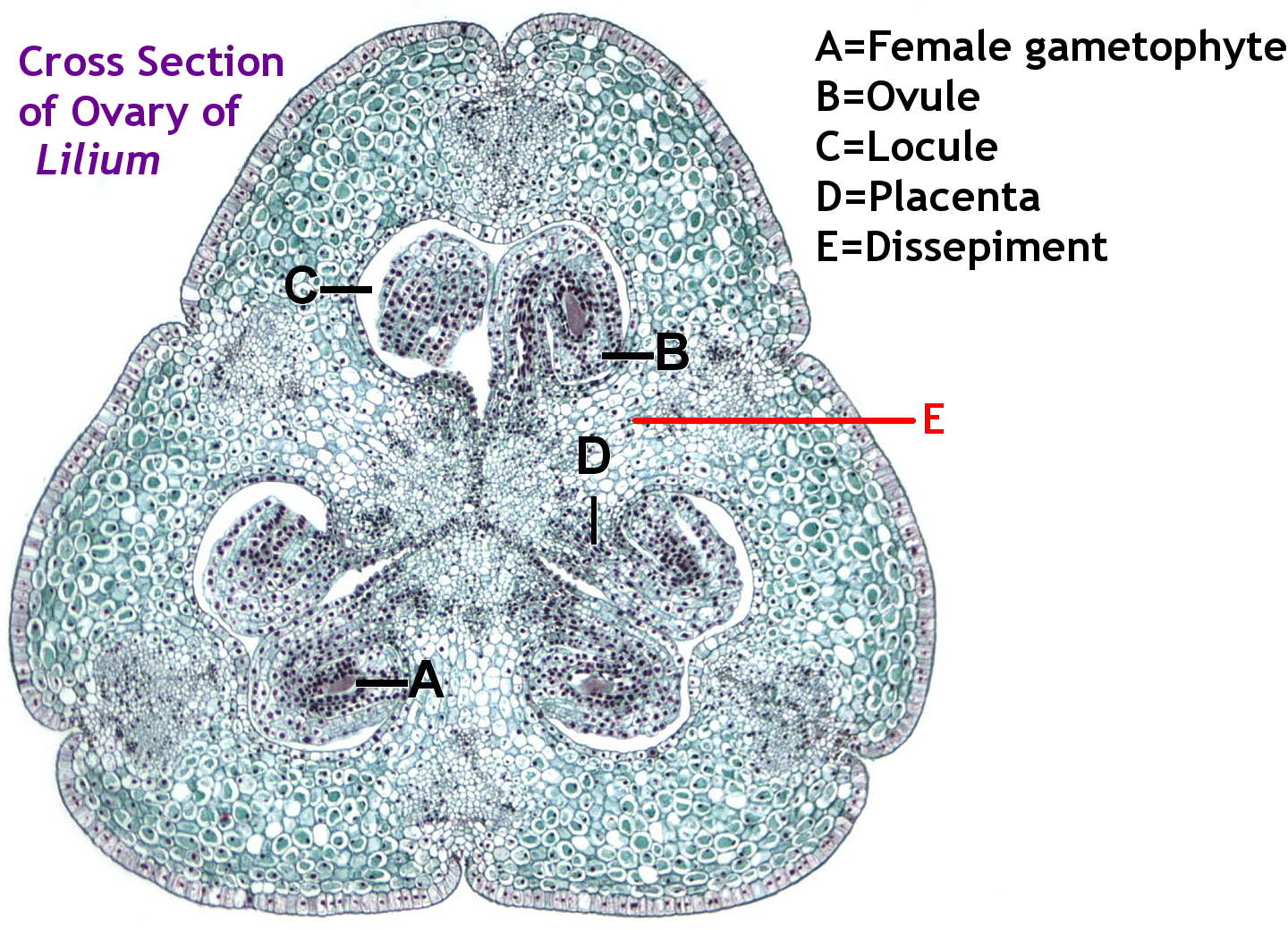
    Lilium ovary cross section
    Figure \(\PageIndex{1}\): Cross section of Lilium ovary, showing A=Female gametophyte, B=Ovule, C=Locule, D=Placenta, E=Dissepiment. The label in red added to original contribution of File:Lilium ovary L.jpg provided by Jon Houseman and Matthew Ford. JonRichfield, CC BY-SA 4.0, via Wikimedia Commons.
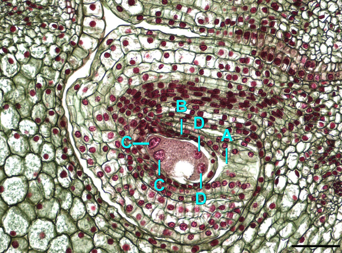
    A cross section of a Lilium ovary, showing just the ovule
    Figure \(\PageIndex{2}\): A Lilium ovary during the second stage of the four nucleate embryo. A-Micropyle, B-Integument, C-Antipodal cells, D-Synergid cells. Scale=0.1mm. Jon Houseman, CC BY-SA 4.0, via Wikimedia Commons.
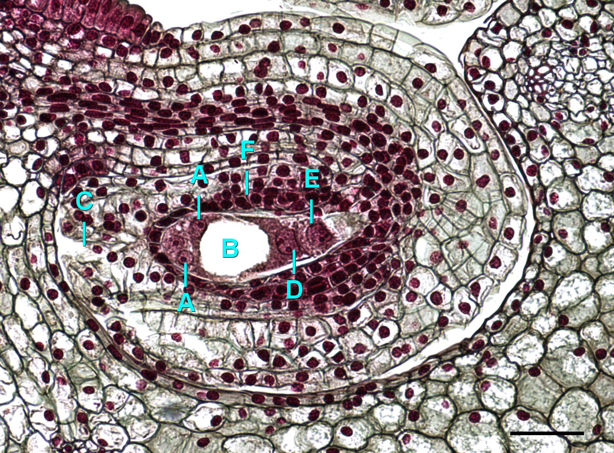
    A cross section of a Lilium ovary, showing just the ovule, at a later stage of development
    Figure \(\PageIndex{3}\): A Lilium ovary at the eight nucleate stage. A-Synergid cells, B-Vacuole, C-Micropyle, D-Polar nucleus, E-Antipodal cell, F-Integument. Scale=0.1mm. Jon Houseman, CC BY-SA 4.0, via Wikimedia Commons.

    Transitioning from Flower to Fruit

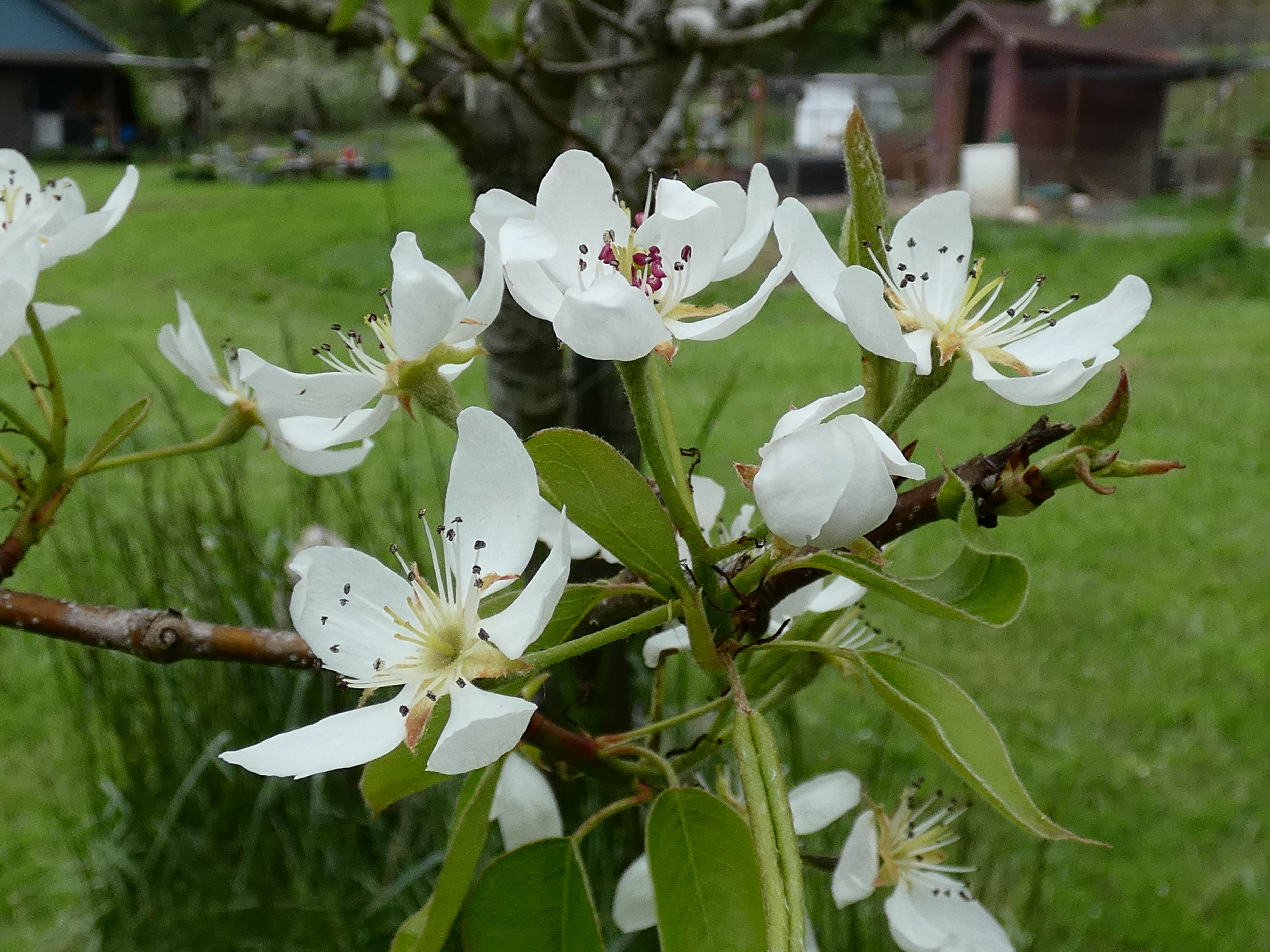
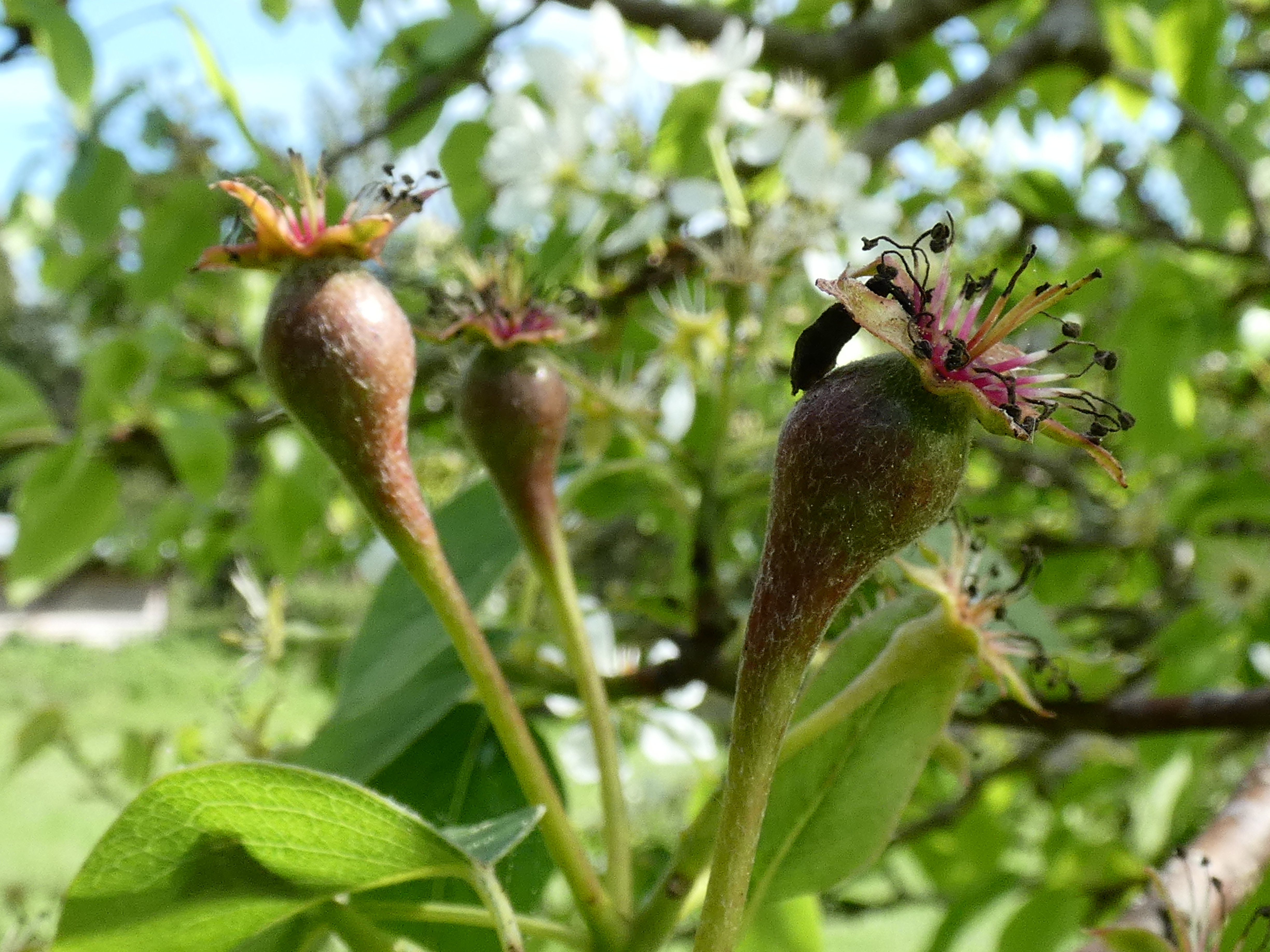
    A cluster of pear blossoms A cluster of pear blossoms from the same tree that has lost its perianth and the ovaries are beginning to swell
    Figure \(\PageIndex{4}\): These two images show clusters of pear blossoms from the same tree. The cluster on the left is in an earlier stage of development. The corolla is still attached and the stamens are radiating outward to disperse pollen. In the image on the right, the corolla has fallen off and the anthers have shriveled. The calyx is still visible and, just below it, the semi-inferior ovary and hypanthium are beginning to swell. Eventually, these will swell to many times their current size and form the fruits we know as pears. Photos by Maria Morrow, CC-BY 4.0.
    Developing salal berries
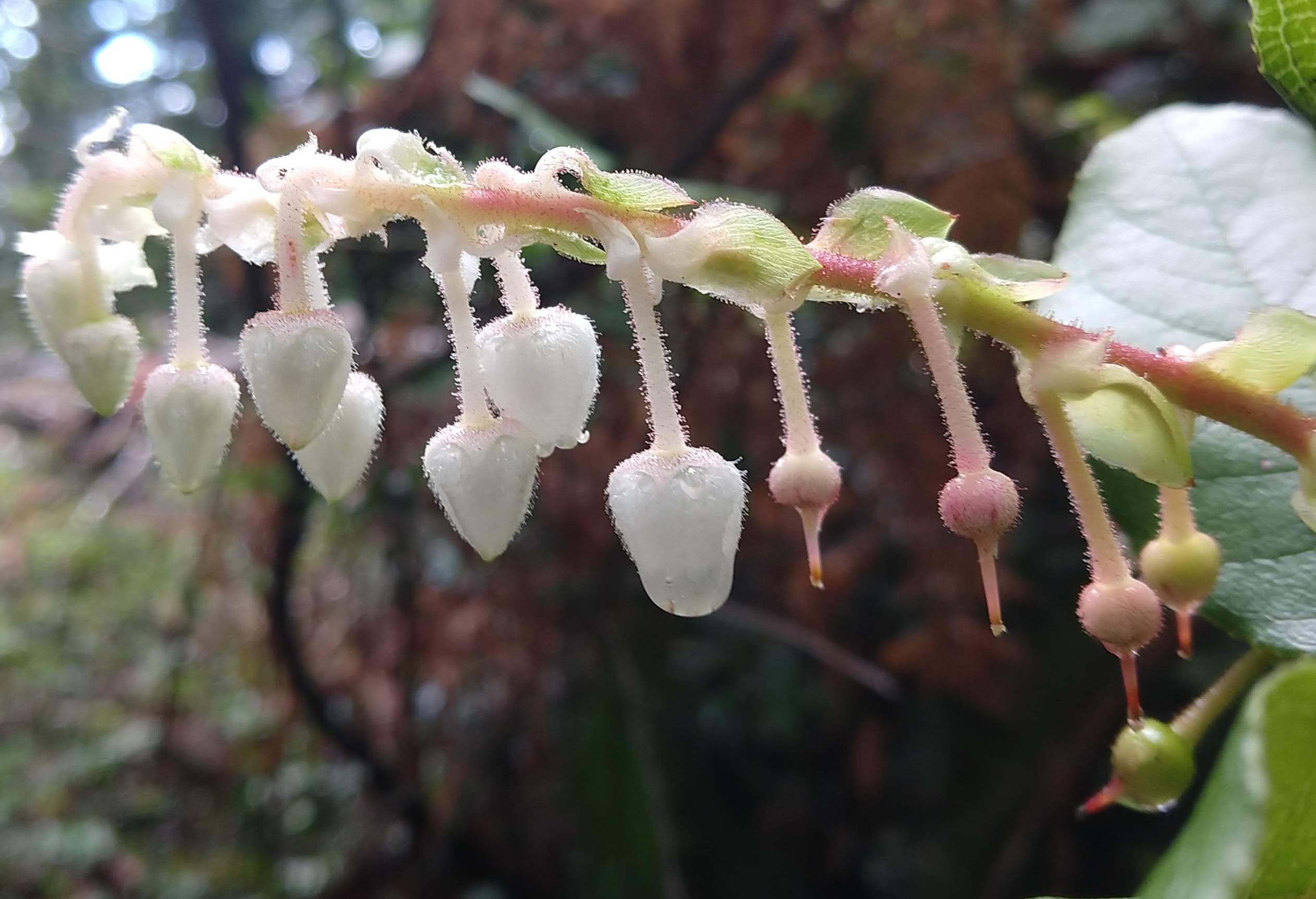
    Figure \(\PageIndex{5}\): Salal (Gaultheria shallon) is a shrub in the same family as blueberries and madrone trees. Each pendant, urn-shaped corolla will fall off, leaving the calyx behind. The inferior ovary will develop into a berry. In this image, younger flowers are being produced on the left side at the end of the inflorescence, while older flowers develop into fruits on the right side. Photo by Maria Morrow, CC-BY 4.0.

    Fruit Anatomy

    A longitudinal section of a corn kernel showing the growing embryo
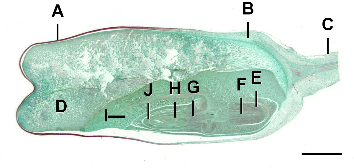
    Figure \(\PageIndex{6}\): A Zea mays kernel. This fruit is a caryopsis, the seed coat is fused to the pericarp. Labels are as follows: A=Pericarp, B=Aleurone, C=Tip cap, D=Endosperm, E=Coleorhiza, F=Radicle, G=Hypocotyl, H=Plumule, I=Scutellum, J=Coleoptile. Scale=1.4mm. Jon Houseman, CC BY-SA 4.0, via Wikimedia Commons.
    Layers of pericarp in a peach
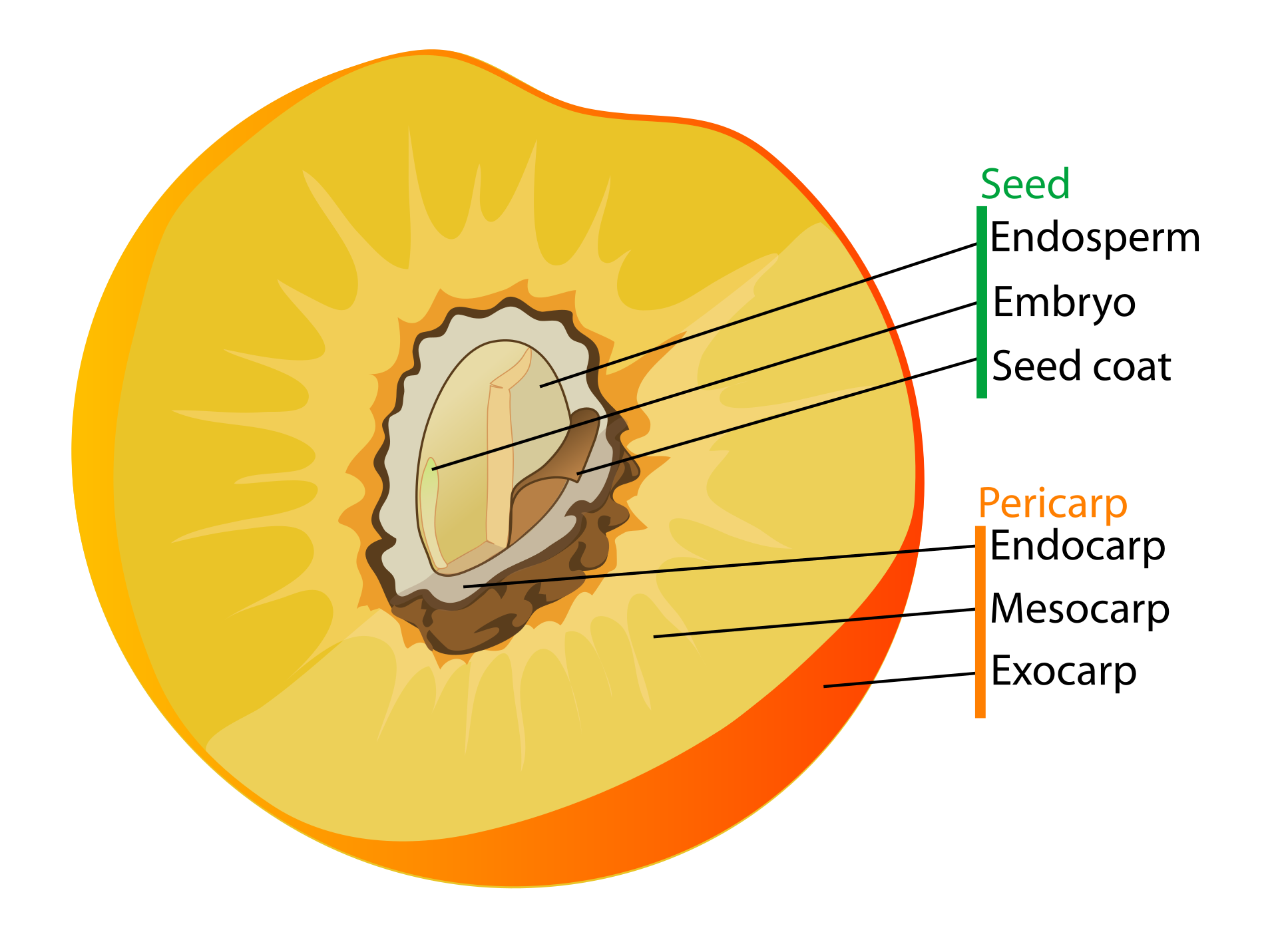
    Figure \(\PageIndex{7}\): A diagram of a drupe. The pericarp of a fruit can be completely fused (as in Figure \(\PageIndex{7}\)) or separated into distinct layers: the endocarp (innermost), mesocarp (middle), and exocarp (outermost). A drupe is a fruit type with a stony endocarp enclosing a single seed, a fleshy mesocarp, and a thin exocarp. Image by LadyofHats, Public domain, via Wikimedia Commons.

    Some Common Fruit Types

    Achene

    Dandelion fruits produced from a head inflorescence Dandelion fruits up close, showing the achene and the modified calyx
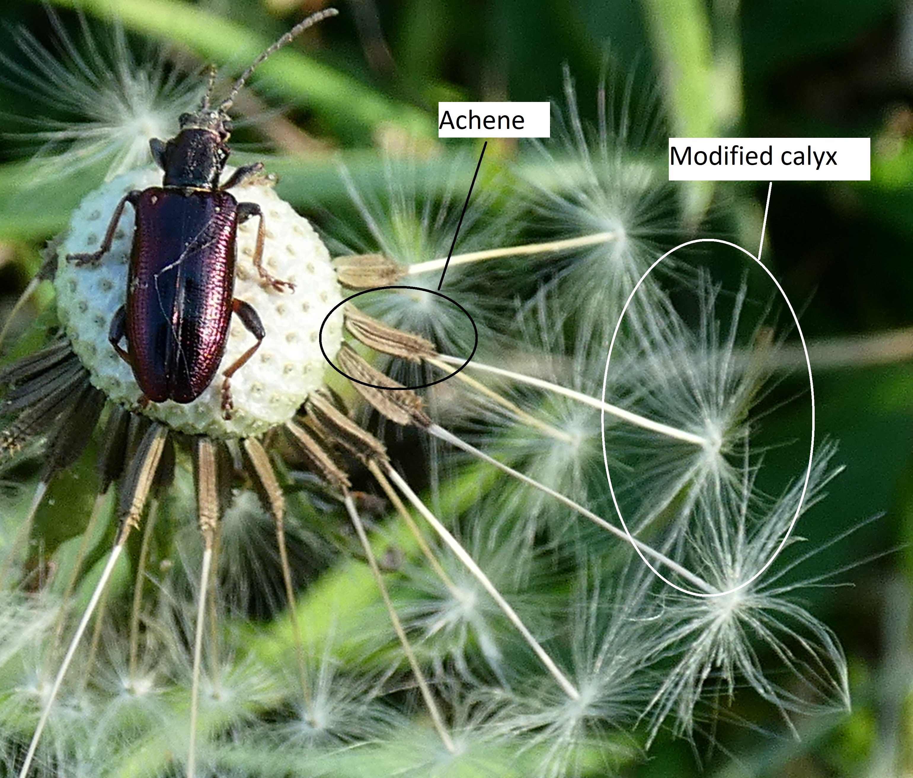
    Figure \(\PageIndex{8}\): These two images show the fruits produced from the head inflorescence of a dandelion. On the left, they are difficult to distinguish, forming a sphere around the receptacle. On the right, some of the fruits have been carried away on the wind, making the others easier to see. Each floret has produced an achene at the base (from an inferior ovary). Only the calyx remains, of the other floral whorls, and has formed into an umbrella-like structure for wind dispersal. Photos by Maria Morrow, CC-BY 4.0.

    Berries

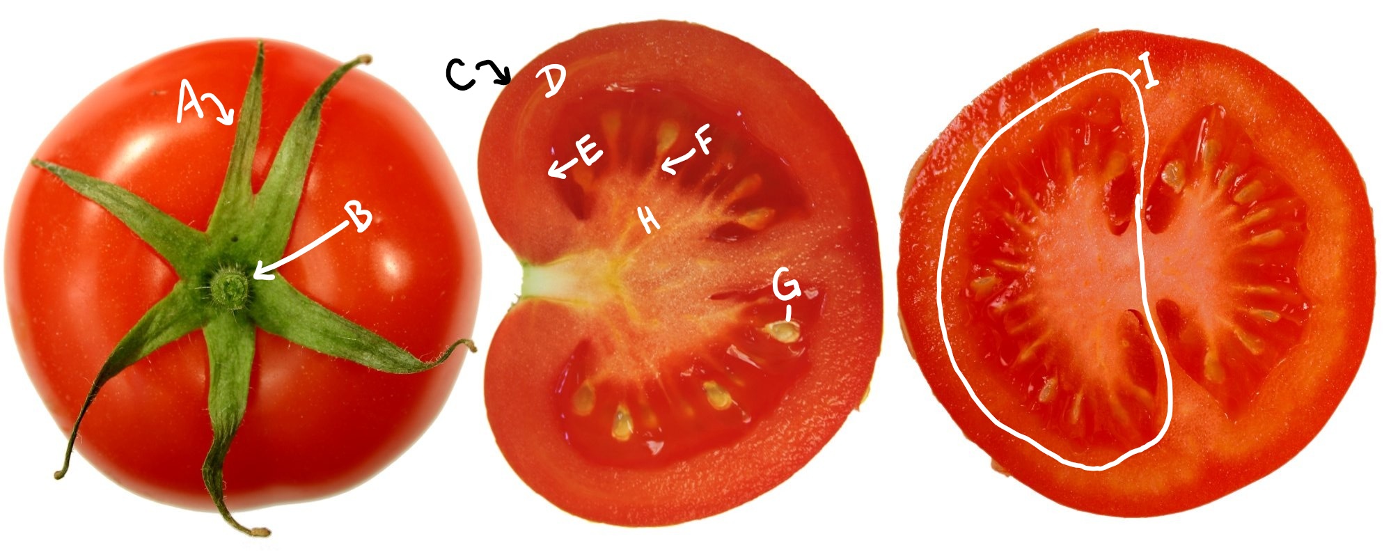
    Three tomatoes in different views and sections
    Figure \(\PageIndex{9}\): A tomato is a berry. The pericarp has distinct layers,but all are fleshy. The exocarp forms a thin skin around a fleshy mesocarp. There are many seeds. Labels are as follows: A=Calyx, B=Peduncle, C=Exocarp, D=Mesocarp, E=Endocarp, F=Funiculus, G=Seed, H=Placenta, I=Locule. Image of the tomatoes from © 2005 User:FoeNyx, CC BY-SA, via Wikimedia Commons with labels added by Maria Morrow.
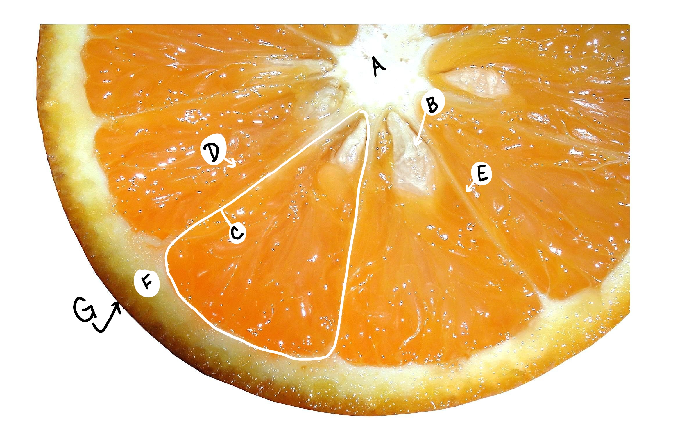
    A cross section of an orange
    Figure \(\PageIndex{10}\): A hesperidium (citrus fruits) is a modified berry. The endocarp forms the skin around the segments, containing seeds and juice-filled trichomes. The exocarp forms an oily rind. A=Pith, B=Seed, C=Locule, D=Juice-filled trichomes, E=Endocarp, F=Mesocarp, G=Exocarp. Image of the orange slice from Paolo Neo, Public domain, via Wikimedia Commons with labels added by Maria Morrow.
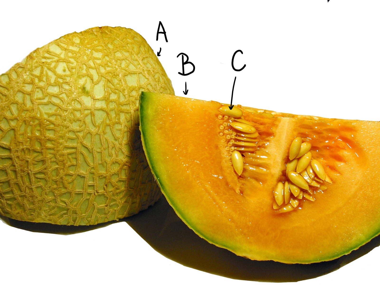
    A sliced cantaloupe with many seeds.
    Figure \(\PageIndex{11}\): A pepo is another modified berry. The exocarp forms a (somewhat) tough rind (A). The mesocarp (B) is still fleshy. It contains many seeds (C). Image from User:Tomia, CC BY-SA, via Wikimedia Commons with labels added by Maria Morrow.

    Accessory Fruits

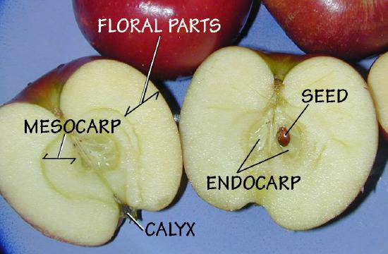
    an apple sliced in half. The fleshy part of the apple is labeled as "floral parts"
    Figure \(\PageIndex{12}\): A pome is an accessory fruit made from a fleshy hypanthium. The papery core is the pericarp, which contains the seeds. Eric Guinther, CC BY-SA, via Wikimedia Commons.
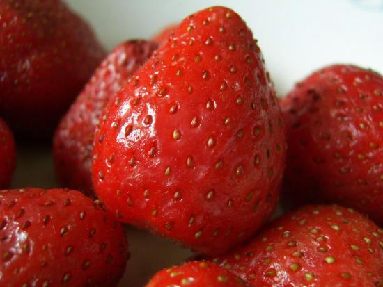
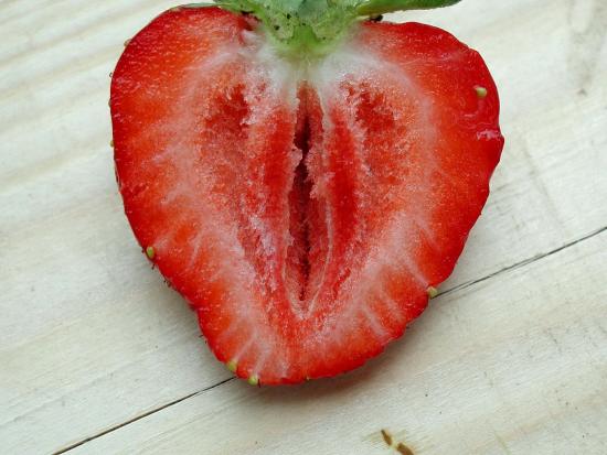
    A strawberry with many seed-like structures stuck to the outside A strawberry, sliced longitudinally
    Figure \(\PageIndex{13}\): Strawberries are accessory fruits formed from a swollen receptacle. The receptacle reddens and swells, pushing the developing fruits outward. The fruits are achenes that look like seeds, stuck to the outside of the receptacle. The calyx of the flower is the green, leafy part. If you look closely, you can see the styles attached to the achenes. Full strawberry photo author unknown, licensed under the Creative Commons Attribution-Share Alike 3.0 Unported license. Sliced strawberry photo by Paolo Neo, Public domain, via Wikimedia Commons.

    This page titled 15.2: Atlas- Fruits is shared under a CC BY 4.0 license and was authored, remixed, and/or curated by Valeria Hochman Adler.

    • Was this article helpful?