
13: Identification of White Blood Cells
- Page ID
- 110869

\( \newcommand{\vecs}[1]{\overset { \scriptstyle \rightharpoonup} {\mathbf{#1}} } \)
\( \newcommand{\vecd}[1]{\overset{-\!-\!\rightharpoonup}{\vphantom{a}\smash {#1}}} \)
\( \newcommand{\dsum}{\displaystyle\sum\limits} \)
\( \newcommand{\dint}{\displaystyle\int\limits} \)
\( \newcommand{\dlim}{\displaystyle\lim\limits} \)
\( \newcommand{\id}{\mathrm{id}}\) \( \newcommand{\Span}{\mathrm{span}}\)
( \newcommand{\kernel}{\mathrm{null}\,}\) \( \newcommand{\range}{\mathrm{range}\,}\)
\( \newcommand{\RealPart}{\mathrm{Re}}\) \( \newcommand{\ImaginaryPart}{\mathrm{Im}}\)
\( \newcommand{\Argument}{\mathrm{Arg}}\) \( \newcommand{\norm}[1]{\| #1 \|}\)
\( \newcommand{\inner}[2]{\langle #1, #2 \rangle}\)
\( \newcommand{\Span}{\mathrm{span}}\)
\( \newcommand{\id}{\mathrm{id}}\)
\( \newcommand{\Span}{\mathrm{span}}\)
\( \newcommand{\kernel}{\mathrm{null}\,}\)
\( \newcommand{\range}{\mathrm{range}\,}\)
\( \newcommand{\RealPart}{\mathrm{Re}}\)
\( \newcommand{\ImaginaryPart}{\mathrm{Im}}\)
\( \newcommand{\Argument}{\mathrm{Arg}}\)
\( \newcommand{\norm}[1]{\| #1 \|}\)
\( \newcommand{\inner}[2]{\langle #1, #2 \rangle}\)
\( \newcommand{\Span}{\mathrm{span}}\) \( \newcommand{\AA}{\unicode[.8,0]{x212B}}\)
\( \newcommand{\vectorA}[1]{\vec{#1}} % arrow\)
\( \newcommand{\vectorAt}[1]{\vec{\text{#1}}} % arrow\)
\( \newcommand{\vectorB}[1]{\overset { \scriptstyle \rightharpoonup} {\mathbf{#1}} } \)
\( \newcommand{\vectorC}[1]{\textbf{#1}} \)
\( \newcommand{\vectorD}[1]{\overrightarrow{#1}} \)
\( \newcommand{\vectorDt}[1]{\overrightarrow{\text{#1}}} \)
\( \newcommand{\vectE}[1]{\overset{-\!-\!\rightharpoonup}{\vphantom{a}\smash{\mathbf {#1}}}} \)
\( \newcommand{\vecs}[1]{\overset { \scriptstyle \rightharpoonup} {\mathbf{#1}} } \)
\(\newcommand{\longvect}{\overrightarrow}\)
\( \newcommand{\vecd}[1]{\overset{-\!-\!\rightharpoonup}{\vphantom{a}\smash {#1}}} \)
\(\newcommand{\avec}{\mathbf a}\) \(\newcommand{\bvec}{\mathbf b}\) \(\newcommand{\cvec}{\mathbf c}\) \(\newcommand{\dvec}{\mathbf d}\) \(\newcommand{\dtil}{\widetilde{\mathbf d}}\) \(\newcommand{\evec}{\mathbf e}\) \(\newcommand{\fvec}{\mathbf f}\) \(\newcommand{\nvec}{\mathbf n}\) \(\newcommand{\pvec}{\mathbf p}\) \(\newcommand{\qvec}{\mathbf q}\) \(\newcommand{\svec}{\mathbf s}\) \(\newcommand{\tvec}{\mathbf t}\) \(\newcommand{\uvec}{\mathbf u}\) \(\newcommand{\vvec}{\mathbf v}\) \(\newcommand{\wvec}{\mathbf w}\) \(\newcommand{\xvec}{\mathbf x}\) \(\newcommand{\yvec}{\mathbf y}\) \(\newcommand{\zvec}{\mathbf z}\) \(\newcommand{\rvec}{\mathbf r}\) \(\newcommand{\mvec}{\mathbf m}\) \(\newcommand{\zerovec}{\mathbf 0}\) \(\newcommand{\onevec}{\mathbf 1}\) \(\newcommand{\real}{\mathbb R}\) \(\newcommand{\twovec}[2]{\left[\begin{array}{r}#1 \\ #2 \end{array}\right]}\) \(\newcommand{\ctwovec}[2]{\left[\begin{array}{c}#1 \\ #2 \end{array}\right]}\) \(\newcommand{\threevec}[3]{\left[\begin{array}{r}#1 \\ #2 \\ #3 \end{array}\right]}\) \(\newcommand{\cthreevec}[3]{\left[\begin{array}{c}#1 \\ #2 \\ #3 \end{array}\right]}\) \(\newcommand{\fourvec}[4]{\left[\begin{array}{r}#1 \\ #2 \\ #3 \\ #4 \end{array}\right]}\) \(\newcommand{\cfourvec}[4]{\left[\begin{array}{c}#1 \\ #2 \\ #3 \\ #4 \end{array}\right]}\) \(\newcommand{\fivevec}[5]{\left[\begin{array}{r}#1 \\ #2 \\ #3 \\ #4 \\ #5 \\ \end{array}\right]}\) \(\newcommand{\cfivevec}[5]{\left[\begin{array}{c}#1 \\ #2 \\ #3 \\ #4 \\ #5 \\ \end{array}\right]}\) \(\newcommand{\mattwo}[4]{\left[\begin{array}{rr}#1 \amp #2 \\ #3 \amp #4 \\ \end{array}\right]}\) \(\newcommand{\laspan}[1]{\text{Span}\{#1\}}\) \(\newcommand{\bcal}{\cal B}\) \(\newcommand{\ccal}{\cal C}\) \(\newcommand{\scal}{\cal S}\) \(\newcommand{\wcal}{\cal W}\) \(\newcommand{\ecal}{\cal E}\) \(\newcommand{\coords}[2]{\left\{#1\right\}_{#2}}\) \(\newcommand{\gray}[1]{\color{gray}{#1}}\) \(\newcommand{\lgray}[1]{\color{lightgray}{#1}}\) \(\newcommand{\rank}{\operatorname{rank}}\) \(\newcommand{\row}{\text{Row}}\) \(\newcommand{\col}{\text{Col}}\) \(\renewcommand{\row}{\text{Row}}\) \(\newcommand{\nul}{\text{Nul}}\) \(\newcommand{\var}{\text{Var}}\) \(\newcommand{\corr}{\text{corr}}\) \(\newcommand{\len}[1]{\left|#1\right|}\) \(\newcommand{\bbar}{\overline{\bvec}}\) \(\newcommand{\bhat}{\widehat{\bvec}}\) \(\newcommand{\bperp}{\bvec^\perp}\) \(\newcommand{\xhat}{\widehat{\xvec}}\) \(\newcommand{\vhat}{\widehat{\vvec}}\) \(\newcommand{\uhat}{\widehat{\uvec}}\) \(\newcommand{\what}{\widehat{\wvec}}\) \(\newcommand{\Sighat}{\widehat{\Sigma}}\) \(\newcommand{\lt}{<}\) \(\newcommand{\gt}{>}\) \(\newcommand{\amp}{&}\) \(\definecolor{fillinmathshade}{gray}{0.9}\)By the end of this lab, you will be able to
- Prepare a blood smear
- Perform a Wright’s stain
- Identify red blood cells, neutrophils, eosinophils, basophils, platelets, monocytes, and lymphocytes under the microscope
- Perform Wright’s stain of a blood smear.
- Conduct a differential blood count to determine the percentage of various leukocytes in blood.
Introduction
Today we will be exploring the white blood cells that are so important to the innate and adaptive immune system. Where will we get the blood? From your finger of course! You will be using a lancet to pierce your finger and collect drops for the preparation of a blood smear. You will then stain the smear using a modified Wright’s stain procedure, and then we will be looking at them under the microscope at 1000x.
All of your red and white blood cells originate from a type of stem cell called a hematopoietic stem cell. These cells are found primarily in your bone marrow, but also in your peripheral blood, and throughout your life, they differentiate into any of the red or white blood cells that are needed. Red blood cells have a short life span of about 120 days, so they are constantly being replaced. Many of your white blood cells have even shorter life spans, some of only minutes or days!
.png?revision=1)
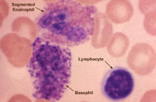
Our Openstax textbook, Chapter 17.3 describes the functions of each of these cells in detail, so these functions are not recapitulated here. However, you will need to be able to recognize them under the microscope. The table below will help you with determining which cells are which.
| Cell type | % found in blood | Images | |
|---|---|---|---|
|
Neutrophil |
55-65% |
User CS99 at German Wikipedia, Public domain, via Wikimedia Commons (left image), CDC Public Health Image Library, Public Domain (right image). |
|
|
Eosinophil |
1-2% |
Davidcsaba Dr. David Csaba L., Public domain, via Wikimedia Commons (left), CDC Public Health Image Library, Public Domain (right image). |
|
|
Basophil |
0.2% |
CDC Public Health Image Library, Public Domain (right image). |
|
|
Monocytes |
3-7% |
CDC Public Health Image Library (left), and CDC Public Health Image Library (right) Public Domain |
|
|
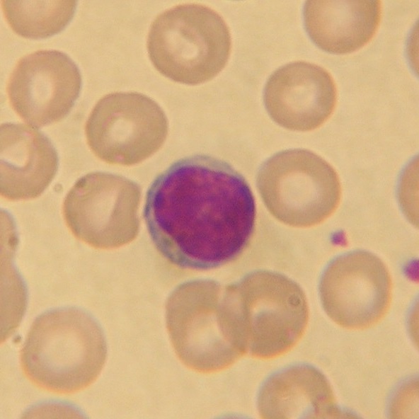
lymphocytes |
25-33% |
Bernanke's Crossbow, CC BY-SA 3.0, via Wikimedia Commons (left image), CDC Public Health Image Library, Public Domain (right) |
|
|
platelet |
N/A |
CDC Public Health Image Library, Public Domain |
|


.jpg?revision=1&size=bestfit&width=312&height=312)


Preparing a blood smear
The procedure for making a blood smear is different from making a bacterial smear. Blood smears are prepared by taking a drop of blood and spreading it across the slide vertically using a second slide. Then the blood is further spread horizontally to cover most of the slide surface.

Wright's Stain
Wright's stain contains the dyes eosin Y and methylene blue. These two stains bind differently to different white blood cell types and allow them to be easily differentiated from one another. Eosin Y stains the cytoplasm of cells a pink or orange color, while methylene blue stains the cellular nuclei a blue color. This makes it fairly straightforward to differentiate the various white blood cells from each other. Additionally, the granules of basophils and eosinophils are stained blue or red, respectively.
We will be using a 1-step Wright's stain in today's lab.
Materials
- Sterile lancets
- Alcohol pad wipes
- Band-aids for finger sticks
- Plastic sharp container
- Wright’s stain
- Microscope slides
Experiment
- Select the finger to puncture, usually the middle or ring finger. Clean the area with an alcohol wipe, and allow it to dry.
- If the room is cold, you may want to try to warm up your hands first and get some circulation going. Use the sterile lancet to puncture the ball of the finger. Wipe away the first drop of blood if you are bleeding enough. For some of us who struggle to get any drops of blood out, you may want to make a slide from the initial drop and then try to make additional slides from subsequent drops if you can. Touch a drop or two of blood with a clean slide.
- Hold a second slide at an angle as illustrated in Figure \(\PageIndex{2}\). While maintaining contact with the bottom slide, pull the top slide back to contact the blood, which will spread by capillary action.
- Maintain firm contact with the bottom slide and push the top slide in one motion to produce the smear. Your instructor will demonstrate!
- Let the blood smear air dry. It is not necessary (or desirable) to heat up the slide before staining.
- Stain the dried smear following the protocol below:
- Dip the smear in Wright's stain for 30 seconds
- Dip the smear in a beaker of distilled water for 45 seconds
- Dip the smear in a second beaker of distilled water for 25 seconds
- DO NOT RINSE BETWEEN DIPS!
- Allow your slide to air dry - do not blot.
- Scan your smear using your 4x objective and try to find a spot where there is a monolayer of blood cells visible. Systematically scan the slide and count at least 50 white blood cells. Calculate the percentage of neutrophils, lymphocytes, monocytes, eosinophils, and basophils out of the total. Do not count platelets of red blood cells.
Data
Count a total of 50 cells (or more!), and fill out the table below.
|
Monocytes |
Lymphocytes |
Segmented Neutrophils |
Eosinophils |
Basophils |
|
|---|---|---|---|---|---|
|
Number |
|||||
|
Percentage |
|||||
|
Expected percentage |
3-7% |
25-33% |
55-65% |
1-3% |
<0.2% |
Questions
- What is the role of each of the cell types you saw today? Include erythrocytes and platelets in your answer.
- Which cells that we saw today have primary roles in the innate immune system? What about the adaptive immune system?
- Which cell type differentiates into innate immune system cells that are “professional” antigen-presenting cells?
- Can you tell the difference between B- and T-cells under the microscope? If not, what would you have to do to differentiate between them?