
26.1: Polyclonal Antibodies
- Page ID
- 144235

\( \newcommand{\vecs}[1]{\overset { \scriptstyle \rightharpoonup} {\mathbf{#1}} } \)
\( \newcommand{\vecd}[1]{\overset{-\!-\!\rightharpoonup}{\vphantom{a}\smash {#1}}} \)
\( \newcommand{\dsum}{\displaystyle\sum\limits} \)
\( \newcommand{\dint}{\displaystyle\int\limits} \)
\( \newcommand{\dlim}{\displaystyle\lim\limits} \)
\( \newcommand{\id}{\mathrm{id}}\) \( \newcommand{\Span}{\mathrm{span}}\)
( \newcommand{\kernel}{\mathrm{null}\,}\) \( \newcommand{\range}{\mathrm{range}\,}\)
\( \newcommand{\RealPart}{\mathrm{Re}}\) \( \newcommand{\ImaginaryPart}{\mathrm{Im}}\)
\( \newcommand{\Argument}{\mathrm{Arg}}\) \( \newcommand{\norm}[1]{\| #1 \|}\)
\( \newcommand{\inner}[2]{\langle #1, #2 \rangle}\)
\( \newcommand{\Span}{\mathrm{span}}\)
\( \newcommand{\id}{\mathrm{id}}\)
\( \newcommand{\Span}{\mathrm{span}}\)
\( \newcommand{\kernel}{\mathrm{null}\,}\)
\( \newcommand{\range}{\mathrm{range}\,}\)
\( \newcommand{\RealPart}{\mathrm{Re}}\)
\( \newcommand{\ImaginaryPart}{\mathrm{Im}}\)
\( \newcommand{\Argument}{\mathrm{Arg}}\)
\( \newcommand{\norm}[1]{\| #1 \|}\)
\( \newcommand{\inner}[2]{\langle #1, #2 \rangle}\)
\( \newcommand{\Span}{\mathrm{span}}\) \( \newcommand{\AA}{\unicode[.8,0]{x212B}}\)
\( \newcommand{\vectorA}[1]{\vec{#1}} % arrow\)
\( \newcommand{\vectorAt}[1]{\vec{\text{#1}}} % arrow\)
\( \newcommand{\vectorB}[1]{\overset { \scriptstyle \rightharpoonup} {\mathbf{#1}} } \)
\( \newcommand{\vectorC}[1]{\textbf{#1}} \)
\( \newcommand{\vectorD}[1]{\overrightarrow{#1}} \)
\( \newcommand{\vectorDt}[1]{\overrightarrow{\text{#1}}} \)
\( \newcommand{\vectE}[1]{\overset{-\!-\!\rightharpoonup}{\vphantom{a}\smash{\mathbf {#1}}}} \)
\( \newcommand{\vecs}[1]{\overset { \scriptstyle \rightharpoonup} {\mathbf{#1}} } \)
\(\newcommand{\longvect}{\overrightarrow}\)
\( \newcommand{\vecd}[1]{\overset{-\!-\!\rightharpoonup}{\vphantom{a}\smash {#1}}} \)
\(\newcommand{\avec}{\mathbf a}\) \(\newcommand{\bvec}{\mathbf b}\) \(\newcommand{\cvec}{\mathbf c}\) \(\newcommand{\dvec}{\mathbf d}\) \(\newcommand{\dtil}{\widetilde{\mathbf d}}\) \(\newcommand{\evec}{\mathbf e}\) \(\newcommand{\fvec}{\mathbf f}\) \(\newcommand{\nvec}{\mathbf n}\) \(\newcommand{\pvec}{\mathbf p}\) \(\newcommand{\qvec}{\mathbf q}\) \(\newcommand{\svec}{\mathbf s}\) \(\newcommand{\tvec}{\mathbf t}\) \(\newcommand{\uvec}{\mathbf u}\) \(\newcommand{\vvec}{\mathbf v}\) \(\newcommand{\wvec}{\mathbf w}\) \(\newcommand{\xvec}{\mathbf x}\) \(\newcommand{\yvec}{\mathbf y}\) \(\newcommand{\zvec}{\mathbf z}\) \(\newcommand{\rvec}{\mathbf r}\) \(\newcommand{\mvec}{\mathbf m}\) \(\newcommand{\zerovec}{\mathbf 0}\) \(\newcommand{\onevec}{\mathbf 1}\) \(\newcommand{\real}{\mathbb R}\) \(\newcommand{\twovec}[2]{\left[\begin{array}{r}#1 \\ #2 \end{array}\right]}\) \(\newcommand{\ctwovec}[2]{\left[\begin{array}{c}#1 \\ #2 \end{array}\right]}\) \(\newcommand{\threevec}[3]{\left[\begin{array}{r}#1 \\ #2 \\ #3 \end{array}\right]}\) \(\newcommand{\cthreevec}[3]{\left[\begin{array}{c}#1 \\ #2 \\ #3 \end{array}\right]}\) \(\newcommand{\fourvec}[4]{\left[\begin{array}{r}#1 \\ #2 \\ #3 \\ #4 \end{array}\right]}\) \(\newcommand{\cfourvec}[4]{\left[\begin{array}{c}#1 \\ #2 \\ #3 \\ #4 \end{array}\right]}\) \(\newcommand{\fivevec}[5]{\left[\begin{array}{r}#1 \\ #2 \\ #3 \\ #4 \\ #5 \\ \end{array}\right]}\) \(\newcommand{\cfivevec}[5]{\left[\begin{array}{c}#1 \\ #2 \\ #3 \\ #4 \\ #5 \\ \end{array}\right]}\) \(\newcommand{\mattwo}[4]{\left[\begin{array}{rr}#1 \amp #2 \\ #3 \amp #4 \\ \end{array}\right]}\) \(\newcommand{\laspan}[1]{\text{Span}\{#1\}}\) \(\newcommand{\bcal}{\cal B}\) \(\newcommand{\ccal}{\cal C}\) \(\newcommand{\scal}{\cal S}\) \(\newcommand{\wcal}{\cal W}\) \(\newcommand{\ecal}{\cal E}\) \(\newcommand{\coords}[2]{\left\{#1\right\}_{#2}}\) \(\newcommand{\gray}[1]{\color{gray}{#1}}\) \(\newcommand{\lgray}[1]{\color{lightgray}{#1}}\) \(\newcommand{\rank}{\operatorname{rank}}\) \(\newcommand{\row}{\text{Row}}\) \(\newcommand{\col}{\text{Col}}\) \(\renewcommand{\row}{\text{Row}}\) \(\newcommand{\nul}{\text{Nul}}\) \(\newcommand{\var}{\text{Var}}\) \(\newcommand{\corr}{\text{corr}}\) \(\newcommand{\len}[1]{\left|#1\right|}\) \(\newcommand{\bbar}{\overline{\bvec}}\) \(\newcommand{\bhat}{\widehat{\bvec}}\) \(\newcommand{\bperp}{\bvec^\perp}\) \(\newcommand{\xhat}{\widehat{\xvec}}\) \(\newcommand{\vhat}{\widehat{\vvec}}\) \(\newcommand{\uhat}{\widehat{\uvec}}\) \(\newcommand{\what}{\widehat{\wvec}}\) \(\newcommand{\Sighat}{\widehat{\Sigma}}\) \(\newcommand{\lt}{<}\) \(\newcommand{\gt}{>}\) \(\newcommand{\amp}{&}\) \(\definecolor{fillinmathshade}{gray}{0.9}\)- Compare the method of development, use, and characteristics of monoclonal and polyclonal antibodies
- Explain the nature of antibody cross-reactivity and why it occurs
- Compare and contrast false-positive vs. false-negative test results
In addition to being crucial for our normal immune response, antibodies provide powerful tools for research and diagnostic purposes. The high specificity of antibodies makes them an excellent tool for detecting and quantifying a broad array of targets, from drugs to serum proteins to microorganisms. With in vitro assays, antibodies can be used to precipitate soluble antigens, agglutinate (clump) cells, opsonize and kill bacteria with the assistance of complement, and neutralize drugs, toxins, and viruses.
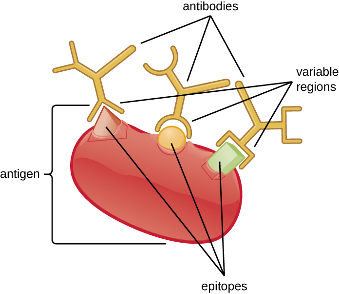
An antibody’s specificity results from the antigen-binding site formed within the variable regions—regions of the antibody that have unique patterns of amino acids that can only bind to target antigens with a molecular sequence that provides complementary charges and noncovalent bonds. There are limitations to antibody specificity, however. Some antigens are so chemically similar that cross-reactivity occurs; in other words, antibodies raised against one antigen bind to a chemically similar but different antigen. Consider an antigen that consists of a single protein with multiple epitopes (Figure \(\PageIndex{1}\)). This single protein may stimulate the production of many different antibodies, some of which may bind to chemically identical epitopes on other proteins.
Cross-reactivity is more likely to occur between antibodies and antigens that have low affinity or avidity. Affinity, which can be determined experimentally, is a measure of the binding strength between an antibody's binding site and an epitope, whereas avidity is the total strength of all the interactions in an antibody-antigen complex (which may have more than one bonding site). Avidity is influenced by affinity as well as the structural arrangements of the epitope and the variable regions of the antibody. If an antibody has a high affinity/avidity for a specific antigen, it is less likely to cross-react with an antigen for which it has a lower affinity/avidity.
Query \(\PageIndex{1}\)
Query \(\PageIndex{1}\)
Query \(\PageIndex{1}\)
Producing Polyclonal Antibodies
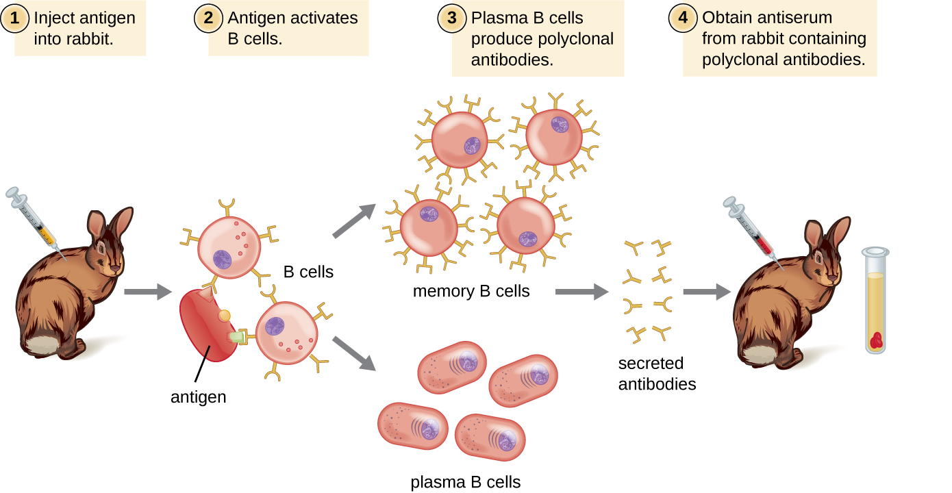
Antibodies used for research and diagnostic purposes are often obtained by injecting a lab animal such as a rabbit or a goat with a specific antigen. Within a few weeks, the animal’s immune system will produce high levels of antibodies specific for the antigen. These antibodies can be harvested in an antiserum, which is whole serum collected from an animal following exposure to an antigen. Because most antigens are complex structures with multiple epitopes, they result in the production of multiple antibodies in the lab animal. This so-called polyclonal antibody response is also typical of the response to infection by the human immune system. Antiserum drawn from an animal will thus contain antibodies from multiple clones of B cells, with each B cell responding to a specific epitope on the antigen (Figure \(\PageIndex{2}\)).
Lab animals are usually injected at least twice with antigen when being used to produce antiserum. The second injection will activate memory cells that make class IgG antibodies against the antigen. The memory cells also undergo affinity maturation, resulting in a pool of antibodies with higher average affinity. Affinity maturation occurs because of mutations in the immunoglobulin gene variable regions, resulting in B cells with slightly altered antigen-binding sites. On re-exposure to the antigen, those B cells capable of producing antibody with higher affinity antigen-binding sites will be stimulated to proliferate and produce more antibody than their lower-affinity peers. An adjuvant, which is a chemical that provokes a generalized activation of the immune system that stimulates greater antibody production, is often mixed with the antigen prior to injection.
Antiserum obtained from animals will not only contain antibodies against the antigen artificially introduced in the laboratory, but it will also contain antibodies to any other antigens to which the animal has been exposed during its lifetime. For this reason, antisera must first be “purified” to remove other antibodies before using the antibodies for research or diagnostic assays.
Query \(\PageIndex{1}\)
Clinical Uses of Polyclonal Antisera
Polyclonal antisera are used in many clinical tests that are designed to determine whether a patient is producing antibodies in response to a particular pathogen. While these tests are certainly powerful diagnostic tools, they have their limitations, because they are an indirect means of determining whether a particular pathogen is present. Tests based on a polyclonal response can sometimes lead to a false-positive result—in other words, a test that confirms the presence of an antigen that is, in fact, not present. Antibody-based tests can also result in a false-negative result, which occurs when the test fails to detect an antibody that is, in fact, present.
The accuracy of antibody tests can be described in terms of test sensitivity and test specificity. Test sensitivity is the probability of getting a positive test result when the patient is indeed infected. If a test has high sensitivity, the probability of a false negative is low. Test specificity, on the other hand, is the probability of getting a negative test result when the patient is not infected. If a test has high specificity, the probability of a false positive is low.
False positives often occur due to cross-reactivity, which can occur when epitopes from a different pathogen are similar to those found on the pathogen being tested for. For this reason, antibody-based tests are often used only as screening tests; if the results are positive, other confirmatory tests are used to make sure that the results were not a false positive.
For example, a blood sample from a patient suspected of having hepatitis C can be screened for the virus using antibodies that bind to antigens on hepatitis C virus. If the patient is indeed infected with hepatitis C virus, the antibodies will bind to the antigens, yielding a positive test result. If the patient is not infected with hepatitic C virus, the antibodies will generally not bind to anything and the test should be negative; however, a false positive may occur if the patient has been previously infected by any of a variety of pathogens that elicit antibodies that cross-react with the hepatitis C virus antigens. Antibody tests for hepatitis C have high sensitivity (a low probability of a false negative) but low specificity (a high probability of a false positive). Thus, patients who test positive must have a second, confirmatory test to rule out the possibility of a false positive. The confirmatory test is a more expensive and time-consuming test that directly tests for the presence of hepatitis C viral RNA in the blood. Only after the confirmatory test comes back positive can the patient be definitively diagnosed with a hepatitis C infection. Antibody-based tests can result in a false negative if, for any reason, the patient’s immune system has not produced detectable levels of antibodies. For some diseases, it may take several weeks following infection before the immune system produces enough antibodies to cross the detection threshold of the assay. In immunocompromised patients, the immune system may not be capable of producing a detectable level of antibodies.
Another limitation of using antibody production as an indicator of disease is that antibodies in the blood will persist long after the infection has been cleared. Depending on the type of infection, antibodies will be present for many months; sometimes, they may be present for the remainder of the patient’s life. Thus, a positive antibody-based test only means that the patient was infected at some point in time; it does not prove that the infection is active.
In addition to their role in diagnosis, polyclonal antisera can activate complement, detect the presence of bacteria in clinical and food industry settings, and perform a wide array of precipitation reactions that can detect and quantify serum proteins, viruses, or other antigens. However, with the many specificities of antibody present in a polyclonal antiserum, there is a significant likelihood that the antiserum will cross-react with antigens to which the individual was never exposed. Therefore, we must always account for the possibility of false-positive results when working with a polyclonal antiserum.
Query \(\PageIndex{1}\)
Case Study Preview: “The Syringe Scandal: A Test of Trust and Immunity”
A hospital reels from a public health crisis when a healthcare worker battling addiction is caught swapping patient syringes - leading to possible HIV exposure for over 1,300 patients. You’ll follow the hospital’s urgent epidemiological response: contact tracing, ELISA testing, confirmatory western blots, and PCR diagnostics. Along the way, you’ll unpack the strengths and pitfalls of each diagnostic method, including false positives, indeterminate results, and the seroconversion window.
This case puts you in the shoes of public health teams navigating patient risk, legal liability, and the science of HIV detection and confirmatory testing. You'll also explore how flow cytometry, CD4 counts, and PCR are used to monitor immune status and guide treatment - reminding us that behind every test result is a person seeking answers.
Can an early test provide peace of mind - or false hope? And what happens when confirmatory results fall into the gray zone?
Chapter 20 Case Study - The Syringe Scandal: A Test of Trust and Immunity
Key Concepts and Summary
- Antibodies bind with high specificity to antigens used to challenge the immune system, but they may also show cross-reactivity by binding to other antigens that share chemical properties with the original antigen.
- Injection of an antigen into an animal will result in a polyclonal antibody response in which different antibodies are produced that react with the various epitopes on the antigen.
- Polyclonal antisera are useful for some types of laboratory assays, but other assays require more specificity. Diagnostic tests that use polyclonal antisera are typically only used for screening because of the possibility of false-positive and false-negative results.