

10.4: Small Intestine
- Page ID
- 208110

\( \newcommand{\vecs}[1]{\overset { \scriptstyle \rightharpoonup} {\mathbf{#1}} } \)
\( \newcommand{\vecd}[1]{\overset{-\!-\!\rightharpoonup}{\vphantom{a}\smash {#1}}} \)
\( \newcommand{\dsum}{\displaystyle\sum\limits} \)
\( \newcommand{\dint}{\displaystyle\int\limits} \)
\( \newcommand{\dlim}{\displaystyle\lim\limits} \)
\( \newcommand{\id}{\mathrm{id}}\) \( \newcommand{\Span}{\mathrm{span}}\)
( \newcommand{\kernel}{\mathrm{null}\,}\) \( \newcommand{\range}{\mathrm{range}\,}\)
\( \newcommand{\RealPart}{\mathrm{Re}}\) \( \newcommand{\ImaginaryPart}{\mathrm{Im}}\)
\( \newcommand{\Argument}{\mathrm{Arg}}\) \( \newcommand{\norm}[1]{\| #1 \|}\)
\( \newcommand{\inner}[2]{\langle #1, #2 \rangle}\)
\( \newcommand{\Span}{\mathrm{span}}\)
\( \newcommand{\id}{\mathrm{id}}\)
\( \newcommand{\Span}{\mathrm{span}}\)
\( \newcommand{\kernel}{\mathrm{null}\,}\)
\( \newcommand{\range}{\mathrm{range}\,}\)
\( \newcommand{\RealPart}{\mathrm{Re}}\)
\( \newcommand{\ImaginaryPart}{\mathrm{Im}}\)
\( \newcommand{\Argument}{\mathrm{Arg}}\)
\( \newcommand{\norm}[1]{\| #1 \|}\)
\( \newcommand{\inner}[2]{\langle #1, #2 \rangle}\)
\( \newcommand{\Span}{\mathrm{span}}\) \( \newcommand{\AA}{\unicode[.8,0]{x212B}}\)
\( \newcommand{\vectorA}[1]{\vec{#1}} % arrow\)
\( \newcommand{\vectorAt}[1]{\vec{\text{#1}}} % arrow\)
\( \newcommand{\vectorB}[1]{\overset { \scriptstyle \rightharpoonup} {\mathbf{#1}} } \)
\( \newcommand{\vectorC}[1]{\textbf{#1}} \)
\( \newcommand{\vectorD}[1]{\overrightarrow{#1}} \)
\( \newcommand{\vectorDt}[1]{\overrightarrow{\text{#1}}} \)
\( \newcommand{\vectE}[1]{\overset{-\!-\!\rightharpoonup}{\vphantom{a}\smash{\mathbf {#1}}}} \)
\( \newcommand{\vecs}[1]{\overset { \scriptstyle \rightharpoonup} {\mathbf{#1}} } \)
\(\newcommand{\longvect}{\overrightarrow}\)
\( \newcommand{\vecd}[1]{\overset{-\!-\!\rightharpoonup}{\vphantom{a}\smash {#1}}} \)
\(\newcommand{\ket}[1]{\left| #1 \right>}\)
\(\newcommand{\bra}[1]{\left< #1 \right|}\)
\(\newcommand{\braket}[2]{\left< #1 \vphantom{#2} \right| \left. #2 \vphantom{#1} \right>}\)
\(\newcommand{\braopket}[3]{\left< #1 \vphantom{#2}\vphantom{#3} \right| #2 \vphantom{#1}\vphantom{#3} \left| #3 \vphantom{#1}\vphantom{#2} \right>}\)
\(\newcommand{\qmvec}[1]{\mathbf{\vec{#1}}}\)
\(\newcommand{\op}[1]{\hat{\mathbf{#1}}}\)
\(\newcommand{\expect}[1]{\langle #1 \rangle}\)
\(\newcommand{\dfn}[1]{\emph{\textbf{#1}}}\)
The process of digestion does not always go as it should. Many people suffer from indigestion, or dyspepsia, a condition of impaired digestion. Symptoms may include upper abdominal fullness or pain, heartburn, nausea, belching, or some combination of these symptoms. The majority of cases of indigestion occur without evidence of an organic disease that is likely to explain the symptoms. Anxiety or certain foods or medications (such as aspirin) may be contributing factors in these cases. In other cases, indigestion is a symptom of an organic disease, most often gastroesophageal reflux disease (GERD) or gastritis. In a small minority of cases, indigestion is a symptom of a peptic ulcer of the stomach or duodenum, usually caused by a bacterial infection. Very rarely, indigestion is a sign of cancer.
An occasional bout of indigestion is usually nothing to worry about, especially in people less than 55 years of age. However, if you suffer frequent or chronic indigestion, it’s a good idea to see a doctor. If an underlying disorder such as GERD or an ulcer is causing indigestion, this can and should be treated. If no organic disease is discovered, the doctor can recommend lifestyle changes or treatments to help prevent or soothe the symptoms of acute indigestion. Lifestyle changes might include modifications in eating habits, such as eating more slowly, eating smaller meals, or avoiding fatty foods. You also might be advised to refrain from taking certain medications, especially on an empty stomach. The use of antacids or other medications to relieve symptoms may also be recommended
Small Intestine
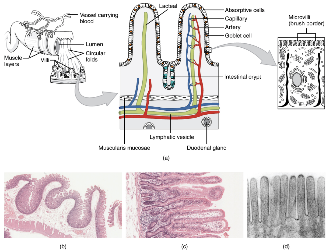
The small intestine is a narrow but very long tubular organ. Its average length in adults is 4.6 m (15 ft) in females and 6.9 m (22 ft 8 in.) in males. It is approximately 2.5 to 3.0 cm (1.0 to 1.2 in.) in diameter (it is called “small” because it is much smaller in diameter than the large intestine). It is the site of most chemical digestion and virtually all absorption of nutrients. Many digestive enzymes are active in the small intestine, some of which are produced by the small intestine itself, and some of which are produced by the pancreas, an accessory organ of the digestive system. Much of the inner lining of the small intestine is covered by tiny finger-like projections called villi, each of which in turn is covered by even tinier projections called microvilli. These projections, shown in Figure \(\PageIndex{2}\), greatly increase the surface area through which nutrients can be absorbed from the small intestine. Because of these projections, the internal surface area of the small intestine totals an average of about 30 m2 (323 ft2).
.jpg?revision=1&size=bestfit&width=520&height=283)
The first part of the small intestine receives partially digested, semi-liquid chyme from the stomach. It receives digestive enzymes and alkaline bicarbonate from the pancreas through the pancreatic duct, and it receives bile from the liver via the gallbladder through the common bile duct (Figure \(\PageIndex{3}\)). The bicarbonate from the pancreas as well as bile from the liver neutralize the highly acidic chyme after it enters the small intestine from the stomach. This is necessary because the digestive enzymes in the small intestine require a nearly neutral environment in order to work (pH of about 7). The three major classes of compounds that undergo chemical digestion in the small intestine are carbohydrates, proteins, and lipids.
Chemical Digestion
Carbohydrates
Most carbohydrates humans consume are in the form of starch, a polysaccharide. Carbohydrates digestion begins in the mouth with salivary amylase and is completed in the small intestine. Polysaccharides are broken down to their monomer form, monosaccharides. In addition, food often contains cellulose, a plant polysaccharide that cannot be digested by humans. This undigestible polysaccharide is usually referred to as fiber, and ultimately passes undigested to the large intestine, where it may be digested by intestinal bacteria.
Other sugars are digested with the help of different enzymes produced by the small intestine. For example, sucrose, or table sugar, is a disaccharide that is broken down by the enzyme sucrase to form glucose and fructose, which are readily absorbed by the small intestine. Digestion of the sugar lactose, which is found in milk, requires the enzyme lactase, which breaks down lactose into glucose and galactose, which are then absorbed by the small intestine. Fewer than half of all adults produce sufficient lactase to be able to digest lactose. Those who cannot are said to be lactose intolerant.
Proteins
Protein digestion begins in the stomach with pepsin and is completed in the small intestine. Like polysaccharides, proteins are also broken down to their monomer form, amino acids.
Lipids
Lipid digestion is more complicated because of their chemical composition. Bile is required for the digestion of lipids because lipids are oily and do not dissolve in the watery chyme. Bile emulsifies, or breaks up, large globules of food lipids into much smaller ones, much as dish detergent breaks up grease (Figure \(\PageIndex{4}\)). The smaller droplets provide a great deal more surface area to be acted upon by lipase and also point the hydrophilic (“water-loving”) heads of the fatty acids outward into the watery chyme. Lipase, a lipid digesting enzyme, can then access and break down the small droplets into individual fatty acid molecules.

Absorption
When digestion is finished, it results in many simple nutrient molecules that must go through the process of absorption from the GI tract by blood or lymph so they can be used by cells throughout the body. Water is absorbed in the large intestine. However, about 95 percent of nutrient molecules are absorbed in the small intestine, including sugars, amino acids, and fatty acids. The epithelial tissue lining the small intestine is specialized for absorption. It has many wrinkles and is covered with villi and microvilli, creating an enormous surface area for absorption. As shown in Figure \(\PageIndex{5}\), each villus also has a network of blood capillaries and fine lymphatic vessels called lacteals close to its surface. The thin surface layer of epithelial cells of the villi transports nutrients from the lumen of the small intestine into these capillaries and lacteals. Blood in the capillaries absorbs most of the molecules, including simple sugars, amino acids, glycerol, salts, and water-soluble vitamins (vitamin C and the many B vitamins). Lymph in the lacteals absorbs fatty acids and fat-soluble vitamins (vitamins A, D, E, and K). Absorption occurs by simple diffusion (water and fatty acids), facilitated diffusion (the simple sugar fructose), or active transport (amino acids, small peptides, water-soluble vitamins, and most glucose).
After being absorbed in the small intestine, nutrient molecules are transported to other parts of the body for storage or further chemical modification. For example, amino acids are transported to the liver to be used for protein synthesis. All substances in chyme that remain undigested or unabsorbed by the time they reach the distal end of the small intestine pass into the large intestine.

Review
- How is the lining of the small intestine specialized for digestion and absorption?
- What digestive substances are secreted into the small intestine, and what compounds in food do they help digest?
- Where does absorption of nutrients take place?
- Name the parts of the large intestine.
- Identify the main functions of the large intestine.
- How do beneficial bacteria in the large intestine help the human organism?
- True or False. The first part of the large intestine is where most chemical digestion takes place.
- True or False. The small intestine is actually longer than the large intestine.
- Define digestion. Where does it occur?
- What is the role of enzymes in chemical digestion?
- What is absorption? When does it occur?
- a. Where does most absorption occur in the digestive system?
b. Why does most of the absorption occur in this organ and not earlier in the GI tract?
- a. Where is bile produced?
b. What is the function of bile?
- The pH of the stomach ___________ .
A. is neutral B. is alkaline
C. is acidic D. depends only on what you eat
- Lymph absorbs __________ .
A. fatty acids B. sugars
C. amino acids D. vitamin C
Explore More
Did you know you have about 100 million neurons in your intestines? In this fascinating TED talk, food scientist Heribert Watzke talks about the "hidden brain" in our gut and the surprising things it makes us feel.
Attributions
- Dyspepsia wafers, Public Domain via Flickr.com
- Small Intestines from Microbiology by Open Stax CC BY 3.0
- biliary tract medium by www.6xc.com.au6XC, CC BY-NC-ND 4.0
- bile emulsification by Cenveo licensed CC BY 3.0
- Intestinal villus simplified by Snow93, Public Domain via Wikimedia Commons
- Text adapted from Human Biology by CK-12 licensed CC BY-NC 3.0