
11.7: Human Senses
- Page ID
- 22525

\( \newcommand{\vecs}[1]{\overset { \scriptstyle \rightharpoonup} {\mathbf{#1}} } \)
\( \newcommand{\vecd}[1]{\overset{-\!-\!\rightharpoonup}{\vphantom{a}\smash {#1}}} \)
\( \newcommand{\id}{\mathrm{id}}\) \( \newcommand{\Span}{\mathrm{span}}\)
( \newcommand{\kernel}{\mathrm{null}\,}\) \( \newcommand{\range}{\mathrm{range}\,}\)
\( \newcommand{\RealPart}{\mathrm{Re}}\) \( \newcommand{\ImaginaryPart}{\mathrm{Im}}\)
\( \newcommand{\Argument}{\mathrm{Arg}}\) \( \newcommand{\norm}[1]{\| #1 \|}\)
\( \newcommand{\inner}[2]{\langle #1, #2 \rangle}\)
\( \newcommand{\Span}{\mathrm{span}}\)
\( \newcommand{\id}{\mathrm{id}}\)
\( \newcommand{\Span}{\mathrm{span}}\)
\( \newcommand{\kernel}{\mathrm{null}\,}\)
\( \newcommand{\range}{\mathrm{range}\,}\)
\( \newcommand{\RealPart}{\mathrm{Re}}\)
\( \newcommand{\ImaginaryPart}{\mathrm{Im}}\)
\( \newcommand{\Argument}{\mathrm{Arg}}\)
\( \newcommand{\norm}[1]{\| #1 \|}\)
\( \newcommand{\inner}[2]{\langle #1, #2 \rangle}\)
\( \newcommand{\Span}{\mathrm{span}}\) \( \newcommand{\AA}{\unicode[.8,0]{x212B}}\)
\( \newcommand{\vectorA}[1]{\vec{#1}} % arrow\)
\( \newcommand{\vectorAt}[1]{\vec{\text{#1}}} % arrow\)
\( \newcommand{\vectorB}[1]{\overset { \scriptstyle \rightharpoonup} {\mathbf{#1}} } \)
\( \newcommand{\vectorC}[1]{\textbf{#1}} \)
\( \newcommand{\vectorD}[1]{\overrightarrow{#1}} \)
\( \newcommand{\vectorDt}[1]{\overrightarrow{\text{#1}}} \)
\( \newcommand{\vectE}[1]{\overset{-\!-\!\rightharpoonup}{\vphantom{a}\smash{\mathbf {#1}}}} \)
\( \newcommand{\vecs}[1]{\overset { \scriptstyle \rightharpoonup} {\mathbf{#1}} } \)
\( \newcommand{\vecd}[1]{\overset{-\!-\!\rightharpoonup}{\vphantom{a}\smash {#1}}} \)
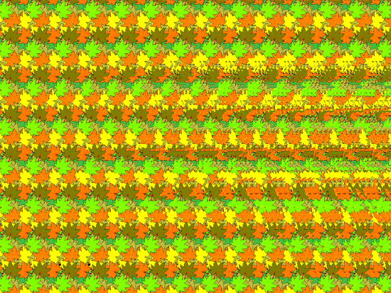
\(\newcommand{\avec}{\mathbf a}\) \(\newcommand{\bvec}{\mathbf b}\) \(\newcommand{\cvec}{\mathbf c}\) \(\newcommand{\dvec}{\mathbf d}\) \(\newcommand{\dtil}{\widetilde{\mathbf d}}\) \(\newcommand{\evec}{\mathbf e}\) \(\newcommand{\fvec}{\mathbf f}\) \(\newcommand{\nvec}{\mathbf n}\) \(\newcommand{\pvec}{\mathbf p}\) \(\newcommand{\qvec}{\mathbf q}\) \(\newcommand{\svec}{\mathbf s}\) \(\newcommand{\tvec}{\mathbf t}\) \(\newcommand{\uvec}{\mathbf u}\) \(\newcommand{\vvec}{\mathbf v}\) \(\newcommand{\wvec}{\mathbf w}\) \(\newcommand{\xvec}{\mathbf x}\) \(\newcommand{\yvec}{\mathbf y}\) \(\newcommand{\zvec}{\mathbf z}\) \(\newcommand{\rvec}{\mathbf r}\) \(\newcommand{\mvec}{\mathbf m}\) \(\newcommand{\zerovec}{\mathbf 0}\) \(\newcommand{\onevec}{\mathbf 1}\) \(\newcommand{\real}{\mathbb R}\) \(\newcommand{\twovec}[2]{\left[\begin{array}{r}#1 \\ #2 \end{array}\right]}\) \(\newcommand{\ctwovec}[2]{\left[\begin{array}{c}#1 \\ #2 \end{array}\right]}\) \(\newcommand{\threevec}[3]{\left[\begin{array}{r}#1 \\ #2 \\ #3 \end{array}\right]}\) \(\newcommand{\cthreevec}[3]{\left[\begin{array}{c}#1 \\ #2 \\ #3 \end{array}\right]}\) \(\newcommand{\fourvec}[4]{\left[\begin{array}{r}#1 \\ #2 \\ #3 \\ #4 \end{array}\right]}\) \(\newcommand{\cfourvec}[4]{\left[\begin{array}{c}#1 \\ #2 \\ #3 \\ #4 \end{array}\right]}\) \(\newcommand{\fivevec}[5]{\left[\begin{array}{r}#1 \\ #2 \\ #3 \\ #4 \\ #5 \\ \end{array}\right]}\) \(\newcommand{\cfivevec}[5]{\left[\begin{array}{c}#1 \\ #2 \\ #3 \\ #4 \\ #5 \\ \end{array}\right]}\) \(\newcommand{\mattwo}[4]{\left[\begin{array}{rr}#1 \amp #2 \\ #3 \amp #4 \\ \end{array}\right]}\) \(\newcommand{\laspan}[1]{\text{Span}\{#1\}}\) \(\newcommand{\bcal}{\cal B}\) \(\newcommand{\ccal}{\cal C}\) \(\newcommand{\scal}{\cal S}\) \(\newcommand{\wcal}{\cal W}\) \(\newcommand{\ecal}{\cal E}\) \(\newcommand{\coords}[2]{\left\{#1\right\}_{#2}}\) \(\newcommand{\gray}[1]{\color{gray}{#1}}\) \(\newcommand{\lgray}[1]{\color{lightgray}{#1}}\) \(\newcommand{\rank}{\operatorname{rank}}\) \(\newcommand{\row}{\text{Row}}\) \(\newcommand{\col}{\text{Col}}\) \(\renewcommand{\row}{\text{Row}}\) \(\newcommand{\nul}{\text{Nul}}\) \(\newcommand{\var}{\text{Var}}\) \(\newcommand{\corr}{\text{corr}}\) \(\newcommand{\len}[1]{\left|#1\right|}\) \(\newcommand{\bbar}{\overline{\bvec}}\) \(\newcommand{\bhat}{\widehat{\bvec}}\) \(\newcommand{\bperp}{\bvec^\perp}\) \(\newcommand{\xhat}{\widehat{\xvec}}\) \(\newcommand{\vhat}{\widehat{\vvec}}\) \(\newcommand{\uhat}{\widehat{\uvec}}\) \(\newcommand{\what}{\widehat{\wvec}}\) \(\newcommand{\Sighat}{\widehat{\Sigma}}\) \(\newcommand{\lt}{<}\) \(\newcommand{\gt}{>}\) \(\newcommand{\amp}{&}\) \(\definecolor{fillinmathshade}{gray}{0.9}\)Figure \(\PageIndex{1}\) appears at first glance to be just a pattern of colored leaves, but hidden within it is the three-dimensional shape of an ant. Can you see the ant among the leaves? This figure is an example of a stereogram, which is a two-dimensional picture that reveals a three-dimensional object when viewed correctly. If you can’t see the hidden image, it doesn’t mean that there is anything wrong with your eyes. It’s all in how your brain interprets what your eyes are sensing. The eyes are special sensory organs, and vision is one of our special senses.
Special and General Senses
The human body has two basic types of senses, called special senses and general senses. Special senses have specialized sense organs that gather sensory information and change it into nerve impulses. Special senses include the vision for which the eyes are the specialized sense organs, hearing (ears), balance (ears), taste (tongue), and smell (nasal passages). General senses, in contrast, are all associated with the sense of touch and lack special sense organs. Instead, sensory information about touch is gathered by the skin and other body tissues, all of which have important functions besides gathering sensory information. Whether the senses are special or general, however, all of them depend on cells called sensory receptors.
Sensory Receptors
A sensory receptor is a specialized nerve cell that responds to a stimulus in the internal or external environment by generating a nerve impulse. The nerve impulse then travels along with the sensory (afferent) nerve to the central nervous system for processing and to form a response.
There are several different types of sensory receptors that respond to different kinds of stimuli:
- Mechanoreceptors respond to mechanical forces such as pressure, roughness, vibration, and stretching. Most mechanoreceptors are found in the skin and are needed for the sense of touch. Mechanoreceptors are also found in the inner ear where they are needed for the senses of hearing and balance.
- Thermoreceptors respond to variations in temperature. They are found mostly in the skin and detect temperatures that are above or below body temperature.
- Nociceptors respond to potentially damaging stimuli, which are generally perceived as pain. They are found in internal organs as well as on the surface of the body. Different nociceptors are activated depending on the particular stimulus. For example, some detect damaging heat or cold, others detect excessive pressure, and still, others detect painful chemicals such as very hot spices in food.
- Photoreceptors detect and respond to light. Most photoreceptors are found in the eyes and are needed for the sense of vision.
- Chemoreceptors respond to certain chemicals. They are found mainly in taste buds on the tongue, where they are needed for the sense of taste; and in nasal passages, where they are needed for the sense of smell.
Touch
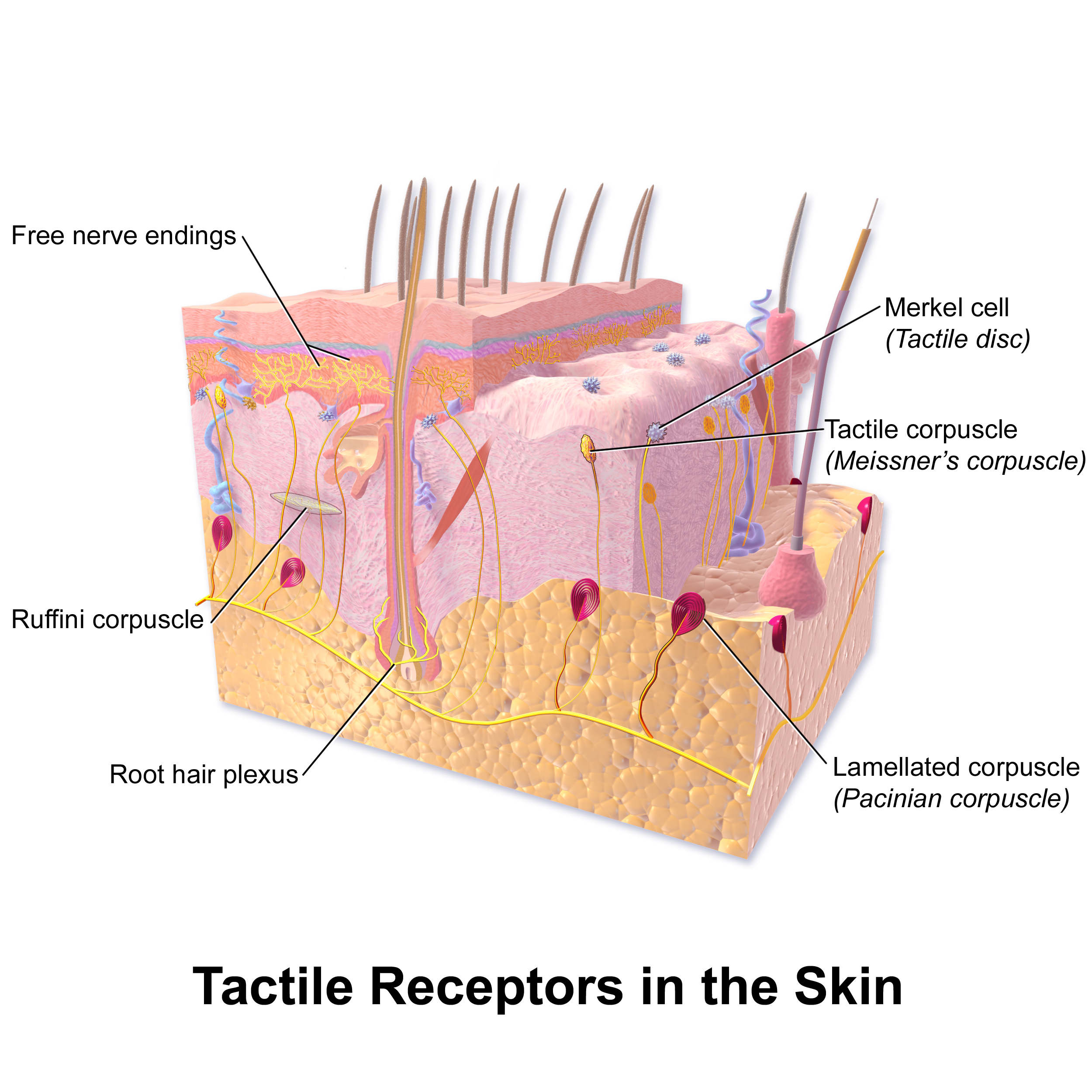
Touch is the ability to sense pressure, vibration, temperature, pain, and other tactile stimuli. These types of stimuli are detected by mechanoreceptors, thermoreceptors, and nociceptors all over the body, but most noticeably in the skin. These receptors are especially concentrated on the tongue, lips, face, palms of the hands, and soles of the feet. Various types of tactile receptors in the skin are shown in Figure \(\PageIndex{2}\).
Vision
Vision, or sight, is the ability to sense light and see. The eye is the special sensory organ that collects and focuses light and forms images. However, the eye is not sufficient for us to see. The brain also plays a necessary role in vision.
How the Eye Works
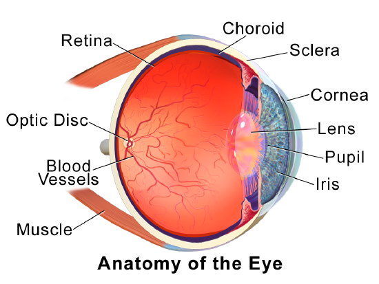
Figure \(\PageIndex{3}\) shows the anatomy of the human eye in cross-section. The eye gathers and focuses light to form an image and then changes the image to nerve impulses that travel to the brain. How the eye performs these functions is summarized in the following steps.
- Light passes first through the cornea, which is a clear outer layer that protects the eye and helps to focus the light by refracting, or bending, it.
- Light next enters the interior of the eye through an opening called the pupil. The size of this opening is controlled by the colored part of the eye, called the iris, which adjusts the size based on the brightness of the light. The iris causes the pupil to narrow in bright light and widen in dim light.
- The light then passes through the lens, which refracts the light even more and focuses it on the retina at the back of the eye as an inverted image.
- The retina contains photoreceptor cells of two types, called rods and cones. Rods, which are found mainly in all areas of the retina other than the very center, are particularly sensitive to low levels of light. Cones, which are found mainly in the center of the retina, are sensitive to light of different colors and allow color vision. The rods and cones convert the light that strikes them to nerve impulses.
- The nerve impulses from the rods and cones travel to the optic nerve via the optic disc, which is a circular area at the back of the eye where the optic nerve connects to the retina.
Role of the Brain in Vision
The optic nerves from both eyes meet and cross just below the bottom of the hypothalamus in the brain. The information from both eyes is sent to the visual cortex in the occipital lobe of the cerebrum, which is part of the cerebral cortex. The visual cortex is the largest system in the human brain and is responsible for processing visual images. It interprets messages from both eyes and “tells” us what we are seeing.
Vision Problems
Vision problems are very common. Two of the most common are myopia and hyperopia, and they often start in childhood or adolescence. Another common problem, called presbyopia, occurs in most people beginning in middle adulthood. All three problems result in blurred vision due to the failure of the eyes to focus images correctly on the retina.
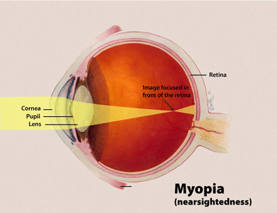
Myopia
Myopia, or nearsightedness, occurs when the light that comes into the eye does not directly focus on the retina but in front of it, as shown in Figure \(\PageIndex{4}\). This causes the image of distant objects to be out of focus but does not affect the focus of close objects. Myopia may occur because the eyeball is elongated from front to back or because the cornea is too curved. Myopia can be corrected through the use of corrective lenses, either eyeglasses or contact lenses. Myopia can also be corrected by refractive surgery performed with a laser.
Hyperopia
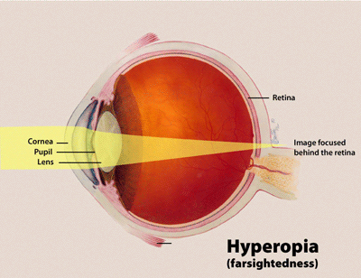
Hyperopia, or farsightedness, occurs when the light that comes into the eye does not directly focus on the retina but behind it, as shown in Figure \(\PageIndex{5}\). This causes the image of close objects to be out of focus but does not affect the focus of distant objects. Hyperopia may occur because the eyeball is too short from front to back or because the lens is not curved enough. Hyperopia can be corrected through the use of corrective lenses or laser surgery.
Presbyopia
Presbyopia is a vision problem associated with aging in which the eye gradually loses its ability to focus on close objects. The precise cause of presbyopia is not known for certain, but evidence suggests that the lens may become less elastic with age, and the muscles that control the lens may lose power as people grow older. The first signs of presbyopia – eyestrain, difficulty seeing in dim light, problems focusing on small objects, and fine print – are usually first noticed between the ages of 40 and 50. Most older people with this problem use corrective lenses to focus on close objects because surgical procedures to correct presbyopia have not been as successful as those for myopia and hyperopia.
Hearing
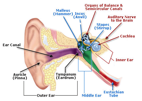
Hearing is the ability to sense sound waves, and the ear is the organ that senses sound. Sound waves enter the ear through the ear canal and travel to the eardrum (see the diagram of the ear in Figure \(\PageIndex{6}\)). The sound waves strike the eardrum and make it vibrate. The vibrations then travel through the three tiny bones (hammer, anvil, and stirrup) of the middle ear, which amplify the vibrations. From the middle ear, the vibrations pass to the cochlea in the inner ear. The cochlea is a coiled tube filled with liquid. The liquid moves in response to the vibrations, causing tiny hair cells (which are mechanoreceptors) lining the cochlea to bend. In response, the hair cells send nerve impulses to the auditory nerve, which carries the impulses to the brain. The brain interprets the impulses and “tells” us what we are hearing.
Taste and Smell
Taste and smell are both abilities to sense chemicals, so taste and olfactory (odor) receptors are chemoreceptors. Both types of chemoreceptors send nerve impulses to the brain along sensory nerves, and the brain “tells” us what we are tasting or smelling.
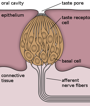
Taste receptors are found in tiny bumps on the tongue called taste buds. You can see a diagram of a taste receptor cell and related structures in Figure \(\PageIndex{7}\). Taste receptor cells make contact with chemicals in food through tiny openings called taste pores. When certain chemicals bind with taste receptor cells, it generates nerve impulses that travel through afferent nerves to the CNS. There are separate taste receptors for sweet, salty, sour, bitter, and meaty tastes. The meaty or savory taste is called umami.
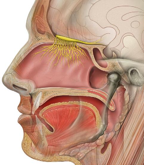
Olfactory receptors line the passages inside the nasal passages (Figure \(\PageIndex{8}\)). There are millions of olfactory receptors, which sense chemicals in the air. Unlike taste receptors, which can sense only five different tastes, olfactory receptors can sense hundreds of different odors and send signals to the olfactory bulb of the brain. Did you ever notice that food seems to have less taste when you have a stuffy nose? This occurs because the sense of smell contributes to the sense of taste, and a stuffy nose interferes with the ability to smell.
The most common cause of blindness in the Western hemisphere is age-related macular degeneration (AMD). About 15 million people in the United States have this type of blindness, and 30 million people are affected worldwide. At present, there is no cure for AMD. The disease occurs with the death of a layer of cells called retinal pigment epithelium, which normally provides nutrients and other support to the macula of the eye. The macula is an oval-shaped pigmented area near the center of the retina that is specialized for high visual acuity and has the retina’s greatest concentration of cones. When the epithelial cells die and the macula is no longer supported or nourished, the macula also starts to die. Patients experience a black spot in the center of their vision, and as the disease progresses, the black spot grows outward. Patients eventually lose the ability to read and even to recognize familiar faces before developing total blindness.
In 2016, a landmark surgery was performed as a trial on a patient with severe AMD. In the first-ever operation of its kind, Dr. Pete Coffey of the University of London implanted a tiny patch of cells behind the retina in each of the patient’s eyes. The cells were retinal pigmented epithelial cells that had been grown in a lab from stem cells, which are undifferentiated cells that have the ability to develop into other cell types. By six months out from the operation, the new cells were still surviving, and the doctor was hopeful that the patient’s vision loss would stop and even be reversed. At that point, several other operations had already been planned to test the new procedure. If these cases are a success, Dr. Coffey predicts that the surgery will become as routine as cataract surgery and prevent millions of patients from losing their vision.
Review
- Compare and contrast special senses and general senses.
- What are sensory receptors?
- List five types of sensory receptors and the type of stimulus each detects.
- Describe the range of tactile stimuli that are detected in the sense of touch.
- Explain how the eye collects and focuses light to form an image and converts it to nerve impulses.
- Identify two common vision problems, including both their causes and their effects on vision.
- Explain how the structures of the ear collect and amplify sound waves and transform them into nerve impulses.
- What role does the ear play in balance? Which structures of the ear are involved in balance?
- Describe two ways that the body senses chemicals and the special sense organs that are involved in these senses.
- Explain why your skin can detect different types of stimuli, such as pressure and temperature.
- Choose one. Sensory information is sent to the central nervous system via (efferent/afferent) nerves.
- Identify a mechanoreceptor used in two different human senses, and describe the type of mechanical stimuli that each one detects.
- If a person is blind but their retina is functioning properly, where do you think the damage might be? Explain your answer.
- When you see colors, what receptor cells are activated? Where are these receptors located? What lobe of the brain is primarily used to process visual information?
- The auditory nerve carries:
- Smell information
- Taste information
- Balance information
- Sound information
Explore More
Some people “see” sounds, “hear” colors, or “taste” words. This rare ability is called synesthesia, and it is thought to be caused by cross-wiring of the senses in the brain. To learn more about this intriguing phenomenon, watch this fascinating TED animation:
Many people experience the dizzying effects of vertigo at some point in their lives. Learn more here:
Attributions
- Bigant by GifTagger assumed CC BY 3.0 via Wikimedia Commons
- Skin tactile receptors by Blausen.com staff (2014). "Medical gallery of Blausen Medical 2014". WikiJournal of Medicine 1 (2). DOI:10.15347/wjm/2014.010. ISSN 2002-4436. CC BY 3.0 via Wikimedia Commons
- Eye anatomy by Blausen.com staff (2014). "Medical gallery of Blausen Medical 2014". WikiJournal of Medicine 1 (2). DOI:10.15347/wjm/2014.010. ISSN 2002-4436. CC BY 3.0 via Wikimedia Commons
- Myopia by National Eye Institute, public domain via Wikimedia Commons
- Hyperopia by National Eye Institute, public domain via Wikimedia Commons
- Human ear public domain via Wikimedia Commons
- Taste buds by Jonas Töle dedicated CC0 via Wikimedia Commons
- Head olfactory nerve by Patrick J. Lynch, medical illustrator, CC BY 2.5 via Wikimedia Commons
- Text adapted from Human Biology by CK-12 licensed CC BY-NC 3.0