
11.5: Central Nervous System
- Page ID
- 22523

\( \newcommand{\vecs}[1]{\overset { \scriptstyle \rightharpoonup} {\mathbf{#1}} } \)
\( \newcommand{\vecd}[1]{\overset{-\!-\!\rightharpoonup}{\vphantom{a}\smash {#1}}} \)
\( \newcommand{\id}{\mathrm{id}}\) \( \newcommand{\Span}{\mathrm{span}}\)
( \newcommand{\kernel}{\mathrm{null}\,}\) \( \newcommand{\range}{\mathrm{range}\,}\)
\( \newcommand{\RealPart}{\mathrm{Re}}\) \( \newcommand{\ImaginaryPart}{\mathrm{Im}}\)
\( \newcommand{\Argument}{\mathrm{Arg}}\) \( \newcommand{\norm}[1]{\| #1 \|}\)
\( \newcommand{\inner}[2]{\langle #1, #2 \rangle}\)
\( \newcommand{\Span}{\mathrm{span}}\)
\( \newcommand{\id}{\mathrm{id}}\)
\( \newcommand{\Span}{\mathrm{span}}\)
\( \newcommand{\kernel}{\mathrm{null}\,}\)
\( \newcommand{\range}{\mathrm{range}\,}\)
\( \newcommand{\RealPart}{\mathrm{Re}}\)
\( \newcommand{\ImaginaryPart}{\mathrm{Im}}\)
\( \newcommand{\Argument}{\mathrm{Arg}}\)
\( \newcommand{\norm}[1]{\| #1 \|}\)
\( \newcommand{\inner}[2]{\langle #1, #2 \rangle}\)
\( \newcommand{\Span}{\mathrm{span}}\) \( \newcommand{\AA}{\unicode[.8,0]{x212B}}\)
\( \newcommand{\vectorA}[1]{\vec{#1}} % arrow\)
\( \newcommand{\vectorAt}[1]{\vec{\text{#1}}} % arrow\)
\( \newcommand{\vectorB}[1]{\overset { \scriptstyle \rightharpoonup} {\mathbf{#1}} } \)
\( \newcommand{\vectorC}[1]{\textbf{#1}} \)
\( \newcommand{\vectorD}[1]{\overrightarrow{#1}} \)
\( \newcommand{\vectorDt}[1]{\overrightarrow{\text{#1}}} \)
\( \newcommand{\vectE}[1]{\overset{-\!-\!\rightharpoonup}{\vphantom{a}\smash{\mathbf {#1}}}} \)
\( \newcommand{\vecs}[1]{\overset { \scriptstyle \rightharpoonup} {\mathbf{#1}} } \)
\( \newcommand{\vecd}[1]{\overset{-\!-\!\rightharpoonup}{\vphantom{a}\smash {#1}}} \)
\(\newcommand{\avec}{\mathbf a}\) \(\newcommand{\bvec}{\mathbf b}\) \(\newcommand{\cvec}{\mathbf c}\) \(\newcommand{\dvec}{\mathbf d}\) \(\newcommand{\dtil}{\widetilde{\mathbf d}}\) \(\newcommand{\evec}{\mathbf e}\) \(\newcommand{\fvec}{\mathbf f}\) \(\newcommand{\nvec}{\mathbf n}\) \(\newcommand{\pvec}{\mathbf p}\) \(\newcommand{\qvec}{\mathbf q}\) \(\newcommand{\svec}{\mathbf s}\) \(\newcommand{\tvec}{\mathbf t}\) \(\newcommand{\uvec}{\mathbf u}\) \(\newcommand{\vvec}{\mathbf v}\) \(\newcommand{\wvec}{\mathbf w}\) \(\newcommand{\xvec}{\mathbf x}\) \(\newcommand{\yvec}{\mathbf y}\) \(\newcommand{\zvec}{\mathbf z}\) \(\newcommand{\rvec}{\mathbf r}\) \(\newcommand{\mvec}{\mathbf m}\) \(\newcommand{\zerovec}{\mathbf 0}\) \(\newcommand{\onevec}{\mathbf 1}\) \(\newcommand{\real}{\mathbb R}\) \(\newcommand{\twovec}[2]{\left[\begin{array}{r}#1 \\ #2 \end{array}\right]}\) \(\newcommand{\ctwovec}[2]{\left[\begin{array}{c}#1 \\ #2 \end{array}\right]}\) \(\newcommand{\threevec}[3]{\left[\begin{array}{r}#1 \\ #2 \\ #3 \end{array}\right]}\) \(\newcommand{\cthreevec}[3]{\left[\begin{array}{c}#1 \\ #2 \\ #3 \end{array}\right]}\) \(\newcommand{\fourvec}[4]{\left[\begin{array}{r}#1 \\ #2 \\ #3 \\ #4 \end{array}\right]}\) \(\newcommand{\cfourvec}[4]{\left[\begin{array}{c}#1 \\ #2 \\ #3 \\ #4 \end{array}\right]}\) \(\newcommand{\fivevec}[5]{\left[\begin{array}{r}#1 \\ #2 \\ #3 \\ #4 \\ #5 \\ \end{array}\right]}\) \(\newcommand{\cfivevec}[5]{\left[\begin{array}{c}#1 \\ #2 \\ #3 \\ #4 \\ #5 \\ \end{array}\right]}\) \(\newcommand{\mattwo}[4]{\left[\begin{array}{rr}#1 \amp #2 \\ #3 \amp #4 \\ \end{array}\right]}\) \(\newcommand{\laspan}[1]{\text{Span}\{#1\}}\) \(\newcommand{\bcal}{\cal B}\) \(\newcommand{\ccal}{\cal C}\) \(\newcommand{\scal}{\cal S}\) \(\newcommand{\wcal}{\cal W}\) \(\newcommand{\ecal}{\cal E}\) \(\newcommand{\coords}[2]{\left\{#1\right\}_{#2}}\) \(\newcommand{\gray}[1]{\color{gray}{#1}}\) \(\newcommand{\lgray}[1]{\color{lightgray}{#1}}\) \(\newcommand{\rank}{\operatorname{rank}}\) \(\newcommand{\row}{\text{Row}}\) \(\newcommand{\col}{\text{Col}}\) \(\renewcommand{\row}{\text{Row}}\) \(\newcommand{\nul}{\text{Nul}}\) \(\newcommand{\var}{\text{Var}}\) \(\newcommand{\corr}{\text{corr}}\) \(\newcommand{\len}[1]{\left|#1\right|}\) \(\newcommand{\bbar}{\overline{\bvec}}\) \(\newcommand{\bhat}{\widehat{\bvec}}\) \(\newcommand{\bperp}{\bvec^\perp}\) \(\newcommand{\xhat}{\widehat{\xvec}}\) \(\newcommand{\vhat}{\widehat{\vvec}}\) \(\newcommand{\uhat}{\widehat{\uvec}}\) \(\newcommand{\what}{\widehat{\wvec}}\) \(\newcommand{\Sighat}{\widehat{\Sigma}}\) \(\newcommand{\lt}{<}\) \(\newcommand{\gt}{>}\) \(\newcommand{\amp}{&}\) \(\definecolor{fillinmathshade}{gray}{0.9}\)Figure \(\PageIndex{1}\) is a very odd-looking drawing and is called a homunculus. The mass represents a cross-sectional wedge of the human brain. The drawing shows some areas of the brain associated with different parts of the body. As you can see, larger areas of the brain in this region are associated with the hands, face, and tongue than the legs and feet. Given the importance of speech, manual dexterity, and face-to-face social interactions in human beings, it is not surprising that relatively large areas of the brain are needed to control these body parts. The brain is the most complex organ in the human body and part of the central nervous system.

What Is the Central Nervous System?
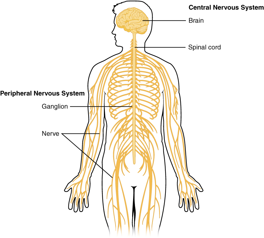
The central nervous system (CNS) is the part of the nervous system that includes the brain and spinal cord. Figure \(\PageIndex{2}\) shows the central nervous system as one of the two main divisions of the total nervous system. The other main division is the peripheral nervous system (PNS). The CNS and PNS work together to control virtually all body functions. You can read much more about the PNS in the concept Peripheral Nervous System.
The delicate nervous tissues of the central nervous system are protected by major physical and chemical barriers. Physically, the brain and spinal cord are surrounded by tough meninges, a three-layer protective sheath that also contains cushioning cerebrospinal fluid. The bones of the skull and spinal vertebrae also contribute to physically protecting the brain and spinal cord. Chemically, the brain and spinal cord are isolated from the circulation — and most toxins or pathogens in the blood — by the blood-brain barrier. The blood-brain barrier is a highly selective membrane formed of endothelial cells (a type of glial cells) that separates the circulating blood from the extracellular fluid in the CNS. The barrier allows water, certain gases, glucose, and some other molecules needed by the brain and spinal cord to cross from the blood into the CNS while keeping out potentially harmful substances. These physical and chemical barriers make the CNS less susceptible to injury. However, damage to the CNS is likely to have more serious consequences.
The Brain
The brain is the control center not only of the rest of the nervous system but of the entire organism. The adult brain makes up only about 2 percent of the body’s weight, but it uses about 20 percent of the body’s total energy. The brain contains an estimated one hundred billion neurons, and each neuron has thousands of synaptic connections to other neurons. The brain also has about the same number of glial cells as neurons. No wonder the brain uses so much energy! In addition, the brain uses mostly glucose for energy. As a result, if the brain is deprived of glucose, it can lead to unconsciousness. The brain is able to store some glucose in the form of glycogen, but in much smaller amounts than are found in the liver and skeletal muscles.
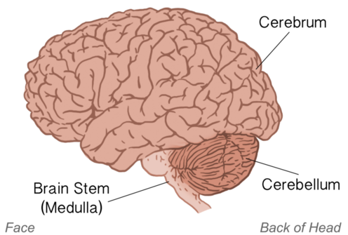
The brain controls such mental processes as reasoning, imagination, memory, and language. It also interprets information from the senses and commands the body how to respond. It controls basic physical processes such as breathing and heartbeat as well as voluntary activities such as walking and writing. The brain has three major parts: the cerebrum, cerebellum, and brain stem (Figure \(\PageIndex{3}\)). The figure shows the brain from the left side of the head. It shows how the brain would appear if the skull and meninges were removed. The brain stem via its medulla links to the spinal cord. The cerebellum is a small section at the back of the brain. The largest part of the brain is the cerebrum.
Cerebrum
The cerebrum is the largest part of the brain. It controls conscious, intellectual functions. For example, it controls reasoning, language, memory, sight, touch, and hearing. When you read a book, play a video game, or recognize a classmate, you are using your cerebrum.
Hemispheres and Lateralization of the Cerebrum
The cerebrum is divided from front to back into two halves called the left and right hemispheres. The two hemispheres are connected by a thick bundle of axons, known as the corpus callosum, which lies deep within the brain. The corpus callosum is the main avenue of communication between the two hemispheres. It connects each point in the cerebrum to the mirror-image point in the opposite hemisphere.
The right and left hemispheres of the cerebrum are similar in shape, and most areas of the cerebrum are found in both hemispheres. Some areas, however, show lateralization, or a concentration in one hemisphere or the other. For example, in most people, language functions are more concentrated in the left hemisphere, whereas abstract reasoning and visual-spatial abilities are more concentrated in the right hemisphere.
For reasons that are not yet clear, each hemisphere of the brain interacts primarily with the opposite side of the body. The left side of the brain receives messages from and sends commands to the right side of the body, and the right side of the brain receives messages from and sends commands to the left side of the body. Sensory nerves from the spinal cord to the brain and motor nerves from the brain to the spinal cord both cross the midline of the body at the level of the brain stem.
Cerebral Cortex
Most of the information processing in the brain actually takes place in the cerebral cortex. This is a rind of gray matter and other tissues just a few millimeters thick that makes up the outer surface of the cerebrum in both hemispheres of the brain. The cerebral cortex has many folds in it that greatly increase the amount of surface area of the brain that can fit within the skull. Because of all the folds in the human cerebral cortex, it has a surface area of about 2,500 cm2(2.5 ft2). The size and importance of the cerebral cortex are far greater in the human brain than the brains of any other vertebrates including nonhuman primates.
Lobes of the Cerebral Cortex
Each hemisphere of the cerebrum is further divided into the four lobes shown in Figure \(\PageIndex{4}\) and described below.
1. The frontal lobes are located at the front of the brain behind the forehead. The frontal lobes are associated with executive functions such as attention, self-control, planning, problem-solving, reasoning, abstract thought, language, and personality.
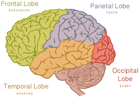
2. The parietal lobes are located behind the frontal lobes at the top of the head. The parietal lobes are involved in sensation, including temperature, touch, and taste. Reading and arithmetic are also functions of the parietal lobes.
3. The temporal lobes are located at the sides of the head below the frontal and parietal lobes. The temporal lobes enable hearing, the formation and retrieval of memories, and the integration of memories and sensations.
4. The occipital lobes are located at the back of the head below the parietal lobes. The occipital lobes are the smallest of the four pairs of lobes. They are dedicated almost solely to vision.
Inner Structures of the Brain
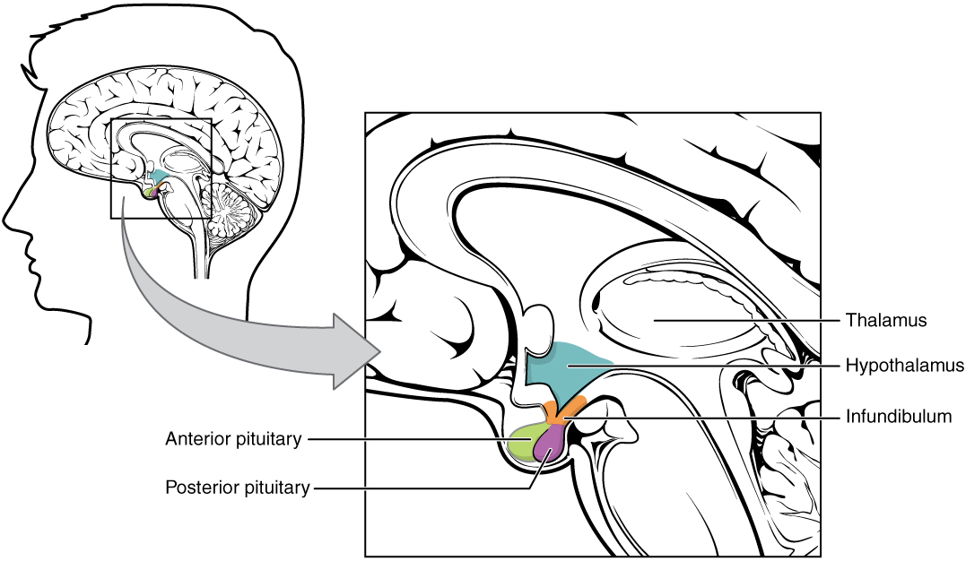
Several structures are located deep within the brain and are important for communication between the brain and spinal cord or the rest of the body. These structures include the hypothalamus and thalamus. Figure \(\PageIndex{5}\) shows where these structures are located in the brain. The cerebrum, hypothalamus, and thalamus exist in two halves, one in each hemisphere.
Hypothalamus
The hypothalamus is located just above the brain stem and is about the size of an almond. The hypothalamus is responsible for certain metabolic processes and other activities of the autonomic nervous system, including body temperature, heart rate, hunger, thirst, fatigue, sleep, wakefulness, and circadian (24-hour) rhythms. The hypothalamus is also an important emotional center of the brain. The hypothalamus can regulate so many body functions because it responds to many different internal and external signals, including messages from the brain, light, steroid hormones, stress, and invading pathogens, among others.
One way the hypothalamus influences body functions is by synthesizing hormones that directly influence body processes. For example, it synthesizes the hormone oxytocin, which stimulates uterine contractions during childbirth and the letdown of milk during lactation. It also synthesizes the hormone vasopressin (also called antidiuretic hormone), which stimulates the kidneys to reabsorb more water and excrete more concentrated urine. These two hormones are sent from the hypothalamus via a stalk-like structure called the infundibulum (see diagram above) directly to the posterior (back) portion of the pituitary gland, which secretes them into the blood.
The main way the hypothalamus influences body functions is by controlling the pituitary gland, known as the master gland of the endocrine system. The hypothalamus synthesizes neurohormones called releasing factors that travel through the infundibulum directly to the anterior (front) part of the pituitary gland. The releasing factors generally either stimulate or inhibit the secretion of anterior pituitary hormones, most of which control other glands of the endocrine system.
Thalamus
The thalamus, which is located near the hypothalamus (Figure \(\PageIndex{5}\)), is a major hub for information traveling back and forth between the spinal cord and cerebrum. It filters sensory information traveling to the cerebrum. It relays sensory signals to the cerebral cortex and motor signals to the spinal cord. It is also involved in the regulation of consciousness, sleep, and alertness.
Cerebellum
The cerebellum is just below the cerebrum and at the back of the brain behind the brain stem (Figure \(\PageIndex{3}\)). It coordinates body movements and is involved in movements that are learned with repeated practice. For example, when you hit a softball with a bat or touch type on a keyboard you are using the cerebellum. Many nerve pathways link the cerebellum with motor neurons throughout the body.
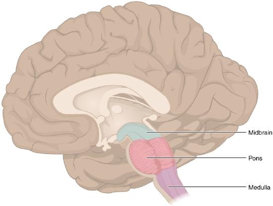
Brain Stem
Sometimes called the “lower brain,” the brain stem is the lower part of the brain that is joined to the spinal cord. There are three parts to the brainstem: the midbrain, the pons, and the medulla oblongata, which are shown in Figure \(\PageIndex{6}\) below. The brain stem is primarily involved in the unconscious autonomic functions as well as several types of sensory information. It also helps coordinate large body movements such as walking and running. The midbrain deals with sight and sound information and translates these inputs before sending them to the forebrain. The pons relays messages to other parts of the brain (primarily the cerebrum and cerebellum) and helps regulate breathing. Some researchers have hypothesized that the pons plays a role in dreaming. Some of the functions of the Pons are shared by the medulla oblongata, also called the medulla. The medulla controls several subconscious homeostatic functions such as breathing, heart and blood vessel activity, swallowing, and digestion.
One of the brain stem’s most important roles is that of an “information highway.” That is, all of the information coming from the body to the brain and the information from the cerebrum to the body go through the brain stem. Sensory pathways for such things as pain, temperature, touch, and pressure sensation go upward to the cerebrum, and motor pathways for movement and other body processes go downward to the spinal cord. Most of the axons in the motor pathways cross from one side of the CNS to the other as they pass through the medulla oblongata. As a result, the right side of the brain controls much of the movement on the left side of the body, and the left side of the brain controls much of the movement on the right side of the body.
Spinal Cord
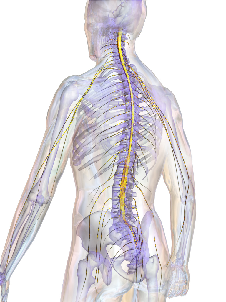
The spinal cord is a long, thin, tubular bundle of nervous tissues that extends from the brain stem and continues down the center of the back to the pelvis. It is highlighted in yellow in Figure \(\PageIndex{7}\). The spinal cord is enclosed within but is shorter than, the vertebral column.
Structure of the Spinal Cord
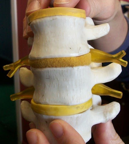
The center of the spinal cord consists of gray matter, which is made up mainly of cell bodies of neurons, including interneurons and motor neurons. The gray matter is surrounded by white matter that consists mainly of myelinated axons of motor and sensory neurons. Spinal nerves, which connect the spinal cord to the PNS, exit from the spinal cord between vertebrae (Figure \(\PageIndex{8}\)).
Functions of the Spinal Cord
The spinal cord serves as an information superhighway. It passes messages from the body to the brain and from the brain to the body. Sensory (afferent) nerves carry nerve impulses to the brain from sensory receptor cells everywhere in and on the body. Motor (efferent) nerves carry nerve impulses away from the brain to glands, organs, or muscles throughout the body.
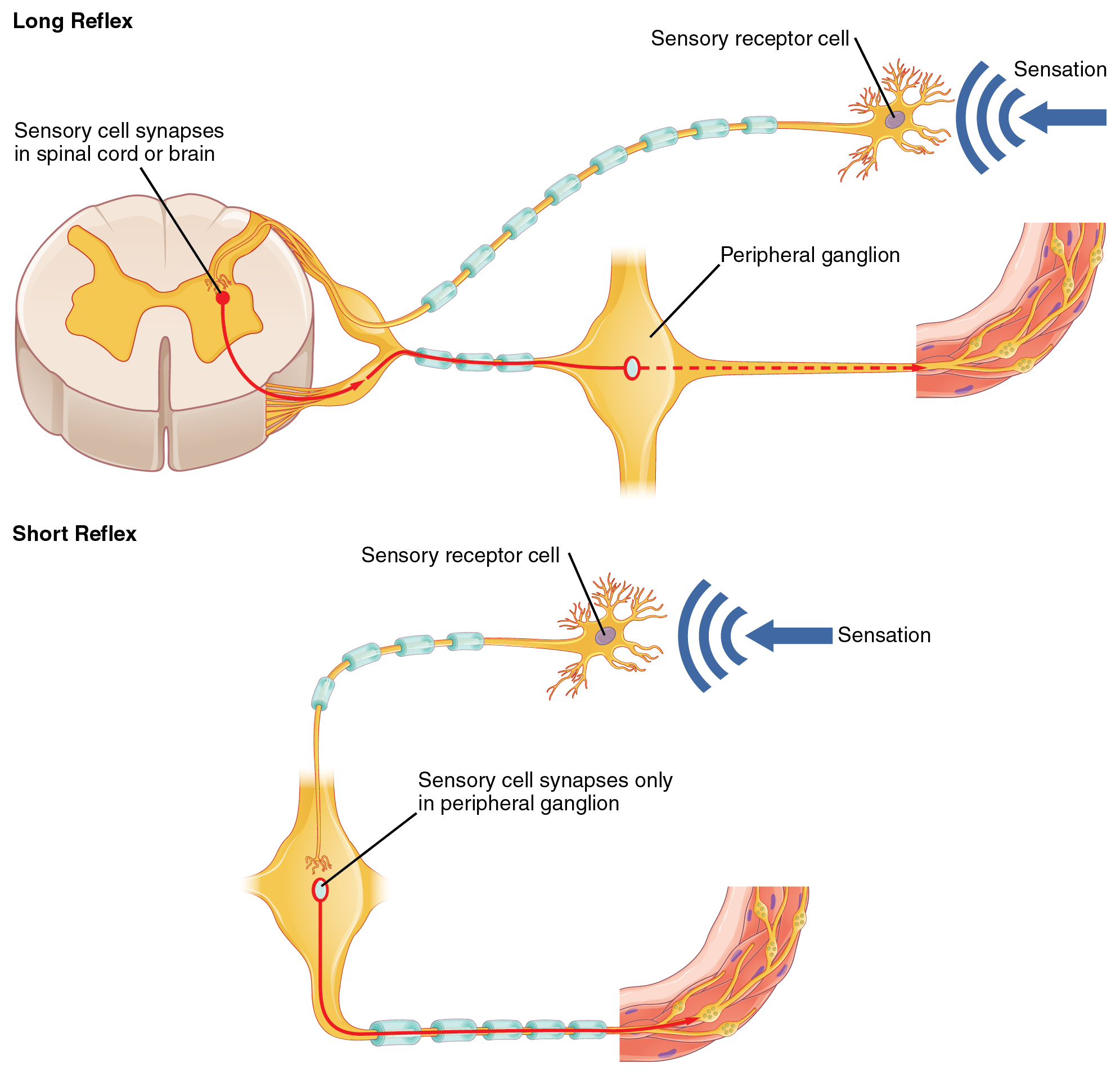
The spinal cord also independently controls certain rapid responses called reflexes without any input from the brain. You can see how this may happen in Figure \(\PageIndex{9}\). A sensory receptor responds to a sensation and sends a nerve impulse along a sensory nerve to the spinal cord. In the spinal cord, the message passes to an interneuron and from the interneuron to a motor nerve, which carries the impulse to a muscle. The muscle contracts in response. These neuron connections form a reflex arc, which requires no input from the brain. No doubt you have experienced such reflex actions yourself. For example, you may have reached out to touch a pot on the stove, not realizing that it was very hot. Virtually at the same moment that you feel the burning heat, you jerk your arm back and remove your hand from the pot.
Injuries to the Spinal Cord
Physical damage to the spinal cord may result in paralysis, which is a loss of sensation and movement in part of the body. Paralysis generally affects all the areas of the body below the level of the injury because nerve impulses are interrupted and can no longer travel back and forth between the brain and body beyond that point. If an injury to the spinal cord produces nothing more than swelling, the symptoms may be transient. However, if nerve fibers (axons) in the spinal cord are badly damaged, the loss of function may be permanent. Experimental studies have shown that spinal nerve fibers attempt to regrow, but tissue destruction usually produces scar tissue that cannot be penetrated by the regrowing nerves, as well as other factors that inhibit nerve fiber regrowth in the central nervous system.
Each year, many millions of people have a stroke, and stroke is the second leading cause of death in adults. Stroke, also known as cerebrovascular accident, occurs when poor blood flow to the brain results in the death of brain cells. There are two main types of strokes:
- Ischemic strokes occur due to a lack of blood flow because of a blood clot in an artery going to the brain.
- Hemorrhagic strokes occur due to bleeding from a broken blood vessel in the brain.
Either type of stroke may result in paralysis, loss of the ability to speak or comprehend speech, loss of bladder control, personality changes, and many other potential effects, depending on the part of the brain that is injured. The effects of a stroke may be mild and transient or more severe and permanent. A stroke may even be fatal. It generally depends on the type of stroke and how extensive it is.
Are you at risk of stroke? The main risk factor for stroke is age: about two-thirds of strokes occur in people over the age of 65. There is nothing you can do about your age, but most other stroke risk factors can be reduced with lifestyle changes or medications. The risk factors include high blood pressure, tobacco smoking, obesity, high blood cholesterol, diabetes mellitus, and atrial fibrillation.
Chances are good that you or someone you know is at risk of a stroke, so it is important to recognize a stroke if one occurs. Stoke is a medical emergency, and the more quickly treatment is given, the better the outcome is likely to be. In the case of ischemic strokes, the use of clot-busting drugs may prevent permanent brain damage if administered within 3 or 4 hours of the stroke. Remembering the signs of a stroke is easy.
They are summed up by the acronym FAST, as explained in the chart below.
Review
- What is the central nervous system?
- How is the central nervous system protected?
- What is the overall function of the brain?
- Identify the three main parts of the brain and one function of each part.
- Describe the hemispheres of the brain.
- Explain and give examples of lateralization of the brain.
- Identify one function of each of the four lobes of the cerebrum.
- Summarize the structure and function of the cerebral cortex.
- Explain how the hypothalamus controls the endocrine system.
- Describe the spinal cord.
- What is the main function of the spinal cord?
- Explain how reflex actions occur.
- Why do severe spinal cord injuries usually cause paralysis?
- What do you think are some possible consequences of severe damage to the brain stem? How might this compare to the consequences of severe damage to the frontal lobe? Explain your answer.
- Information travels very quickly in the nervous system, but generally, the longer the path between areas, the longer it takes. Based on this, explain why you think reflexes often occur at the spinal cord level and do not require input from the brain.
Explore More
More than 40 million people worldwide suffer from Alzheimer’s disease, a brain disorder, and the number is expected to grow dramatically in the coming decades. The disease was discovered more than a century ago, but little progress has been made in finding a cure. Watch this exciting TED talk in which scientist Samuel Cohen shares a new breakthrough in Alzheimer's research as well as a message of hope that a cure for Alzheimer’s will be found.
Attributions
- Sensory Homunculus by Popadius adapted from OpenStax, licensed CC BY 3.0 via Wikimedia Commons
- Overview of nervous system by OpenStax, licensed CC BY 4.0 via Wikimedia Commons
- Brain by Laura Guerin, CC BY-NC 3.0 via CK-12
- Brain lobes by Laura Guerin, CC BY-NC 3.0 via CK-12
- Hypothalamus-Pituitary Complex by OpenStax, licensed CC BY 4.0 via Wikimedia Commons
- Brain stem by OpenStax, licensed CC BY 4.0 via Wikimedia Commons
- Spinal cord by BruceBlaus licensed CC BY 3.0 via Wikimedia Commons
- Spinal readjustment by Tomwsulcer dedicated CC0 via Wikimedia Commons
- Short and long reflexes by OpenStax, licensed CC BY 4.0 via Wikimedia Commons
- Stroke Communications Kit by CDC, public domain
- Text adapted from Human Biology by CK-12 licensed CC BY-NC 3.0