
23.4: Fetal Stage
- Page ID
- 17806

\( \newcommand{\vecs}[1]{\overset { \scriptstyle \rightharpoonup} {\mathbf{#1}} } \)
\( \newcommand{\vecd}[1]{\overset{-\!-\!\rightharpoonup}{\vphantom{a}\smash {#1}}} \)
\( \newcommand{\dsum}{\displaystyle\sum\limits} \)
\( \newcommand{\dint}{\displaystyle\int\limits} \)
\( \newcommand{\dlim}{\displaystyle\lim\limits} \)
\( \newcommand{\id}{\mathrm{id}}\) \( \newcommand{\Span}{\mathrm{span}}\)
( \newcommand{\kernel}{\mathrm{null}\,}\) \( \newcommand{\range}{\mathrm{range}\,}\)
\( \newcommand{\RealPart}{\mathrm{Re}}\) \( \newcommand{\ImaginaryPart}{\mathrm{Im}}\)
\( \newcommand{\Argument}{\mathrm{Arg}}\) \( \newcommand{\norm}[1]{\| #1 \|}\)
\( \newcommand{\inner}[2]{\langle #1, #2 \rangle}\)
\( \newcommand{\Span}{\mathrm{span}}\)
\( \newcommand{\id}{\mathrm{id}}\)
\( \newcommand{\Span}{\mathrm{span}}\)
\( \newcommand{\kernel}{\mathrm{null}\,}\)
\( \newcommand{\range}{\mathrm{range}\,}\)
\( \newcommand{\RealPart}{\mathrm{Re}}\)
\( \newcommand{\ImaginaryPart}{\mathrm{Im}}\)
\( \newcommand{\Argument}{\mathrm{Arg}}\)
\( \newcommand{\norm}[1]{\| #1 \|}\)
\( \newcommand{\inner}[2]{\langle #1, #2 \rangle}\)
\( \newcommand{\Span}{\mathrm{span}}\) \( \newcommand{\AA}{\unicode[.8,0]{x212B}}\)
\( \newcommand{\vectorA}[1]{\vec{#1}} % arrow\)
\( \newcommand{\vectorAt}[1]{\vec{\text{#1}}} % arrow\)
\( \newcommand{\vectorB}[1]{\overset { \scriptstyle \rightharpoonup} {\mathbf{#1}} } \)
\( \newcommand{\vectorC}[1]{\textbf{#1}} \)
\( \newcommand{\vectorD}[1]{\overrightarrow{#1}} \)
\( \newcommand{\vectorDt}[1]{\overrightarrow{\text{#1}}} \)
\( \newcommand{\vectE}[1]{\overset{-\!-\!\rightharpoonup}{\vphantom{a}\smash{\mathbf {#1}}}} \)
\( \newcommand{\vecs}[1]{\overset { \scriptstyle \rightharpoonup} {\mathbf{#1}} } \)
\(\newcommand{\longvect}{\overrightarrow}\)
\( \newcommand{\vecd}[1]{\overset{-\!-\!\rightharpoonup}{\vphantom{a}\smash {#1}}} \)
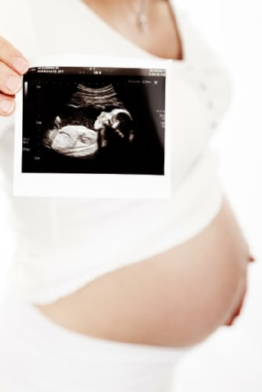
\(\newcommand{\avec}{\mathbf a}\) \(\newcommand{\bvec}{\mathbf b}\) \(\newcommand{\cvec}{\mathbf c}\) \(\newcommand{\dvec}{\mathbf d}\) \(\newcommand{\dtil}{\widetilde{\mathbf d}}\) \(\newcommand{\evec}{\mathbf e}\) \(\newcommand{\fvec}{\mathbf f}\) \(\newcommand{\nvec}{\mathbf n}\) \(\newcommand{\pvec}{\mathbf p}\) \(\newcommand{\qvec}{\mathbf q}\) \(\newcommand{\svec}{\mathbf s}\) \(\newcommand{\tvec}{\mathbf t}\) \(\newcommand{\uvec}{\mathbf u}\) \(\newcommand{\vvec}{\mathbf v}\) \(\newcommand{\wvec}{\mathbf w}\) \(\newcommand{\xvec}{\mathbf x}\) \(\newcommand{\yvec}{\mathbf y}\) \(\newcommand{\zvec}{\mathbf z}\) \(\newcommand{\rvec}{\mathbf r}\) \(\newcommand{\mvec}{\mathbf m}\) \(\newcommand{\zerovec}{\mathbf 0}\) \(\newcommand{\onevec}{\mathbf 1}\) \(\newcommand{\real}{\mathbb R}\) \(\newcommand{\twovec}[2]{\left[\begin{array}{r}#1 \\ #2 \end{array}\right]}\) \(\newcommand{\ctwovec}[2]{\left[\begin{array}{c}#1 \\ #2 \end{array}\right]}\) \(\newcommand{\threevec}[3]{\left[\begin{array}{r}#1 \\ #2 \\ #3 \end{array}\right]}\) \(\newcommand{\cthreevec}[3]{\left[\begin{array}{c}#1 \\ #2 \\ #3 \end{array}\right]}\) \(\newcommand{\fourvec}[4]{\left[\begin{array}{r}#1 \\ #2 \\ #3 \\ #4 \end{array}\right]}\) \(\newcommand{\cfourvec}[4]{\left[\begin{array}{c}#1 \\ #2 \\ #3 \\ #4 \end{array}\right]}\) \(\newcommand{\fivevec}[5]{\left[\begin{array}{r}#1 \\ #2 \\ #3 \\ #4 \\ #5 \\ \end{array}\right]}\) \(\newcommand{\cfivevec}[5]{\left[\begin{array}{c}#1 \\ #2 \\ #3 \\ #4 \\ #5 \\ \end{array}\right]}\) \(\newcommand{\mattwo}[4]{\left[\begin{array}{rr}#1 \amp #2 \\ #3 \amp #4 \\ \end{array}\right]}\) \(\newcommand{\laspan}[1]{\text{Span}\{#1\}}\) \(\newcommand{\bcal}{\cal B}\) \(\newcommand{\ccal}{\cal C}\) \(\newcommand{\scal}{\cal S}\) \(\newcommand{\wcal}{\cal W}\) \(\newcommand{\ecal}{\cal E}\) \(\newcommand{\coords}[2]{\left\{#1\right\}_{#2}}\) \(\newcommand{\gray}[1]{\color{gray}{#1}}\) \(\newcommand{\lgray}[1]{\color{lightgray}{#1}}\) \(\newcommand{\rank}{\operatorname{rank}}\) \(\newcommand{\row}{\text{Row}}\) \(\newcommand{\col}{\text{Col}}\) \(\renewcommand{\row}{\text{Row}}\) \(\newcommand{\nul}{\text{Nul}}\) \(\newcommand{\var}{\text{Var}}\) \(\newcommand{\corr}{\text{corr}}\) \(\newcommand{\len}[1]{\left|#1\right|}\) \(\newcommand{\bbar}{\overline{\bvec}}\) \(\newcommand{\bhat}{\widehat{\bvec}}\) \(\newcommand{\bperp}{\bvec^\perp}\) \(\newcommand{\xhat}{\widehat{\xvec}}\) \(\newcommand{\vhat}{\widehat{\vvec}}\) \(\newcommand{\uhat}{\widehat{\uvec}}\) \(\newcommand{\what}{\widehat{\wvec}}\) \(\newcommand{\Sighat}{\widehat{\Sigma}}\) \(\newcommand{\lt}{<}\) \(\newcommand{\gt}{>}\) \(\newcommand{\amp}{&}\) \(\definecolor{fillinmathshade}{gray}{0.9}\)This mother-to-be is holding an ultrasound image of her fetus. She is nearly nine months pregnant, so the fetus is fully developed and almost ready to be born. The fetus has grown tremendously and changed in many other ways since it was a tiny embryo seven months previously.
Defining the Fetal Stage
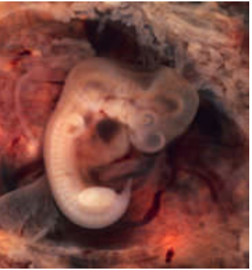
A fetus is a prenatal human being between the embryonic stage and birth. The fetal stage extends from the beginning of the ninth week after fertilization to about 38 weeks after fertilization, which is the average time of birth. The fetal stage lasts a total of approximately 30 weeks. Figure \(\PageIndex{2}\) shows a seven-week-old embryo that is just getting ready to begin the fetal stage of development. At 7 weeks the embryo is about 10 mm long and has a big forehead. It is developing the inner ear but not the outer ear. The limb buds are visible.
Fetal Development
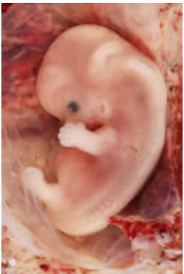
The image in Figure \(\PageIndex{3}\) shows a fetus at the start of week 9, the first week of the fetal stage. The fetus is shown larger than its actual size, which from crown to rump is only about 3.2 cm (1.3 in.) long. Even at this early age, however, the fetus has developed to the point of being recognizable as a human being. It possesses virtually all of the major body organs. However, most of the organs are not yet fully developed and functional, and some are not yet situated in their final anatomical locations. These final developments will occur during the remainder of the fetal stage.
Weeks 9 to 15
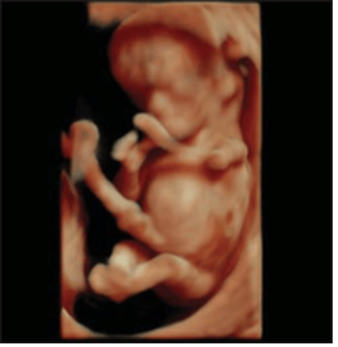
During weeks 9 to 15, the fetus’s reproductive organs develop rapidly. The external genitals of male and female fetuses are rather similar in appearance at first, but they will be clearly differentiated by week 12. At that point, the biological sex of the fetus can be determined with almost 100 percent accuracy using obstetric ultrasound. Figure \(\PageIndex{4}\) s shows a fetus at 11 weeks.
Other developments that usually occur in the fetus during weeks 9 to 15 after fertilization include the following:
- Facial development continues. The eyelids form, the ears move toward their final position on the sides of the head, and tooth buds appear.
- Fine, colorless hair called lanugo starts to grow on the fetus’s face. It will eventually cover most of the body until it is shed close to the time of birth.
- The thyroid gland matures and starts producing thyroid hormones. The liver and pancreas also start producing their secretions.
- The kidneys start functioning. The amniotic fluid the fetus swallows can now pass out of the body as urine.
- The fetus is very active. This is the result of uncontrolled movements and twitches that occur as the muscles, brain, and nervous pathways develop. The fetus may move its limb, make a fist with its fingers, and make sucking motions with its mouth. Generally, brain activity can be detected during these early weeks of the fetal stage.
Weeks 16 to 26
Many important changes occur in the fetus during weeks 16 to 26 after fertilization. Some of the specific developments that occur during weeks 16 to 26 include the following.
- The brain and sensory nerves develop to the point that the fetus has a sense of touch. This may lead to the fetus stroking its face, touching its limbs, and even sucking its thumb.
- The eyes and ears continue to develop. The eyes move to a forward-facing position, and the retinas develop. The ears also move to their final position, and the outer ears are now elevated above the surface of the head. Development of the middle ear and auditory nerve allows the fetus to hear. It can hear both internal sounds (such as the mother’s heartbeat) and external sounds (such as voices). The fetus may even be startled by loud noises.
- The fetus’s bones have already been developing, but they now start to ossify, beginning with the clavicles and bones in the legs. The bone marrow also develops and starts producing blood cells. Prior to this time, blood cells were produced by the liver and spleen.
- Alveoli form in the lungs. These functional units of the lungs must be fully developed before an infant can breathe air after birth.
- Considerable muscle development occurs. The fetus’s movements become more forceful, and the movements stimulate further development of the muscles.
- The intestines develop sufficiently that small amounts of sugars can be absorbed from the amniotic fluid that is swallowed. Virtually all of the fetus’s nutrients, however, still come from the mother’s blood via the placenta.
- The fetus develops a thick waxy coating called vernix. This coating protects the fetus’s skin from becoming chapped or irritated by amniotic fluid.
Weeks 27 to 38
During weeks 27 to 38 after fertilization, the bones of the fetus complete their development. The fetus also grows rapidly during these final weeks, and its body fat increases substantially. Its formerly wrinkled skin starts to plump out as layers of subcutaneous fat are deposited.
Additional changes that occur in the fetus during weeks 27 to 38 include the following:
- The fetus’s head hair grows thicker and coarser while the lanugo is shed. The waxy vernix covering the fetus becomes thicker at first, but most of it will disappear by birth.
- In preparation for breathing after birth, the fetus will repeatedly mimic breathing by moving the diaphragm. By about week 32, the lungs are likely to be fully developed so the fetus can breathe on its own, should it be born this early.
- The fetus can not only hear and feel touch, but its eyes can now detect light. In fact, the pupils can constrict and dilate in response to light.
- During this phase, the fetus sleeps much of the time. Its brain, however, is continuously active.
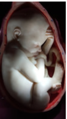
By the end of week 38, the fetus measures about 51 cm (20 in.) long. A 38-week fetus is pictured in Figure \(\PageIndex{5}\).
Fetal Circulation
The heart and blood vessels that form the cardiovascular system are among the earliest organs to develop in the embryo. They continue to develop in complexity and grow in size during the fetal stage. However, until birth, the circulation of blood in the fetus is different than the postnatal circulation will be, primarily because the lungs are not yet in use. The fetus cannot breathe the air because it is floating in amniotic fluid. Instead, the fetus obtains oxygen from the mother’s blood via the placenta and umbilical cord.
Prenatal Circulation
The fetal circulation before birth is illustrated in Figure \(\PageIndex{6}\). Oxygen-rich blood from the placenta is carried to the fetus by the umbilical vein. Some of the blood flows through a fetal vein called the ductus venosus, which carries the blood to the inferior vena cava. In turn, the vena cava carries blood to the right atrium of the heart. Throughout the fetal stage, there is an opening between the right and left atria, called the foramen ovale, which allows most of the blood reaching the right atrium to flow directly into the left atrium, thus bypassing the pulmonary circulation. Blood that enters the left atrium is pumped into the left ventricle, and from there through the aorta, the major artery that carries blood to the rest of the body. Blood that reaches the umbilical arteries flows back through the umbilical cord to the placenta, where carbon dioxide and other waste products from the fetus enter the maternal circulation.
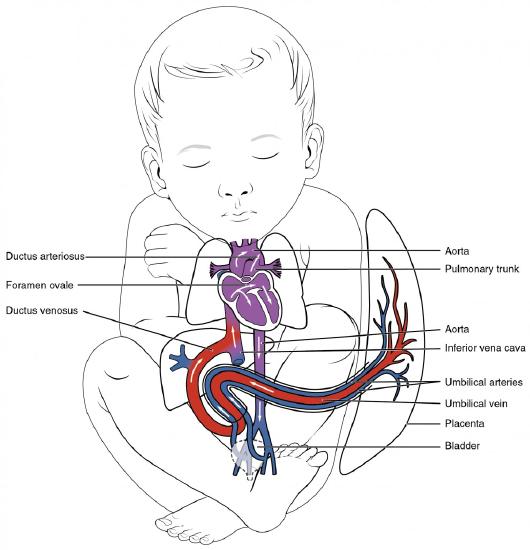
Not all of the blood reaching the right atrium through the ductus venosus passes directly into the left atrium via the foramen ovale. A small amount of blood is pumped from the right atrium into the right ventricle, and from the right ventricle into the pulmonary arteries. A fetal artery called the ductus arteriosus directs most of this blood away from the nonfunctioning lungs by shunting blood from the pulmonary trunk to the aorta.
Postnatal Circulation
After birth, as the newborn takes the first breath, the blood circulation suddenly changes. There is decreased resistance in the lungs now that the infant is surrounded by air instead of amniotic fluid. The lowered resistance allows more blood to flow into the pulmonary arteries from the right atrium and ventricle, and less to flow through the foramen ovale into the left atrium. Blood now travels to the lungs through the pulmonary arteries and then back to the heart through the pulmonary veins to the left atrium. This produces an increase in pressure in the left atrium that forces the foramen ovale to close. Once the foramen ovale closes, blood can no longer flow through it and bypass the pulmonary circulation. The ductus arteriosus is no longer needed to shunt blood away from the lungs, and it normally closes within a day or two of birth. The ductus venosus usually closes within another couple of days.
Birth Weight
The fetal growth rate is one of two major factors that determine the weight of the fetus at birth, or birth weight, which averages about 3.4 kg (7.5 lb.) in a full-term infant. The other factor that determines birthweight is the length of gestation. Infants born before full term, which is defined as 36-40 weeks after fertilization, are usually smaller than full-term infants because they have spent less time growing in the uterus. Pre-term birth is one of the major causes of low birth weight, which is defined as a birth weight lower than 2.5 kg (5.5 lb.), regardless of gestational age. Low birth weight increases the risk of death shortly after birth. As many as 30 percent of deaths in the first month of life occur in preterm infants. Holding the length of gestation constant, a newborn may be classified as small for gestational age, appropriate for gestation age, or large for gestational age. Fetuses that did not grow adequately before birth may end up being small for gestational age, even when they are born at full term.
Viability of the Fetus
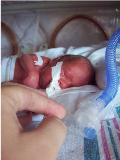
Fetal viability refers to the point in fetal development at which the fetus is likely to be able to survive outside the uterus. When babies are born too early, they have an elevated risk of dying within the first few hours to weeks of life. The main causes of early mortality in pre-term infants are inadequately developed respiratory and nervous systems. For babies born at 23 weeks of gestation, the chances of surviving are only between 20 and 35 percent, and survival is possible only with intensive, advanced medical care. For babies born at 25 weeks of gestation, the survival chances are much greater — as high as 70 percent — but again, intensive medical intervention is needed (see the newborn infant in Figure \(\PageIndex{7}\). The chances of survival are much better after 26 weeks of gestation. More than 90 percent of babies survive if they are born after 26 weeks and receive any necessary medical care. What a difference just three weeks makes!
Review
- Define fetus. Delineate the fetal stage.
- Describe the fetus at the beginning of the fetal stage.
- ist some of the fetal developments that occur between weeks 9 and 15 after fertilization.
- Give examples of fetal changes that occur during weeks 16 through 26 after fertilization.
- Identify several developments that take place in the fetus between week 27 and birth.
- How and why is fetal blood circulation different from postnatal circulation?
- Compare and contrast fetal and adult hemoglobin.
- Outline the typical pattern of fetal growth in size.
- What is IUGR? What is its leading cause?
- What is the average weight of a full-term infant at birth? How is low birth weight defined, and what are the two major causes of low birth weight?
- Define fetal viability. At what age is a fetus likely to be viable?
- Put the following events in order of when they occur, from earliest to latest:
- The kidneys start functioning.
- The ductus arteriosis closes.
- The fetus begins to detect light.
- The fetus begins to hear.
- True or False: A fetus can produce urine.
- True or False: The umbilical artery carries oxygenated blood to the fetus.
- If a baby is born at 30 weeks, what is one type of medical intervention that might be necessary to keep the baby alive? Explain your answer.
Explore More
Attributions
- Ultrasound by Public Domain Images, Pixabay license
- Human Embryo (7th week of pregnancy) by Ed Uthman, licensed CC BY 2.0 via Flickr
- 9-Week Human Embryo from Ectopic Pregnancy by Ed Uthman, licensed CC BY 2.0 via Flickr
- fetus at the 11th gestational week - 3D HD live rendered image by Araujo Júnior E, Santana EF, Nardozza LM, Moron AF - Radiol Bras (2015 Jan-Feb) CC BY-NC 3.0 via SciELO
- Eesti Tervishoiu Museum Estonian Health Care Museum Tallinn Estonia 2016 by A. Currell, licensed CC BY-NC 2.0 via Flickr
- Fetal Circulation by OpenStax College, CC BY 3.0 via Wikimedia Commons
- Premature infant by ceejayoz, licensed CC BY 2.0 via Wikimedia Commons
- Text adapted from Human Biology by CK-12 licensed CC BY-NC 3.0