21.7: Cancer
- Page ID
- 17750
\( \newcommand{\vecs}[1]{\overset { \scriptstyle \rightharpoonup} {\mathbf{#1}} } \)
\( \newcommand{\vecd}[1]{\overset{-\!-\!\rightharpoonup}{\vphantom{a}\smash {#1}}} \)
\( \newcommand{\dsum}{\displaystyle\sum\limits} \)
\( \newcommand{\dint}{\displaystyle\int\limits} \)
\( \newcommand{\dlim}{\displaystyle\lim\limits} \)
\( \newcommand{\id}{\mathrm{id}}\) \( \newcommand{\Span}{\mathrm{span}}\)
( \newcommand{\kernel}{\mathrm{null}\,}\) \( \newcommand{\range}{\mathrm{range}\,}\)
\( \newcommand{\RealPart}{\mathrm{Re}}\) \( \newcommand{\ImaginaryPart}{\mathrm{Im}}\)
\( \newcommand{\Argument}{\mathrm{Arg}}\) \( \newcommand{\norm}[1]{\| #1 \|}\)
\( \newcommand{\inner}[2]{\langle #1, #2 \rangle}\)
\( \newcommand{\Span}{\mathrm{span}}\)
\( \newcommand{\id}{\mathrm{id}}\)
\( \newcommand{\Span}{\mathrm{span}}\)
\( \newcommand{\kernel}{\mathrm{null}\,}\)
\( \newcommand{\range}{\mathrm{range}\,}\)
\( \newcommand{\RealPart}{\mathrm{Re}}\)
\( \newcommand{\ImaginaryPart}{\mathrm{Im}}\)
\( \newcommand{\Argument}{\mathrm{Arg}}\)
\( \newcommand{\norm}[1]{\| #1 \|}\)
\( \newcommand{\inner}[2]{\langle #1, #2 \rangle}\)
\( \newcommand{\Span}{\mathrm{span}}\) \( \newcommand{\AA}{\unicode[.8,0]{x212B}}\)
\( \newcommand{\vectorA}[1]{\vec{#1}} % arrow\)
\( \newcommand{\vectorAt}[1]{\vec{\text{#1}}} % arrow\)
\( \newcommand{\vectorB}[1]{\overset { \scriptstyle \rightharpoonup} {\mathbf{#1}} } \)
\( \newcommand{\vectorC}[1]{\textbf{#1}} \)
\( \newcommand{\vectorD}[1]{\overrightarrow{#1}} \)
\( \newcommand{\vectorDt}[1]{\overrightarrow{\text{#1}}} \)
\( \newcommand{\vectE}[1]{\overset{-\!-\!\rightharpoonup}{\vphantom{a}\smash{\mathbf {#1}}}} \)
\( \newcommand{\vecs}[1]{\overset { \scriptstyle \rightharpoonup} {\mathbf{#1}} } \)
\(\newcommand{\longvect}{\overrightarrow}\)
\( \newcommand{\vecd}[1]{\overset{-\!-\!\rightharpoonup}{\vphantom{a}\smash {#1}}} \)
\(\newcommand{\avec}{\mathbf a}\) \(\newcommand{\bvec}{\mathbf b}\) \(\newcommand{\cvec}{\mathbf c}\) \(\newcommand{\dvec}{\mathbf d}\) \(\newcommand{\dtil}{\widetilde{\mathbf d}}\) \(\newcommand{\evec}{\mathbf e}\) \(\newcommand{\fvec}{\mathbf f}\) \(\newcommand{\nvec}{\mathbf n}\) \(\newcommand{\pvec}{\mathbf p}\) \(\newcommand{\qvec}{\mathbf q}\) \(\newcommand{\svec}{\mathbf s}\) \(\newcommand{\tvec}{\mathbf t}\) \(\newcommand{\uvec}{\mathbf u}\) \(\newcommand{\vvec}{\mathbf v}\) \(\newcommand{\wvec}{\mathbf w}\) \(\newcommand{\xvec}{\mathbf x}\) \(\newcommand{\yvec}{\mathbf y}\) \(\newcommand{\zvec}{\mathbf z}\) \(\newcommand{\rvec}{\mathbf r}\) \(\newcommand{\mvec}{\mathbf m}\) \(\newcommand{\zerovec}{\mathbf 0}\) \(\newcommand{\onevec}{\mathbf 1}\) \(\newcommand{\real}{\mathbb R}\) \(\newcommand{\twovec}[2]{\left[\begin{array}{r}#1 \\ #2 \end{array}\right]}\) \(\newcommand{\ctwovec}[2]{\left[\begin{array}{c}#1 \\ #2 \end{array}\right]}\) \(\newcommand{\threevec}[3]{\left[\begin{array}{r}#1 \\ #2 \\ #3 \end{array}\right]}\) \(\newcommand{\cthreevec}[3]{\left[\begin{array}{c}#1 \\ #2 \\ #3 \end{array}\right]}\) \(\newcommand{\fourvec}[4]{\left[\begin{array}{r}#1 \\ #2 \\ #3 \\ #4 \end{array}\right]}\) \(\newcommand{\cfourvec}[4]{\left[\begin{array}{c}#1 \\ #2 \\ #3 \\ #4 \end{array}\right]}\) \(\newcommand{\fivevec}[5]{\left[\begin{array}{r}#1 \\ #2 \\ #3 \\ #4 \\ #5 \\ \end{array}\right]}\) \(\newcommand{\cfivevec}[5]{\left[\begin{array}{c}#1 \\ #2 \\ #3 \\ #4 \\ #5 \\ \end{array}\right]}\) \(\newcommand{\mattwo}[4]{\left[\begin{array}{rr}#1 \amp #2 \\ #3 \amp #4 \\ \end{array}\right]}\) \(\newcommand{\laspan}[1]{\text{Span}\{#1\}}\) \(\newcommand{\bcal}{\cal B}\) \(\newcommand{\ccal}{\cal C}\) \(\newcommand{\scal}{\cal S}\) \(\newcommand{\wcal}{\cal W}\) \(\newcommand{\ecal}{\cal E}\) \(\newcommand{\coords}[2]{\left\{#1\right\}_{#2}}\) \(\newcommand{\gray}[1]{\color{gray}{#1}}\) \(\newcommand{\lgray}[1]{\color{lightgray}{#1}}\) \(\newcommand{\rank}{\operatorname{rank}}\) \(\newcommand{\row}{\text{Row}}\) \(\newcommand{\col}{\text{Col}}\) \(\renewcommand{\row}{\text{Row}}\) \(\newcommand{\nul}{\text{Nul}}\) \(\newcommand{\var}{\text{Var}}\) \(\newcommand{\corr}{\text{corr}}\) \(\newcommand{\len}[1]{\left|#1\right|}\) \(\newcommand{\bbar}{\overline{\bvec}}\) \(\newcommand{\bhat}{\widehat{\bvec}}\) \(\newcommand{\bperp}{\bvec^\perp}\) \(\newcommand{\xhat}{\widehat{\xvec}}\) \(\newcommand{\vhat}{\widehat{\vvec}}\) \(\newcommand{\uhat}{\widehat{\uvec}}\) \(\newcommand{\what}{\widehat{\wvec}}\) \(\newcommand{\Sighat}{\widehat{\Sigma}}\) \(\newcommand{\lt}{<}\) \(\newcommand{\gt}{>}\) \(\newcommand{\amp}{&}\) \(\definecolor{fillinmathshade}{gray}{0.9}\)During his many months on the campaign trail in 2015 and 2016, U.S. Democratic presidential candidate Bernie Sanders was feeling the burn — sunburn, that is. There’s no doubt that candidate Sanders was exposed to a lot of UV light during all those outdoor rallies in states across the nation. Exposure to UV light is the single greatest risk factor for skin cancer, so it’s perhaps not surprising that Sanders was diagnosed with a small skin cancer on his cheek by December of 2016 (although most of his cumulative UV exposure probably occurred earlier in life). In Sanders's case, the cheek cancer was a basal cell carcinoma. This is the most common type of skin cancer. Fortunately, basal cell carcinoma is also a slowly growing and highly curable cancer. Sanders had his basal cell carcinoma completely removed in an hour-long outpatient procedure, after which he was able to return immediately to work. Unfortunately, most other types of cancer are faster growing and more difficult to cure than basal cell carcinoma, including a relatively rare but potentially deadly form of skin cancer called melanoma.
 Figure \(\PageIndex{1}\): Feel the Bern. Bernie Sanders is shown on fire in a political poster. The caption says "Feel The Bern."
Defining Cancer
Cancer is actually a group of more than 100 diseases, all of which involve abnormal cell growth with the potential to invade or spread to other parts of the body. In general terms, cancer occurs when the cell cycle is no longer regulated due to DNA damage. The number of potential underlying causes of this DNA damage is great, so there are many different risk factors for cancer. Any cells that become cancerous divide more quickly than normal cells. They may form a mass of abnormal cells called a tumor. The rapidly dividing cells take up nutrients and space, damaging the normal cells around them. If the cancer cells spread to other parts of the body, they invade and damage other tissues and organs. They may eventually lead to death.
By far, the most common of the 100-plus types of human cancer is basal cell carcinoma, the type of skin cancer Bernie Sanders had removed in 2016. Basal cell carcinoma makes up 40 percent of all new cancers each year in the United States. Other common types of cancer include lung, colorectal, prostate (in males), and breast (in females) cancers. These cancers are not as common as skin cancer, but they cause the majority of cancer deaths.
How Cancer Occurs
Cancer occurs when a normal cell is transformed into a cancer cell. This happens when the genes that regulate cell growth and differentiation are altered through mutations. The genes involved include proto-oncogenes and tumor-suppressor genes. Proto-oncogenes are genes that normally promote the growth and division of normal cells. Tumor-suppressor genes are genes that normally inhibit the division and survival of abnormal cells. Genetic alterations may result in the formation of cancer-causing oncogenes, over-expression of normal proto-oncogenes, or under-expression or disabling of tumor-suppressor genes.
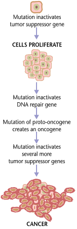
As shown in Figure \(\PageIndex{2}\), changes in multiple genes are typically required to transform a normal cell into a cancer cell. The entire process is similar to a chain reaction. Initial DNA errors are compounded by additional errors, and each error progressively allows the cell to escape more controls on cell growth and cell division.

How Cancer Spreads
Once a normal cell transforms into a cancer cell and starts dividing out of control, cancer cells can spread from the original site (called the primary tumor) to other tissues. This can occur in three different ways. One way is local spread, in which aggressively dividing cancer cells directly invade nearby tissues. Another way involves the lymphatic system. Cancer cells can spread to regional lymph nodes through lymph vessels that pass by the primary tumor.
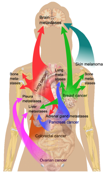
The third way cancer cells can spread is through the blood to distant sites. This is called metastasis, and the new cancers that form are called metastases. Although the blood can carry cancer cells to tissues everywhere in the body, cancer cells generally grow only in certain sites (Figure \(\PageIndex{3}\)). Different types of cancers tend to metastasize to particular organs. The most common places for metastases to occur are the brain, lungs, bones, and liver. Almost all cancers can metastasize, especially during the late stages of the disease. Cancer that has metastasized generally has the worst prognosis and is associated with most cancer deaths.

Risk Factors for Cancer
The direct cause of any cancer is DNA damage in genes that control the cell cycle. In this sense, all cancers are genetic diseases. The DNA damage can be inherited from parents or result spontaneously from environmental exposures to cancer-causing agents, or carcinogens. Having risk factors for cancer increases your chances of getting cancer either way.
Inherited Risk Factors
Certain mutant genes greatly increase the risk of particular cancers, such as the BRCA1 and BRCA2 genes, which increase the risk for breast and ovarian cancer in women to 75 percent. However, environmental factors also play a role even in this example because the genes do not cause cancer in every woman who inherits them. Genes with such a great effect on cancer risk cause fewer than 10 percent of human cancers.
Environmental Risk Factors
Environmental risk factors are the major risk factors for at least 90 percent of human cancers. Environmental risk factors include certain pathogens, such as hepatitis viruses that increase the risk of liver cancer. Other, nonpathogenic environmental factors that increase cancer risk include radon, which increases the risk of lung cancer, especially in smokers; air pollution, which increases the risk of lung cancer and bladder cancer; and UV light, which increases the risk of both carcinoma and melanoma skin cancers.
Avoidable lifestyle choices — especially smoking tobacco, eating an unhealthy diet, and not exercising — are some of the most important but preventable environmental risk factors for cancer. In fact, the majority of cancer deaths could be prevented by making healthy lifestyle choices. Not using tobacco would prevent an estimated 25 percent of cancer deaths. Eating right, getting adequate exercise, and avoiding obesity would prevent another 35 percent of cancer deaths.
Diagnosing Cancer
Early diagnosis and treatment are the keys to curing cancer, although not all cancers can be cured. Many cancers are first detected through routine screening of asymptomatic people. Many other cancers are noticed by early warning signs of cancer. A definitive diagnosis of cancer requires a biopsy before treatment can begin.
Cancer Screening
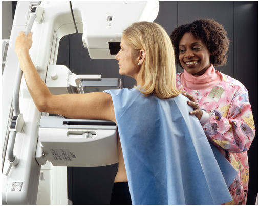
Screening for cancer has the aim of detecting common cancers in people who do not yet have any noticeable symptoms. Examples of cancers for which screening is usually recommended for high-risk groups include colon cancer (older people), breast cancer (older females), prostate cancer (older men), and skin cancer (e.g., light-skinned people, people with excessive UV light exposure). Screening of asymptomatic people for cancer may involve physical examinations (e.g., visual inspection of the skin for skin cancer), medical imaging (mammogram for breast cancer, pictured in Figure \(\PageIndex{4}\)), blood test (PSA test for prostate cancer), other tissue tests (Pap test for cervical cancer), or endoscopy (colonoscopy for colon cancer).

Routine cancer screening is somewhat controversial. Screening has both risks and benefits, and not everyone agrees that the benefits always outweigh the risks. Ideally, potential benefits are early detection, treatment, and cure of the disease in more people. However, if no treatment is available or treatment is unlikely to result in a cure, there is obviously less benefit to screening. Potential risks of cancer screening include false positives, which may lead to unnecessary anxiety and more invasive testing. Other potential risks include harm from excessive radiation and excessive cost or pain from screening procedures. Given the controversial nature of screening, it is not surprising that sometimes-conflicting screening recommendations are made. As a general rule, people should follow the advice of a health care provider who is familiar with their status.
Warning Signs of Cancer
There is no routine screening for many types of cancers. Instead, it is up to patients and healthcare providers to notice signs that might indicate cancer. Most cancers do not initially cause pain, but there may be other early warning signs. A good way to remember the signs is using the mnemonic CAUTION:
- Change in the bowel of bladder habits
- A sore that does not heal
- Unusual bleeding or discharge
- Thickening or lump in the breast or elsewhere
- Indigestion or difficulty in swallowing
- Obvious change in wart or mole
- Nagging cough or hoarseness
Most of the early warning signs of cancer are likely to be caused by the mass of a primary tumor or its ulceration. For example, the mass of a tumor in the rectum might cause a change in bowel habits. If the tumor was ulcerated, it might cause bleeding in the rectum and blood in the stool. A tumor in a breast might produce a detectable lump; a tumor in a lung might interfere with normal breathing and cause a persistent cough.
The early warning signs of cancer are not diagnostic of cancer because they could have many other causes — and chances are they do. However, the presence of one or more of the signs should prompt a visit to the doctor to find out for sure.
Biopsy
A definitive diagnosis of cancer requires a biopsy. In a biopsy, a tissue sample from the patient is examined microscopically by a pathologist (doctor specializing in disease diagnosis based on tissue changes). Cancers are classified and named by the type of tissue where cancer began (Table \(\PageIndex{1}\)). For example, carcinoma — such as Bernie Sanders’ basal cell carcinoma — is cancer derived from epithelial cells. Besides the skin, carcinomas include cancers of the lung, breast, and colon. Often, different types of cancer can be further distinguished on the basis of the size or shape of the cancer cells. For example, carcinomas of the lung include small-cell carcinomas and non-small-cell carcinomas. This is an important distinction because the two types of lung carcinoma have different prognoses and treatments.
| Type of Cancer | Type of Tissue or Cells Where It Originates | Example of this Type of Cancer |
|---|---|---|
| Carcinoma | epithelial system | skin |
| Sarcoma | connective tissue | bone |
| Leukemia | blood-forming tissue | bone marrow |
| Lymphoma | immune system cells | lymph nodes |
Cancer Staging
A cancer diagnosis generally includes cancer staging. Staging is the use of a classification system that reflects the seriousness of a cancer, such as how large a tumor is and the extent to which cancer has spread. These factors are important to consider in determining the prognosis and appropriate treatment for a given cancer. There are several different staging systems in use for different types of cancer. A general staging system commonly used by cancer registries includes the following stages:
- In situ — Abnormal cells are present but have not spread to nearby tissue.
- Localized — Cancer is limited to the place where it started, with no sign that it has spread beyond local tissue.
- Regional — Cancer has spread to nearby lymph nodes, tissues, or organs.
- Distant — Cancer has spread to distant parts of the body.
Treating Cancer
Many treatments for cancer exist. Some of the options for removing or killing cancer cells and potentially curing cancer are surgery, chemotherapy, radiation therapy, and immunotherapy. Which treatment is used generally depends on the type of cancer and its stage. In many cases, two or more types of curative treatments are used. Palliative treatments, such as drugs to relieve pain, are often provided in addition to curative treatments.
Surgery
Surgery is the primary method to treat most isolated, solid cancers. In an isolated tumor, surgery typically attempts to remove the entire mass, often along with local lymph nodes. If the cancer is still localized, surgery is likely to cure it. If not, surgery may at least lessen symptoms and prolong survival.
Chemotherapy
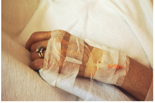
Chemotherapy is the treatment of cancer with one or more drugs that kill cancer cells. The drugs are often delivered directly into the bloodstream rather than orally (Figure \(\PageIndex{5}\)). Chemotherapy may be used alone or, more commonly, in conjunction with other treatments. Most chemotherapy drugs target rapidly dividing cells, not specifically cancerous cells. For example, chemotherapy drugs often target rapidly growing cells in hair roots, causing a temporary loss of hair. A more sophisticated form of chemotherapy, called targeted chemotherapy, targets specific molecules that distinguish cancerous from normal cells. Targeted therapies generally cause fewer adverse side effects. Targeted therapies exist for breast cancer, prostate cancer, melanoma, and several other cancers.

Radiation Therapy
About half of the cancers are treated with radiation therapy, usually in addition to surgery and/or chemotherapy. Radiation therapy is the use of ionizing radiation such as X-rays to kill cancerous tissues. To spare normal tissues through which the radiation must pass to reach cancer, multiple rays are directed toward cancer from different angles. The rays all intersect at the site of cancer, providing a much larger dose than in the surrounding healthy tissue.
Immunotherapy
A variety of newer therapies are directed at helping the immune system fight cancer. This type of therapy is called immunotherapy. Cancer immunotherapy attempts to stimulate the immune system to destroy cancer cells. A variety of such strategies are in use or are undergoing research and testing (see the Feature: Human Biology in the News below). If immunotherapy is used, it is usually in conjunction with other types of treatment, such as surgery or chemotherapy.
Stigma of Cancer
Many other diagnoses have a worse prognosis than most diagnoses of cancer, but a cancer diagnosis usually causes greater fear and dread. There are widespread misconceptions that cancer is always painful or always terminal, and there is a stigma associated with cancer. The stigma is reflected in how people refer to cancer deaths. An obituary is likely to say the deceased “died after a long illness” rather than “died of cancer.” Fear of cancer and cancer stigma may delay cancer diagnoses and treatment and increase the risk of cancer death.
Review
- What is cancer?
- What are some common types of cancer?
- How does a normal cell transform into a cancer cell?
- Describe three ways that cancer cells can spread from the original site of cancer.
- Define metastases.
- Explain why there are many different risk factors for cancer, and identify some common cancer risk factors.
- Why is it important to diagnose cancer early? What are two ways in which cancer can be detected early?
- What is a biopsy, what is its role in a cancer diagnosis, and what else can a biopsy reveal?
- Explain cancer staging and why it is done.
- Discuss types of treatments for cancer.
- Many other diagnoses have a worse prognosis than most diagnoses of cancer, but cancer usually causes greater fear and dread. Why?
- Which stage(s) of cancer, as defined here, are the most likely to be able to be fully cured with surgery? Explain your answer.
- True or False. Activation of tumor-suppressor genes is a common cause of cancer.
- True or False. Lung cancer can commonly spread to the brain.
- Explain why some cancer treatments can damage normal tissue.
Explore More
About 1 in every 3 families is now affected by cancer. Learn more about 2 new treatment options that are giving patients hope again:
Attributions
- Feel the Bern by Carl Glover from Long Beach, California, United States; CC BY 2.0 via Wikimedia Commons
- Cancer requires multiple mutations by National Cancer Institute, NIH, Public Domain via Wikimedia Commons
- Metastasis sites for common cancers by Mikael Häggström, CC0 1.0 via Wikimedia Commons
- Woman receives mammogram by National Cancer Institute, NIH, Public Domain via Wikimedia Commons
- Chemotherapy IV by National Cancer Institute, NIH, Public Domain via Wikimedia Commons
- Text adapted from Human Biology by CK-12 licensed CC BY-NC 3.0


