
21.5: Microscopic Anatomy of the Kidney
- Page ID
- 63097

Nephrons: The Functional Unit
Nephrons take a simple filtrate of the blood and modify it into urine. Many changes take place in the different parts of the nephron before urine is created for disposal. The term forming urine will be used hereafter to describe the filtrate as it is modified into true urine. The principle task of the nephron population is to balance the plasma to homeostatic set points and excrete potential toxins in the urine. They do this by accomplishing three principle functions—filtration, reabsorption, and secretion. They also have additional secondary functions that exert control in three areas: blood pressure (via production of renin), red blood cell production (via the hormone EPO), and calcium absorption (via conversion of calcidiol into calcitriol, the active form of vitamin D).
Renal Corpuscle
As discussed earlier, the renal corpuscle consists of a tuft of capillaries called the glomerulus that is largely surrounded by Bowman’s (glomerular) capsule. The glomerulus is a high-pressure capillary bed between afferent and efferent arterioles. Bowman’s capsule surrounds the glomerulus to form a lumen, and captures and directs this filtrate to the PCT. The outermost part of Bowman’s capsule, the parietal layer, is a simple squamous epithelium. It transitions onto the glomerular capillaries in an intimate embrace to form the visceral layer of the capsule. Here, the cells are not squamous, but uniquely shaped cells (podocytes) extending finger-like arms (pedicels) to cover the glomerular capillaries (Figure \(\PageIndex{1}\)). These projections interdigitate to form filtration slits, leaving small gaps between the digits to form a sieve. As blood passes through the glomerulus, 10 to 20 percent of the plasma filters between these sieve-like fingers to be captured by Bowman’s capsule and funneled to the PCT. Where the fenestrae (windows) in the glomerular capillaries match the spaces between the podocyte “fingers,” the only thing separating the capillary lumen and the lumen of Bowman’s capsule is their shared basement membrane (Figure \(\PageIndex{2}\)). These three features comprise what is known as the filtration membrane. This membrane permits very rapid movement of filtrate from capillary to capsule though pores that are only 70 nm in diameter.
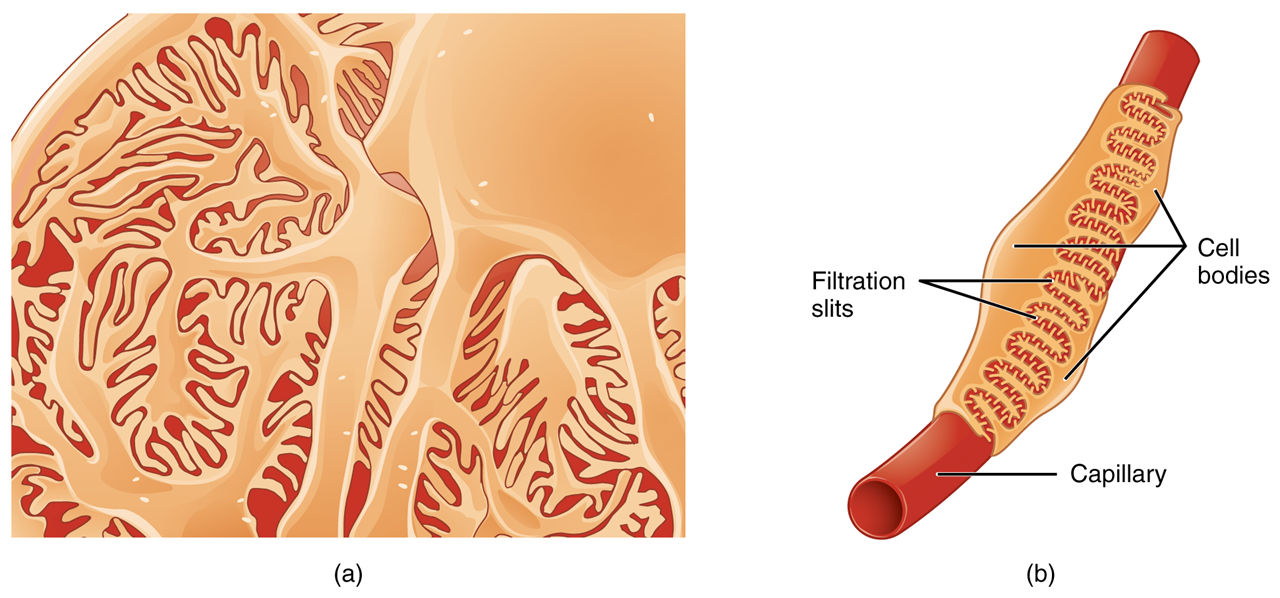
Figure \(\PageIndex{1}\): Podocytes Podocytes interdigitate with structures called pedicels and filter substances in a way similar to fenestrations. In (a), the large cell body can be seen at the top right corner, with branches extending from the cell body. The smallest finger-like extensions are the pedicels. Pedicels on one podocyte always interdigitate with the pedicels of another podocyte. (b) This capillary has three podocytes wrapped around it. (CC-BY-4.0, OpenStax, Human Anatomy)
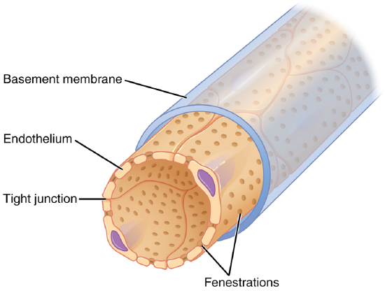
Figure \(\PageIndex{2}\): Fenestrated Capillary Fenestrations allow many substances to diffuse from the blood based primarily on size. (CC-BY-4.0, OpenStax, Human Anatomy)
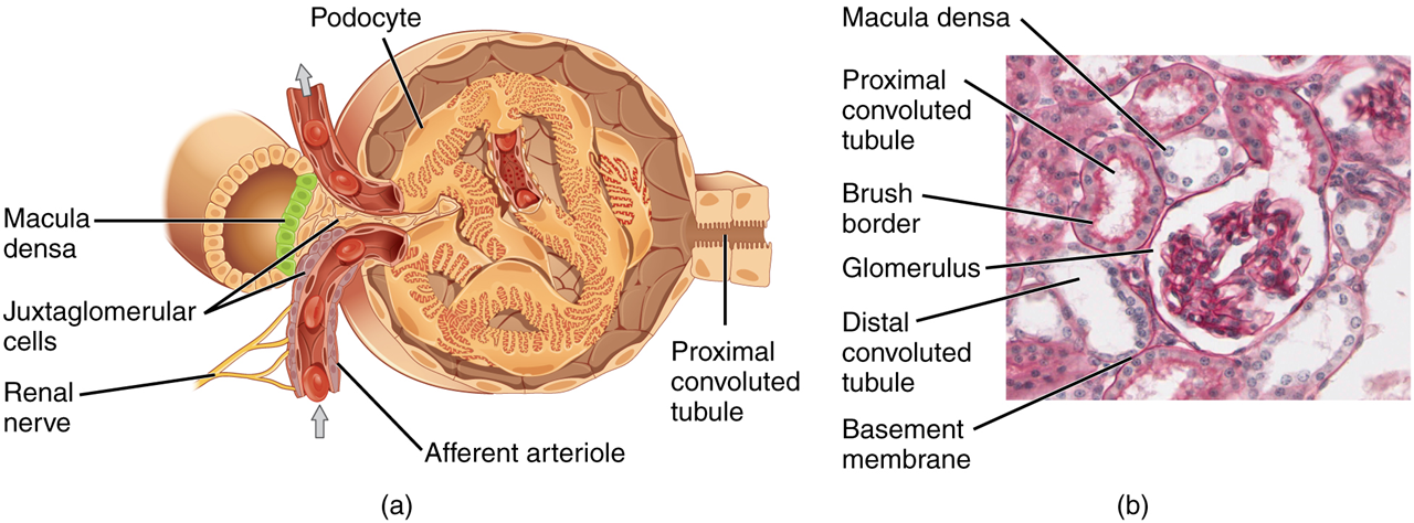
Figure \(\PageIndex{2}\): Juxtaglomerular Apparatus and Glomerulus (a) The JGA allows specialized cells to monitor the composition of the fluid in the DCT and adjust the glomerular filtration rate. (b) This micrograph shows the glomerulus and surrounding structures. LM × 1540. (Micrograph provided by the Regents of University of Michigan Medical School © 2012) (CC-BY-4.0, OpenStax, Human Anatomy)