
9.4: Muscles of the Hips and Thighs
- Page ID
- 59408
\( \newcommand{\vecs}[1]{\overset { \scriptstyle \rightharpoonup} {\mathbf{#1}} } \)
\( \newcommand{\vecd}[1]{\overset{-\!-\!\rightharpoonup}{\vphantom{a}\smash {#1}}} \)
\( \newcommand{\id}{\mathrm{id}}\) \( \newcommand{\Span}{\mathrm{span}}\)
( \newcommand{\kernel}{\mathrm{null}\,}\) \( \newcommand{\range}{\mathrm{range}\,}\)
\( \newcommand{\RealPart}{\mathrm{Re}}\) \( \newcommand{\ImaginaryPart}{\mathrm{Im}}\)
\( \newcommand{\Argument}{\mathrm{Arg}}\) \( \newcommand{\norm}[1]{\| #1 \|}\)
\( \newcommand{\inner}[2]{\langle #1, #2 \rangle}\)
\( \newcommand{\Span}{\mathrm{span}}\)
\( \newcommand{\id}{\mathrm{id}}\)
\( \newcommand{\Span}{\mathrm{span}}\)
\( \newcommand{\kernel}{\mathrm{null}\,}\)
\( \newcommand{\range}{\mathrm{range}\,}\)
\( \newcommand{\RealPart}{\mathrm{Re}}\)
\( \newcommand{\ImaginaryPart}{\mathrm{Im}}\)
\( \newcommand{\Argument}{\mathrm{Arg}}\)
\( \newcommand{\norm}[1]{\| #1 \|}\)
\( \newcommand{\inner}[2]{\langle #1, #2 \rangle}\)
\( \newcommand{\Span}{\mathrm{span}}\) \( \newcommand{\AA}{\unicode[.8,0]{x212B}}\)
\( \newcommand{\vectorA}[1]{\vec{#1}} % arrow\)
\( \newcommand{\vectorAt}[1]{\vec{\text{#1}}} % arrow\)
\( \newcommand{\vectorB}[1]{\overset { \scriptstyle \rightharpoonup} {\mathbf{#1}} } \)
\( \newcommand{\vectorC}[1]{\textbf{#1}} \)
\( \newcommand{\vectorD}[1]{\overrightarrow{#1}} \)
\( \newcommand{\vectorDt}[1]{\overrightarrow{\text{#1}}} \)
\( \newcommand{\vectE}[1]{\overset{-\!-\!\rightharpoonup}{\vphantom{a}\smash{\mathbf {#1}}}} \)
\( \newcommand{\vecs}[1]{\overset { \scriptstyle \rightharpoonup} {\mathbf{#1}} } \)
\( \newcommand{\vecd}[1]{\overset{-\!-\!\rightharpoonup}{\vphantom{a}\smash {#1}}} \)
\(\newcommand{\avec}{\mathbf a}\) \(\newcommand{\bvec}{\mathbf b}\) \(\newcommand{\cvec}{\mathbf c}\) \(\newcommand{\dvec}{\mathbf d}\) \(\newcommand{\dtil}{\widetilde{\mathbf d}}\) \(\newcommand{\evec}{\mathbf e}\) \(\newcommand{\fvec}{\mathbf f}\) \(\newcommand{\nvec}{\mathbf n}\) \(\newcommand{\pvec}{\mathbf p}\) \(\newcommand{\qvec}{\mathbf q}\) \(\newcommand{\svec}{\mathbf s}\) \(\newcommand{\tvec}{\mathbf t}\) \(\newcommand{\uvec}{\mathbf u}\) \(\newcommand{\vvec}{\mathbf v}\) \(\newcommand{\wvec}{\mathbf w}\) \(\newcommand{\xvec}{\mathbf x}\) \(\newcommand{\yvec}{\mathbf y}\) \(\newcommand{\zvec}{\mathbf z}\) \(\newcommand{\rvec}{\mathbf r}\) \(\newcommand{\mvec}{\mathbf m}\) \(\newcommand{\zerovec}{\mathbf 0}\) \(\newcommand{\onevec}{\mathbf 1}\) \(\newcommand{\real}{\mathbb R}\) \(\newcommand{\twovec}[2]{\left[\begin{array}{r}#1 \\ #2 \end{array}\right]}\) \(\newcommand{\ctwovec}[2]{\left[\begin{array}{c}#1 \\ #2 \end{array}\right]}\) \(\newcommand{\threevec}[3]{\left[\begin{array}{r}#1 \\ #2 \\ #3 \end{array}\right]}\) \(\newcommand{\cthreevec}[3]{\left[\begin{array}{c}#1 \\ #2 \\ #3 \end{array}\right]}\) \(\newcommand{\fourvec}[4]{\left[\begin{array}{r}#1 \\ #2 \\ #3 \\ #4 \end{array}\right]}\) \(\newcommand{\cfourvec}[4]{\left[\begin{array}{c}#1 \\ #2 \\ #3 \\ #4 \end{array}\right]}\) \(\newcommand{\fivevec}[5]{\left[\begin{array}{r}#1 \\ #2 \\ #3 \\ #4 \\ #5 \\ \end{array}\right]}\) \(\newcommand{\cfivevec}[5]{\left[\begin{array}{c}#1 \\ #2 \\ #3 \\ #4 \\ #5 \\ \end{array}\right]}\) \(\newcommand{\mattwo}[4]{\left[\begin{array}{rr}#1 \amp #2 \\ #3 \amp #4 \\ \end{array}\right]}\) \(\newcommand{\laspan}[1]{\text{Span}\{#1\}}\) \(\newcommand{\bcal}{\cal B}\) \(\newcommand{\ccal}{\cal C}\) \(\newcommand{\scal}{\cal S}\) \(\newcommand{\wcal}{\cal W}\) \(\newcommand{\ecal}{\cal E}\) \(\newcommand{\coords}[2]{\left\{#1\right\}_{#2}}\) \(\newcommand{\gray}[1]{\color{gray}{#1}}\) \(\newcommand{\lgray}[1]{\color{lightgray}{#1}}\) \(\newcommand{\rank}{\operatorname{rank}}\) \(\newcommand{\row}{\text{Row}}\) \(\newcommand{\col}{\text{Col}}\) \(\renewcommand{\row}{\text{Row}}\) \(\newcommand{\nul}{\text{Nul}}\) \(\newcommand{\var}{\text{Var}}\) \(\newcommand{\corr}{\text{corr}}\) \(\newcommand{\len}[1]{\left|#1\right|}\) \(\newcommand{\bbar}{\overline{\bvec}}\) \(\newcommand{\bhat}{\widehat{\bvec}}\) \(\newcommand{\bperp}{\bvec^\perp}\) \(\newcommand{\xhat}{\widehat{\xvec}}\) \(\newcommand{\vhat}{\widehat{\vvec}}\) \(\newcommand{\uhat}{\widehat{\uvec}}\) \(\newcommand{\what}{\widehat{\wvec}}\) \(\newcommand{\Sighat}{\widehat{\Sigma}}\) \(\newcommand{\lt}{<}\) \(\newcommand{\gt}{>}\) \(\newcommand{\amp}{&}\) \(\definecolor{fillinmathshade}{gray}{0.9}\)Information
There are three layers of gluteal muscles on the posterior hips, just like there are three layers of muscles in the abdominal trunk. The largest of them is the most superficial muscle, the gluteus maximus. Its origin is on the ilium of the coxal bone, and it inserts part-way down the shaft of the femur. It helps maintain erect posture, abducts the thigh, and rotates the thigh outward.
Below the gluteus maximus is the smaller gluteus medius. The gluteus medius muscle helps abducts the thigh along with the gluteus maximus, but can rotate the thigh inward where the gluteus maximus rotates the thigh outward.
Below the gluteus medius are several muscles, one of which is the gluteus minimus, the smallest of the gluteal muscles. It is a synergist for the gluteus medius.
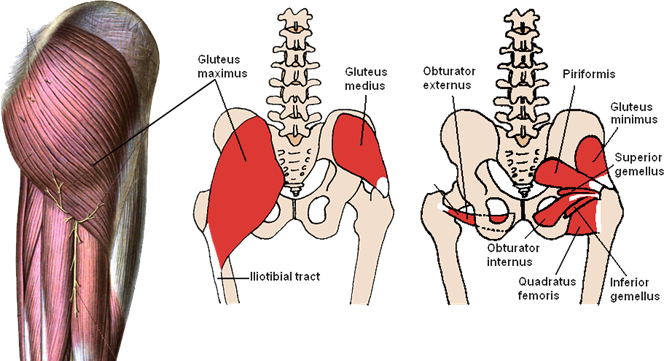
Figure \(\PageIndex{1}\): The three layers of gluteal muscles, gluteus maximus, gluteus medius, gluteus minimus. (CC-BY-SA, Wikimedia)
Like the forearm, the upper leg, or thigh, has a dense arrangement of many muscles. On the anterior side, the most prominent of the muscles are the sartorius muscle and the four muscles that make up quadriceps muscle group (the “quads”.)
The quadriceps sounds like it should be just one muscle, akin to the triceps brachii, but it is a group of four muscles, three visible on the surface, and the fourth obscured. The three surface muscles of the quadriceps are the rectus femoris in the center, the vastus medialis on the medial side, and the vastus lateralis on the lateral side. These three muscles are visible in Figure \(\PageIndex{2}\). Below the rectus femoris and largely hidden by it is the vastus intermedius. This muscle’s position can be seen in Figure 9.9. The four muscle of the quadriceps all extend the lower leg, and the rectus femoris additionally can flex the thigh at the hip.
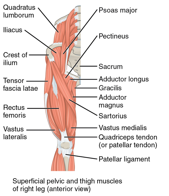
Figure \(\PageIndex{2}\): The superficial muscles of the thigh. ((CC-BY-4.0, OpenStax, Human Anatomy)
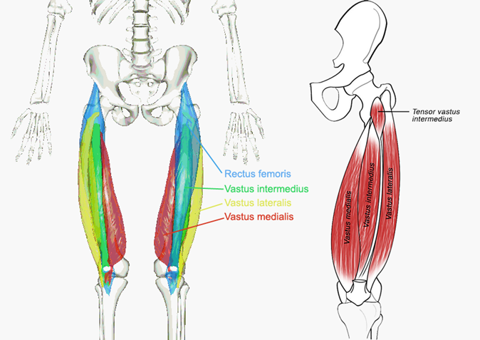
Figure \(\PageIndex{3}\): The quadriceps group of four muscles. The view on the left has the rectus femoris cut away to show the vastus intermedius which is below it. (CC-BY-SA, Wikimedia)
The sartorius muscle is a distinctively long and thin muscle that crosses the thigh diagonally. It is visible in Figure 9.9. Sartorius comes from the Latin for tailor, and this is sometimes called the tailor’s muscle, although the reasons for the nickname are obscure. It may be because the shape of the muscle is thin and long, like a tailor’s measuring tape; it may be because it is close to the inseam a tailor measures when tailoring pants, or it may be because it helps bring about the cross-legged position that tailors often adopt when working.
In the posterior thigh the bulk of the musculature is made up of three long muscles that are collectively called the hamstrings. The origin of this nickname is obscure, but it may have to do with the practice of butchers of hanging the thighs of butchered animals such as pig (the “hams”) by the tendons of these three muscles. Move from the medial edge to the lateral edge of the posterior thigh, the hamstring muscles are the semimembranous muscle, the semitendinosus muscle, and the biceps femorismuscle. Notice the upper leg has a “biceps” muscle just like the upper arm does. This is why you have to indicate which biceps you are taking about when discussing one or other of these muscles. On the medial edge of the posterior thigh is the gracilis muscle. It is also visible on the medial edge of the thigh from the anterior.
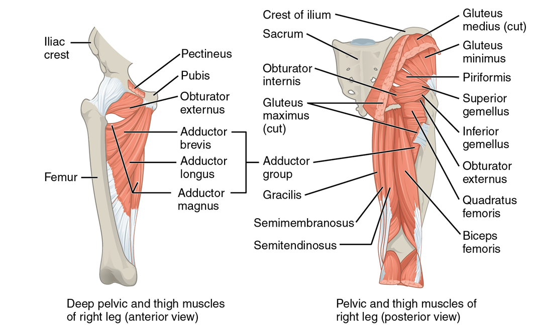
Figure \(\PageIndex{4}\): The muscles of the posterior thigh. (CC-BY-4.0, OpenStax, Human Anatomy)
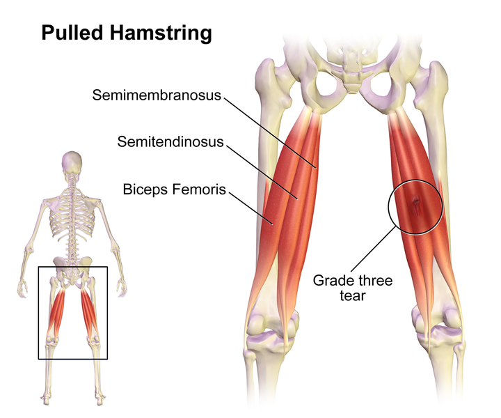
Figure \(\PageIndex{5}\): The hamstring group of muscles of the posterior thigh. (CC-BY-SA, Wikimedia)
LAB 9 EXERCISES \(\PageIndex{1}\)
1. Using the full-scale leg model, locate and identify the muscles of the thigh listed in the table below.
2. Write down the muscles of the thigh in the table below and, for each, give the location of that muscle and what effect contracting that muscle has.
|
Muscle |
Location & description |
Action(s) |
|
Rectus femoris |
|
|
|
Vastus intermedius |
|
|
|
Vastus medialis |
|
|
|
Vastus lateralis |
|
|
|
Sartorius |
|
|
|
Gracilis |
|
|
|
Semimembranosus |
|
|
|
Semitendinosus |
|
|
|
Biceps femoris |
|
|
LICENSES AND ATTRIBUTIONS
CC LICENSED CONTENT, ORIGINAL
A&P Labs. Authored by: Ross Whitwam. Provided by: Mississippi University for Women. Located at: http://www.muw.edu. License: CC BY-SA: Attribution-ShareAlike
CC LICENSED CONTENT, SPECIFIC ATTRIBUTION
Figure \(\PageIndex{1}\). The three layers of gluteal muscles, gluteus maximus, gluteus medius, gluteus minimus. Authored by: Beth ohara~commonswiki. Located at: https://commons.wikimedia.org/wiki/F..._Muscles_3.PNG. License: CC BY- SA: Attribution-ShareAlike
Figure \(\PageIndex{2}\). The superficial muscles of the thigh.. Authored by: OpenStax College. Located at: https://cnx.org/resources/49a26b0c63...e_the_Femur.jp g. License: CC BY-SA: Attribution-ShareAlike
Figure \(\PageIndex{3}\). The quadriceps group of four muscles. The view on the left has the rectus femoris cut away to show the vastus intermedius which is below it.. Authored by: Athikhun.suw. Located at: https://commons.wikimedia.org/wiki/F...ius_muscle.jpg. License: CC BY-SA: Attribution- ShareAlike
Figure \(\PageIndex{4}\) The muscles of the posterior thigh.. Authored by: OpenStax College. Located at: http://cnx.org/resources/49a26b0c6351a2052a16c4fcd339bc092505e492/1122_Gluteal_Muscles_that_Move_the_Femur.jpg. License: CC BY-SA: Attribution-ShareAlike
Figure \(\PageIndex{5}\). The hamstring group of muscles of the posterior thigh.. Authored by: BruceBlaus. Located at: https://commons.wikimedia.org/wiki/F..._Hamstring.png. License: CC BY-SA: Attribution-ShareAlike
PUBLIC DOMAIN CONTENT
Figure \(\PageIndex{1}\). The three layers of gluteal muscles, gluteus maximus, gluteus medius, gluteus minimus.. Authored by: Dr. Johannes Sobotta. Provided by: Sobotta's Human Anatomy 1909. Located at: https://commons.wikimedia.org/wiki/F...Sobo_1909_575- 576.png. License: Public Domain: No Known Copyright