
19.3: Kidneys
- Page ID
- 16840

\( \newcommand{\vecs}[1]{\overset { \scriptstyle \rightharpoonup} {\mathbf{#1}} } \)
\( \newcommand{\vecd}[1]{\overset{-\!-\!\rightharpoonup}{\vphantom{a}\smash {#1}}} \)
\( \newcommand{\dsum}{\displaystyle\sum\limits} \)
\( \newcommand{\dint}{\displaystyle\int\limits} \)
\( \newcommand{\dlim}{\displaystyle\lim\limits} \)
\( \newcommand{\id}{\mathrm{id}}\) \( \newcommand{\Span}{\mathrm{span}}\)
( \newcommand{\kernel}{\mathrm{null}\,}\) \( \newcommand{\range}{\mathrm{range}\,}\)
\( \newcommand{\RealPart}{\mathrm{Re}}\) \( \newcommand{\ImaginaryPart}{\mathrm{Im}}\)
\( \newcommand{\Argument}{\mathrm{Arg}}\) \( \newcommand{\norm}[1]{\| #1 \|}\)
\( \newcommand{\inner}[2]{\langle #1, #2 \rangle}\)
\( \newcommand{\Span}{\mathrm{span}}\)
\( \newcommand{\id}{\mathrm{id}}\)
\( \newcommand{\Span}{\mathrm{span}}\)
\( \newcommand{\kernel}{\mathrm{null}\,}\)
\( \newcommand{\range}{\mathrm{range}\,}\)
\( \newcommand{\RealPart}{\mathrm{Re}}\)
\( \newcommand{\ImaginaryPart}{\mathrm{Im}}\)
\( \newcommand{\Argument}{\mathrm{Arg}}\)
\( \newcommand{\norm}[1]{\| #1 \|}\)
\( \newcommand{\inner}[2]{\langle #1, #2 \rangle}\)
\( \newcommand{\Span}{\mathrm{span}}\) \( \newcommand{\AA}{\unicode[.8,0]{x212B}}\)
\( \newcommand{\vectorA}[1]{\vec{#1}} % arrow\)
\( \newcommand{\vectorAt}[1]{\vec{\text{#1}}} % arrow\)
\( \newcommand{\vectorB}[1]{\overset { \scriptstyle \rightharpoonup} {\mathbf{#1}} } \)
\( \newcommand{\vectorC}[1]{\textbf{#1}} \)
\( \newcommand{\vectorD}[1]{\overrightarrow{#1}} \)
\( \newcommand{\vectorDt}[1]{\overrightarrow{\text{#1}}} \)
\( \newcommand{\vectE}[1]{\overset{-\!-\!\rightharpoonup}{\vphantom{a}\smash{\mathbf {#1}}}} \)
\( \newcommand{\vecs}[1]{\overset { \scriptstyle \rightharpoonup} {\mathbf{#1}} } \)
\(\newcommand{\longvect}{\overrightarrow}\)
\( \newcommand{\vecd}[1]{\overset{-\!-\!\rightharpoonup}{\vphantom{a}\smash {#1}}} \)
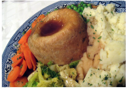
\(\newcommand{\avec}{\mathbf a}\) \(\newcommand{\bvec}{\mathbf b}\) \(\newcommand{\cvec}{\mathbf c}\) \(\newcommand{\dvec}{\mathbf d}\) \(\newcommand{\dtil}{\widetilde{\mathbf d}}\) \(\newcommand{\evec}{\mathbf e}\) \(\newcommand{\fvec}{\mathbf f}\) \(\newcommand{\nvec}{\mathbf n}\) \(\newcommand{\pvec}{\mathbf p}\) \(\newcommand{\qvec}{\mathbf q}\) \(\newcommand{\svec}{\mathbf s}\) \(\newcommand{\tvec}{\mathbf t}\) \(\newcommand{\uvec}{\mathbf u}\) \(\newcommand{\vvec}{\mathbf v}\) \(\newcommand{\wvec}{\mathbf w}\) \(\newcommand{\xvec}{\mathbf x}\) \(\newcommand{\yvec}{\mathbf y}\) \(\newcommand{\zvec}{\mathbf z}\) \(\newcommand{\rvec}{\mathbf r}\) \(\newcommand{\mvec}{\mathbf m}\) \(\newcommand{\zerovec}{\mathbf 0}\) \(\newcommand{\onevec}{\mathbf 1}\) \(\newcommand{\real}{\mathbb R}\) \(\newcommand{\twovec}[2]{\left[\begin{array}{r}#1 \\ #2 \end{array}\right]}\) \(\newcommand{\ctwovec}[2]{\left[\begin{array}{c}#1 \\ #2 \end{array}\right]}\) \(\newcommand{\threevec}[3]{\left[\begin{array}{r}#1 \\ #2 \\ #3 \end{array}\right]}\) \(\newcommand{\cthreevec}[3]{\left[\begin{array}{c}#1 \\ #2 \\ #3 \end{array}\right]}\) \(\newcommand{\fourvec}[4]{\left[\begin{array}{r}#1 \\ #2 \\ #3 \\ #4 \end{array}\right]}\) \(\newcommand{\cfourvec}[4]{\left[\begin{array}{c}#1 \\ #2 \\ #3 \\ #4 \end{array}\right]}\) \(\newcommand{\fivevec}[5]{\left[\begin{array}{r}#1 \\ #2 \\ #3 \\ #4 \\ #5 \\ \end{array}\right]}\) \(\newcommand{\cfivevec}[5]{\left[\begin{array}{c}#1 \\ #2 \\ #3 \\ #4 \\ #5 \\ \end{array}\right]}\) \(\newcommand{\mattwo}[4]{\left[\begin{array}{rr}#1 \amp #2 \\ #3 \amp #4 \\ \end{array}\right]}\) \(\newcommand{\laspan}[1]{\text{Span}\{#1\}}\) \(\newcommand{\bcal}{\cal B}\) \(\newcommand{\ccal}{\cal C}\) \(\newcommand{\scal}{\cal S}\) \(\newcommand{\wcal}{\cal W}\) \(\newcommand{\ecal}{\cal E}\) \(\newcommand{\coords}[2]{\left\{#1\right\}_{#2}}\) \(\newcommand{\gray}[1]{\color{gray}{#1}}\) \(\newcommand{\lgray}[1]{\color{lightgray}{#1}}\) \(\newcommand{\rank}{\operatorname{rank}}\) \(\newcommand{\row}{\text{Row}}\) \(\newcommand{\col}{\text{Col}}\) \(\renewcommand{\row}{\text{Row}}\) \(\newcommand{\nul}{\text{Nul}}\) \(\newcommand{\var}{\text{Var}}\) \(\newcommand{\corr}{\text{corr}}\) \(\newcommand{\len}[1]{\left|#1\right|}\) \(\newcommand{\bbar}{\overline{\bvec}}\) \(\newcommand{\bhat}{\widehat{\bvec}}\) \(\newcommand{\bperp}{\bvec^\perp}\) \(\newcommand{\xhat}{\widehat{\xvec}}\) \(\newcommand{\vhat}{\widehat{\vvec}}\) \(\newcommand{\uhat}{\widehat{\uvec}}\) \(\newcommand{\what}{\widehat{\wvec}}\) \(\newcommand{\Sighat}{\widehat{\Sigma}}\) \(\newcommand{\lt}{<}\) \(\newcommand{\gt}{>}\) \(\newcommand{\amp}{&}\) \(\definecolor{fillinmathshade}{gray}{0.9}\)Pictured here amidst a bed of mixed veggies are a steak and kidney pudding. More often made into a pie, this savory dish is a British favorite. Kidneys on the menu typically come from sheep, pigs, or cows. In these animals as in the human animal, kidneys are the main organs of excretion.
Location of the Kidneys
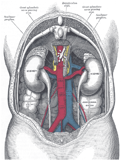
The two bean-shaped kidneys are located high in the back of the abdominal cavity, one on each side of the spine. Both kidneys sit just below the diaphragm, the large breathing muscle that separates the abdominal and thoracic cavities. As you can see in Figure \(\PageIndex{2}\), the right kidney is slightly smaller and lower than the left kidney. The right kidney is behind the liver, and the left kidney is behind the spleen. The location of the liver explains why the right kidney is smaller and lower than the left.
Kidney Anatomy
The shape of each kidney gives it a convex side and a concave side. You can see this clearly in the detailed diagram of kidney anatomy shown in Figure \(\PageIndex{3}\). The concave side is where the renal artery enters the kidney and the renal vein and ureter leave the kidney. This area of the kidney is called the hilum. The entire kidney is surrounded by tough fibrous tissue, called the renal capsule, which in turn is surrounded by two layers of protective, cushioning fat.
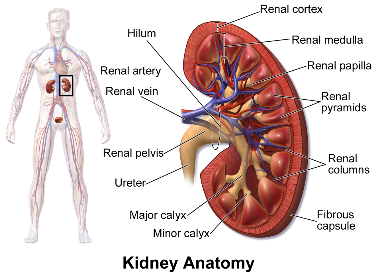
Internally, each kidney is divided into two major layers: the outer renal cortex and the inner renal medulla (see Figure \(\PageIndex{3}\)). These layers take the shape of many cone-shaped renal lobules, each containing a renal cortex surrounding a portion of the medulla called a renal pyramid. Within the renal pyramids are the structural and functional units of the kidneys, the tiny nephrons. Between the renal pyramids are projections of cortex called renal columns. The tip or papilla of each pyramid empties urine into a minor calyx (chamber). Several minor calyces empty into a major calyx, and the latter empties into the funnel-shaped cavity called the renal pelvis, which becomes the ureter as it leaves the kidney.
Renal Circulation
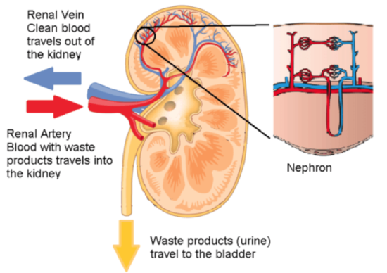
The renal circulation is an important part of the kidney’s main function of filtering waste products from the blood. Blood is supplied to the kidneys via the renal arteries. The right renal artery supplies the right kidney, and the left renal artery supplies the left kidney. These two arteries branch directly from the aorta, which is the largest artery in the body. Each kidney is only about 11 cm (4.4 in.) long and has a mass of just 150 grams (5.3 oz), yet it receives about 10 percent of the total output of blood from the heart. Blood is filtered through the kidneys about 20 times each hour, 24 hours a day, day after day.
As indicated in Figure \(\PageIndex{4}\), each renal artery carries blood with waste products into the kidney. Within the kidney, the renal artery branches into increasingly smaller arteries that extend through the renal columns between the renal pyramids. These arteries, in turn, branch into arterioles that penetrate the renal pyramids. Blood in the arterioles passes through nephrons, the structures that actually filter the blood. After blood passes through the nephrons and is filtered, the clean blood moves through a network of venules that converge into small veins. Small veins merge into increasingly larger ones and ultimately into the renal vein, which carries clean blood away from the kidney to the inferior vena cava.
Nephron Structure and Function
The illustration above gives an indication of the complex structure of a nephron. The nephron is the basic structural and functional unit of the kidney, and each kidney typically contains at least a million of them. As blood flows through a nephron, many materials are filtered out of the blood, needed materials are returned to the blood, and the remaining materials form urine. Most of the waste products removed from the blood and excreted in the urine are byproducts of metabolism. At last half of the waste is urea, a waste product produced by protein catabolism. Another important waste is uric acid, produced in nucleic acid catabolism.
Components of a Nephron
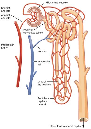
The diagram in Figure \(\PageIndex{5}\) shows in greater detail the components of a nephron. The renal corpuscle is a filtering structure that is consists of a network of capillaries called the glomerulus (plural, glomeruli) and the Bowman's capsule, a space surrounding the glomerulus. Bowman's capsule is the initial structure of a nephron. Extending from Bowman’s capsule is the renal tubule. The proximal end (nearest Bowman’s capsule) of the renal tubule is called the proximal convoluted (coiled) tubule. From here, the renal tubule continues as a loop (known as the loop of Henle), which in turn becomes the distal convoluted tubule. The latter finally joins with a collecting duct. As you can see in the diagram, peritubular capillaries surround the total length of the renal tubule.
The Function of a Nephron
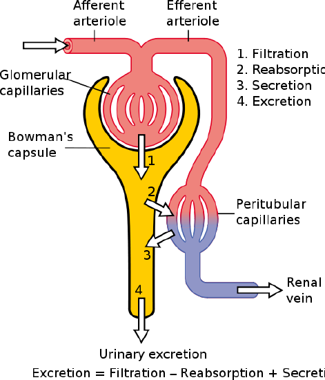
The simplified diagram of a nephron in Figure \(\PageIndex{6}\) shows how the nephron functions. Blood enters the nephron through an arteriole called the afferent arteriole. Some of the blood next passes through the capillaries of the glomerulus. Any blood that doesn’t pass through the glomerulus, as well as blood after it passes through the glomerular capillaries, continues on through an arteriole called the efferent arteriole. The efferent arteriole follows the renal tubule of the nephron, where it continues to play roles in nephron functioning.
Filtration
As blood from the afferent arteriole flows through the glomerular capillaries, it is under pressure. Because of the pressure, water and solutes are filtered out of the blood and into the space made by Bowman’s capsule. This is the filtration stage of nephron function. The filtered substances, called filtrate, pass into Bowman’s capsule and from there into the proximal end of the renal tubule. At this stage, filtrate includes water, salts, organic solids such as nutrients, and waste products of metabolism such as urea.
Reabsorption and Secretion
As filtrate moves through the renal tubule, some of the substances it contains are reabsorbed from the filtrate back into the blood in the efferent arteriole (via peritubular capillaries). This is the reabsorption stage of nephron function. About two-thirds of the filtered salts and water and all of the filtered organic solutes (mainly glucose and amino acids) are reabsorbed from the filtrate by the blood in the peritubular capillaries. Reabsorption occurs mainly in the proximal convoluted tubule and the loop of Henle.
At the distal end of the renal tubule, some additional reabsorption generally occurs. This is also the region of the tubule where other substances from the blood are added to the filtrate in the tubule. The addition of other substances to the filtrate from the blood is called secretion. Both reabsorption and secretion in the distal convoluted tubule are largely under the control of endocrine hormones that maintain homeostasis of water and mineral salts in the blood. These hormones work by controlling what is reabsorbed into the blood from the filtrate and what is secreted from the blood into the filtrate to become urine. For example, the parathyroid hormone causes more calcium to be reabsorbed into the blood and more phosphorus to be secreted into the filtrate.
Collection of Urine and Excretion
By the time the filtrate has passed through the entire renal tubule, it has become the liquid waste known as urine. Urine empties from the distal end of the renal tubule into a collecting duct. From there, the urine flows into increasingly larger collecting ducts. As urine flows through the system of collecting ducts, more water may be reabsorbed from it. This will occur in the presence of antidiuretic hormone from the hypothalamus. This hormone makes the collecting ducts permeable to water, allowing water molecules to pass through them into capillaries by osmosis while preventing the passage of ions or other solutes. As much as three-fourths of the water may be reabsorbed from urine in the collecting ducts, making the urine more concentrated.
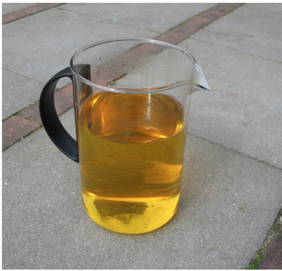
Urine finally exits the largest collecting ducts through the renal papillae. It empties into the renal calyces and finally into the renal pelvis (see Figure \(\PageIndex{3}\). From there, it travels through the ureter to the urinary bladder for eventual excretion from the body. An average of about 1.5 liters of urine is excreted each day. Normally, urine is yellow or amber in color (Figure \(\PageIndex{7}\)). The darker the color, generally the more concentrated the urine is.
Other Functions of the Kidneys
Besides filtering blood and forming urine for the excretion of soluble wastes, the kidneys have several vital functions in maintaining body-wide homeostasis. Most of these functions are related to the composition or volume of urine formed by the kidneys. These functions include maintaining the proper balance of water and salts in the body, normal blood pressure, and the correct range of blood pH. Through the processes of absorption and secretion by nephrons, more or less water, salt ions, acids, or bases are returned to the blood or excreted in urine as needed to maintain homeostasis.
Kidney Hormones
- Aldosterone is secreted by the adrenal cortex. Aldosterone causes the kidneys to increase the reabsorption of sodium ions and water from the filtrate into the blood. This returns the concentration of sodium ions in the blood to normal. The increased water in the blood also increases blood volume and blood pressure.
- Calcitriol is secreted by the kidneys in response to low levels of calcium in the blood. This hormone stimulates the uptake of calcium by the intestine, thus raising blood levels of calcium.
- Erythropoietin is secreted by the kidneys in response to low levels of oxygen in the blood. This hormone stimulates erythropoiesis, which is the production of red blood cells in the bone marrow. Extra red blood cells increase the level of oxygen carried in the blood.
Kidney failure is a complication of common disorders including diabetes mellitus and hypertension. Almost half a million Americans have end-stage kidney disease and need to either receive a donated kidney or have frequent hemodialysis, a medical procedure in which the blood is artificially filtered through a machine. Transplant generally has better outcomes than hemodialysis but demand for organs far outstrips the supply. At any given time, more than 100,000 people in the U.S. are on a waiting list for a kidney transplant, but each year fewer than 20,000 receive them. Every day, 13 Americans die while waiting for a donor's kidney.
For the past decade, Dr. William Fissell, a kidney specialist at Vanderbilt University, has been working to create an implantable part-biological and part-artificial kidney. Using microchips like those used in computers, he has produced an artificial kidney small enough to implant in the patient’s body in place of the failed kidney. According to Dr. Fissell, the artificial kidney is “... a bio-hybrid device that can mimic a kidney to remove enough waste products, salt, and water to keep a patient off [hemo]dialysis.”
The filtration system in the artificial kidney consists of a stack of 15 microchips. Tiny pores in the microchips act as a scaffold for the growth of living kidney cells that can mimic the natural functions of the kidney. The living cells form a membrane to filter the patient’s blood as a biological kidney would, but with less risk of rejection by the patient’s immune system because they are embedded within the device. The new kidney doesn’t need a power source because it uses the natural pressure of blood flowing through arteries to push the blood through the filtration system. A major part of the design of the artificial organ was devoted to fine-tuning the fluid dynamics so blood flows through the device without clotting.
The implantable kidney was given fast-track approval for testing in people by the U.S. Food and Drug Administration because of the potentially life-saving benefits of the device. The artificial kidney is expected to be tested in pilot trials by 2018. Dr. Fissell says he has a long list of patients eager to volunteer for the trials.
Review
- Where are the kidneys located?
- Contrast the renal artery and renal vein.
- Describe the structure of the kidney.
- Identify the functions of a nephron.
- Describe in detail what happens to fluids (blood, filtrate, and urine) as they pass through the parts of a nephron.
- Use the example of the renin-angiotensin-aldosterone system to illustrate how the kidneys control homeostasis with the help of endocrine hormones.
- Identify two endocrine hormones secreted by the kidneys and the functions they control.
- Put the following structures in order of how urine flows out of the kidney, from the earliest to the latest:
collecting ducts; renal tubule; renal pelvis; renal calyces
- Name two regions in the kidney where water is reabsorbed.
- True or False. Once the filtrate enters the renal tubule, no substances are added to it.
- True or False. Some substances are reabsorbed in the distal end of the renal tubule.
- Is the blood in the glomerular capillaries more or less filtered than the blood in the peritubular capillaries? Explain your answer.
- How many nephrons are there per kidney?
A. One
B. Thirteen
C. At least one thousand
D. At least one million
- If blood flow to the kidneys is blocked, what do you think would happen?
- The loop of Henle is part of the:
A. glomerulus
B. renal tubule
C. collecting duct
D. ureter
Explore More
Another researcher working on an implantable kidney is surgeon Anthony Atala. In this fascinating TED talk, he shows how a 3D printer that uses living cells can potentially print out a transplantable kidney. Dr. Atala has already used similar technology to engineer a replacement bladder for a young patient, who is introduced during the talk.
Attributions
- Steak and Kidney Pudding by Annie Mole from London, UK; CC BY 2.0, via Wikimedia Commons
- Vessels of the abdomen by Henry Gray () Anatomy of the Human Body, Public Domain, via Wikimedia Commons
- Kidney Anatomy by Blausen.com staff (2014). "Medical gallery of Blausen Medical 2014". WikiJournal of Medicine 1 (2). DOI:10.15347/wjm/2014.010. ISSN 2002-4436. CC BY 3.0 via Wikimedia Commons
- Kidney by CNX OpenStax, CC BY 4.0, via Wikimedia Commons
- Blood Flow in the Nephron by OpenStax College, CC BY 3.0, via Wikimedia Commons
- Physiology of Nephron by Madhero88, CC BY 3.0, via Wikimedia Commons
- Urine by Sustainable Sanitation Alliance, CC BY 2.0 via Wikimedia Commons
- Text adapted from Human Biology by CK-12 licensed CC BY-NC 3.0