Biology LibreTexts

3.6: Overview of the Cell

  • Page ID
    49666
  • \( \newcommand{\vecs}[1]{\overset { \scriptstyle \rightharpoonup} {\mathbf{#1}} } \) \( \newcommand{\vecd}[1]{\overset{-\!-\!\rightharpoonup}{\vphantom{a}\smash {#1}}} \)\(\newcommand{\id}{\mathrm{id}}\) \( \newcommand{\Span}{\mathrm{span}}\) \( \newcommand{\kernel}{\mathrm{null}\,}\) \( \newcommand{\range}{\mathrm{range}\,}\) \( \newcommand{\RealPart}{\mathrm{Re}}\) \( \newcommand{\ImaginaryPart}{\mathrm{Im}}\) \( \newcommand{\Argument}{\mathrm{Arg}}\) \( \newcommand{\norm}[1]{\| #1 \|}\) \( \newcommand{\inner}[2]{\langle #1, #2 \rangle}\) \( \newcommand{\Span}{\mathrm{span}}\) \(\newcommand{\id}{\mathrm{id}}\) \( \newcommand{\Span}{\mathrm{span}}\) \( \newcommand{\kernel}{\mathrm{null}\,}\) \( \newcommand{\range}{\mathrm{range}\,}\) \( \newcommand{\RealPart}{\mathrm{Re}}\) \( \newcommand{\ImaginaryPart}{\mathrm{Im}}\) \( \newcommand{\Argument}{\mathrm{Arg}}\) \( \newcommand{\norm}[1]{\| #1 \|}\) \( \newcommand{\inner}[2]{\langle #1, #2 \rangle}\) \( \newcommand{\Span}{\mathrm{span}}\)\(\newcommand{\AA}{\unicode[.8,0]{x212B}}\)

    This small section will schematically describe structures of two cells, cell of eukaryote, and cell of prokaryote.

    3.6.01.PNG
    Figure 3.6.1

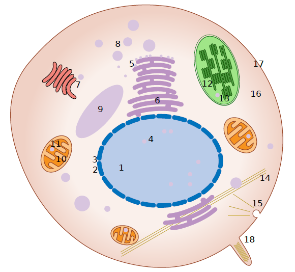
    Cell of eukaryote, second-order cell: 1 nucleus, 2 nuclear envelope, 3 nuclear pore, 4 DNA = chromatin = chromosomes, 5 ribosomes (black), 6 ER, 7 AG, 8 vesicles, 9 vacuole (big vesicle), 10 mitochondria (surrounded with double membrane), 11 mitochondrial DNA, 12 chloroplast (surrounded with double membrane), 13 chloroplast DNA, 14 cytoskeleton (brown), 15 phagocytosis (caught in the middle of process), 16 cytoplasm, 17 cell membrane, 18 eukaryotic flagella.

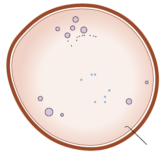
    Prokaryotic cell, cell of Monera, is much smaller, much more rigid and much simpler. Labels not provided because there is not much to label, and what is available, was already shown in the eukaryote. Except the cell wall which is outside of the cell membrane (some eukaryotes have cell wall though), and prokaryotic flagella (right bottom corner) which is just a molecule of protein:

    3.6.02.PNG
    Figure 3.6.2 : Yes, this is all.

    This page titled 3.6: Overview of the Cell is shared under a Public Domain license and was authored, remixed, and/or curated by Alexey Shipunov.

    • Was this article helpful?