
13.2: Mitosis
- Page ID
- 93194

\( \newcommand{\vecs}[1]{\overset { \scriptstyle \rightharpoonup} {\mathbf{#1}} } \)
\( \newcommand{\vecd}[1]{\overset{-\!-\!\rightharpoonup}{\vphantom{a}\smash {#1}}} \)
\( \newcommand{\dsum}{\displaystyle\sum\limits} \)
\( \newcommand{\dint}{\displaystyle\int\limits} \)
\( \newcommand{\dlim}{\displaystyle\lim\limits} \)
\( \newcommand{\id}{\mathrm{id}}\) \( \newcommand{\Span}{\mathrm{span}}\)
( \newcommand{\kernel}{\mathrm{null}\,}\) \( \newcommand{\range}{\mathrm{range}\,}\)
\( \newcommand{\RealPart}{\mathrm{Re}}\) \( \newcommand{\ImaginaryPart}{\mathrm{Im}}\)
\( \newcommand{\Argument}{\mathrm{Arg}}\) \( \newcommand{\norm}[1]{\| #1 \|}\)
\( \newcommand{\inner}[2]{\langle #1, #2 \rangle}\)
\( \newcommand{\Span}{\mathrm{span}}\)
\( \newcommand{\id}{\mathrm{id}}\)
\( \newcommand{\Span}{\mathrm{span}}\)
\( \newcommand{\kernel}{\mathrm{null}\,}\)
\( \newcommand{\range}{\mathrm{range}\,}\)
\( \newcommand{\RealPart}{\mathrm{Re}}\)
\( \newcommand{\ImaginaryPart}{\mathrm{Im}}\)
\( \newcommand{\Argument}{\mathrm{Arg}}\)
\( \newcommand{\norm}[1]{\| #1 \|}\)
\( \newcommand{\inner}[2]{\langle #1, #2 \rangle}\)
\( \newcommand{\Span}{\mathrm{span}}\) \( \newcommand{\AA}{\unicode[.8,0]{x212B}}\)
\( \newcommand{\vectorA}[1]{\vec{#1}} % arrow\)
\( \newcommand{\vectorAt}[1]{\vec{\text{#1}}} % arrow\)
\( \newcommand{\vectorB}[1]{\overset { \scriptstyle \rightharpoonup} {\mathbf{#1}} } \)
\( \newcommand{\vectorC}[1]{\textbf{#1}} \)
\( \newcommand{\vectorD}[1]{\overrightarrow{#1}} \)
\( \newcommand{\vectorDt}[1]{\overrightarrow{\text{#1}}} \)
\( \newcommand{\vectE}[1]{\overset{-\!-\!\rightharpoonup}{\vphantom{a}\smash{\mathbf {#1}}}} \)
\( \newcommand{\vecs}[1]{\overset { \scriptstyle \rightharpoonup} {\mathbf{#1}} } \)
\(\newcommand{\longvect}{\overrightarrow}\)
\( \newcommand{\vecd}[1]{\overset{-\!-\!\rightharpoonup}{\vphantom{a}\smash {#1}}} \)
\(\newcommand{\avec}{\mathbf a}\) \(\newcommand{\bvec}{\mathbf b}\) \(\newcommand{\cvec}{\mathbf c}\) \(\newcommand{\dvec}{\mathbf d}\) \(\newcommand{\dtil}{\widetilde{\mathbf d}}\) \(\newcommand{\evec}{\mathbf e}\) \(\newcommand{\fvec}{\mathbf f}\) \(\newcommand{\nvec}{\mathbf n}\) \(\newcommand{\pvec}{\mathbf p}\) \(\newcommand{\qvec}{\mathbf q}\) \(\newcommand{\svec}{\mathbf s}\) \(\newcommand{\tvec}{\mathbf t}\) \(\newcommand{\uvec}{\mathbf u}\) \(\newcommand{\vvec}{\mathbf v}\) \(\newcommand{\wvec}{\mathbf w}\) \(\newcommand{\xvec}{\mathbf x}\) \(\newcommand{\yvec}{\mathbf y}\) \(\newcommand{\zvec}{\mathbf z}\) \(\newcommand{\rvec}{\mathbf r}\) \(\newcommand{\mvec}{\mathbf m}\) \(\newcommand{\zerovec}{\mathbf 0}\) \(\newcommand{\onevec}{\mathbf 1}\) \(\newcommand{\real}{\mathbb R}\) \(\newcommand{\twovec}[2]{\left[\begin{array}{r}#1 \\ #2 \end{array}\right]}\) \(\newcommand{\ctwovec}[2]{\left[\begin{array}{c}#1 \\ #2 \end{array}\right]}\) \(\newcommand{\threevec}[3]{\left[\begin{array}{r}#1 \\ #2 \\ #3 \end{array}\right]}\) \(\newcommand{\cthreevec}[3]{\left[\begin{array}{c}#1 \\ #2 \\ #3 \end{array}\right]}\) \(\newcommand{\fourvec}[4]{\left[\begin{array}{r}#1 \\ #2 \\ #3 \\ #4 \end{array}\right]}\) \(\newcommand{\cfourvec}[4]{\left[\begin{array}{c}#1 \\ #2 \\ #3 \\ #4 \end{array}\right]}\) \(\newcommand{\fivevec}[5]{\left[\begin{array}{r}#1 \\ #2 \\ #3 \\ #4 \\ #5 \\ \end{array}\right]}\) \(\newcommand{\cfivevec}[5]{\left[\begin{array}{c}#1 \\ #2 \\ #3 \\ #4 \\ #5 \\ \end{array}\right]}\) \(\newcommand{\mattwo}[4]{\left[\begin{array}{rr}#1 \amp #2 \\ #3 \amp #4 \\ \end{array}\right]}\) \(\newcommand{\laspan}[1]{\text{Span}\{#1\}}\) \(\newcommand{\bcal}{\cal B}\) \(\newcommand{\ccal}{\cal C}\) \(\newcommand{\scal}{\cal S}\) \(\newcommand{\wcal}{\cal W}\) \(\newcommand{\ecal}{\cal E}\) \(\newcommand{\coords}[2]{\left\{#1\right\}_{#2}}\) \(\newcommand{\gray}[1]{\color{gray}{#1}}\) \(\newcommand{\lgray}[1]{\color{lightgray}{#1}}\) \(\newcommand{\rank}{\operatorname{rank}}\) \(\newcommand{\row}{\text{Row}}\) \(\newcommand{\col}{\text{Col}}\) \(\renewcommand{\row}{\text{Row}}\) \(\newcommand{\nul}{\text{Nul}}\) \(\newcommand{\var}{\text{Var}}\) \(\newcommand{\corr}{\text{corr}}\) \(\newcommand{\len}[1]{\left|#1\right|}\) \(\newcommand{\bbar}{\overline{\bvec}}\) \(\newcommand{\bhat}{\widehat{\bvec}}\) \(\newcommand{\bperp}{\bvec^\perp}\) \(\newcommand{\xhat}{\widehat{\xvec}}\) \(\newcommand{\vhat}{\widehat{\vvec}}\) \(\newcommand{\uhat}{\widehat{\uvec}}\) \(\newcommand{\what}{\widehat{\wvec}}\) \(\newcommand{\Sighat}{\widehat{\Sigma}}\) \(\newcommand{\lt}{<}\) \(\newcommand{\gt}{>}\) \(\newcommand{\amp}{&}\) \(\definecolor{fillinmathshade}{gray}{0.9}\)By the end of this lesson you will be able to:
- Compare diploid and haploid and identify which cells in the plant are which.
- Understand why cells undergo mitosis.
- Explain how the chromosomes prepare for cell division in the S phase of interphase.
- Recognize what happens to the chromosomes, cell wall, cell membrane, and nuclear membrane in each stage of mitosis.
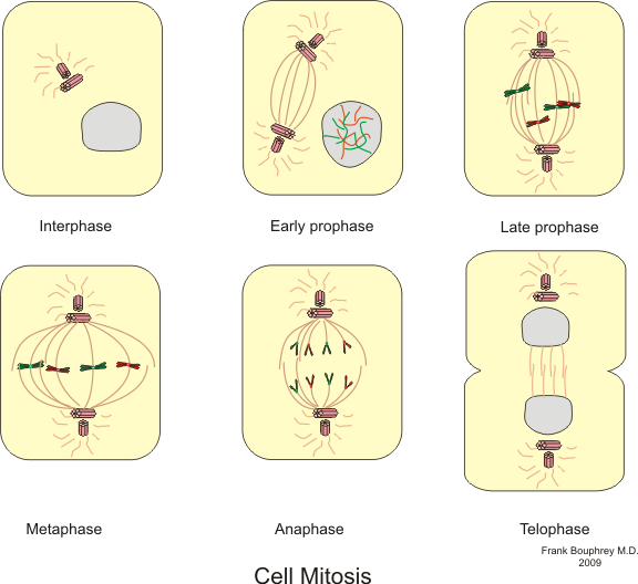
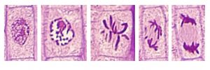
Phases of mitosis. Boumphreyfr. CC BY-SA 3.0
Ploidy
The previous lesson focused on DNA’s ribose-phosphate backbone, on the purine and pyrimidine bases, and on how DNA complexes with protein and coils to form chromatin. Here we’ll look more closely at the synthesis (S) phase of interphase and at the mitosis (M) phase. Recall that the mitosis phase of the cell cycle “pie” is divided into four stages; we’ll look now at what happens in each of those stages and how it contributes to the outcome of mitosis, the equal division of chromosomes into two daughter cells.
Ploidy refers to the number of sets of homologous (identical) chromosomes in a cell.
- In higher organisms like plants (and animals, including humans), gamete cells (egg and sperm) typically each contain one set of each of the chromosomes found in that particular species. When cells contain one set of chromosomes characteristic of the species, this state is called haploid and is abbreviated n.
- When the sperm and egg, each of which are n, unite to form a zygote, the zygote cell now has two sets of chromosomes, one from the male parent’s sperm and one from the female parent’s egg. When cells contain two sets of chromosomes, they are described as diploid, abbreviated 2n.
- Recall that one result of double fertilization in plants is that one sperm cell unites with two female polar bodies to create the endosperm found in seeds. Endosperm cells have three sets of chromosomes, two from the female parent’s polar nuclei (n + n) and one from the male parent’s sperm (n), so this tissue is triploid, abbreviated 3n.
Most of the cells of flowering plants that we have studied so far, like the cells making up the epidermis, cortex, and vascular tissues (but not the sperm and eggs cells), are called somatic cells, and are diploid (2n). Each cell carries two sets of chromosomes: one from the male parent and one from the female parent.
Each species of plant has a characteristic number of chromosomes in its somatic cells. Bur oak has 24. The garden petunia has 14. Green bean has 22. Half of those chromosomes came from the egg and half from the sperm, so the plant has two sets of chromosomes. In the bean, the 22 chromosomes can be numbered from 1 to 11 based on their morphology (chromosomes have different lengths). The numbering only goes to 11, even though there are 22 chromosomes, because each diploid cell has two copies of chromosome 1, two copies of chromosome 2, and so on.
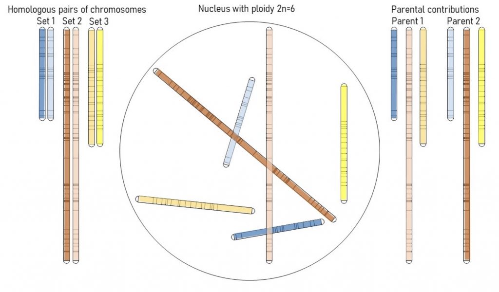
The illustration above shows this for a hypothetical plant’s somatic cell’s nucleus containing 6 chromosomes. On the left side, the chromosomes are rearranged into three pairs of homologs. The matching chromosomes from the two different sets (for instance, the two copies of chromosome 1) are called homologous chromosomes or homologs. Homologs carry, at the same location on the chromosome, the genetic information that affects the same characteristic or function. The version of the information can be different between the homologous chromosomes — that is, the sequence of base pairs may be somewhat different because one homolog came from the female and the other from the male.
The homologs look identical and carry genetic information about particular cell functions at identical places on the chromosome (shown using dark bands at specific locations on the chromosome), but the exact base pair sequences at those locations may differ, resulting in different alleles and gene function. The parental combinations are shown at the right, and are the haploid contribution that resulted from meiosis. The 50% reduction in the sex cells ensures that offspring have the proper diploid chromosome number and matching homologs that are the full compliment of the plants genome.
A cell in the plant’s apical meristem that is preparing to divide is a somatic cell, so it is diploid, and contains two sets of chromosomes. When it undergoes mitosis, the outcome will be two identical diploid sister cells. Each of these sister cells will also be diploid, and will contain exact copies of the two sets of chromosomes that were in the original cell. “Daughter” and “sister” cells refer to the same thing — the new cells that arise as the result of mitosis.
From our study of meristems, you know that growth is the result of the formation of new cells, and the subsequent elongation of those cells. Mitosis is the process that results in the formation of new cells. Cells undergo mitosis, therefore, as part of plant growth.
- Somatic cells of beans have 22 chromosomes. How many chromosomes in a bean sperm cell?
- Corn egg cells have 10 chromosomes. How many chromosomes are found in a corn seed’s endosperm cells?
- Why do cells undergo mitosis? Is it important?
Synthesis
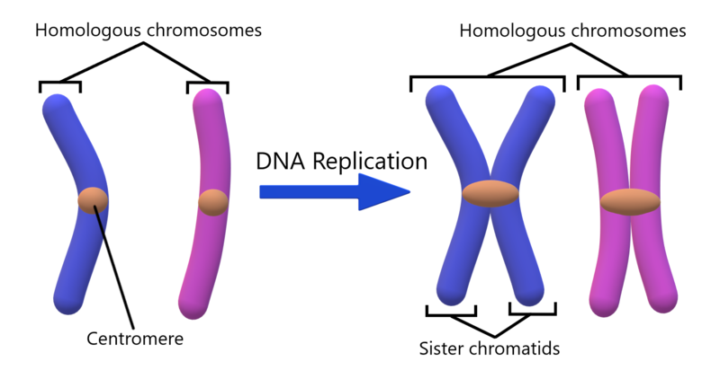
If a cell that undergoes mitosis divides into two cells, how can both of these new cells be identical to each other and to the original cell? Won’t the chromosomes in the original parent cell be divided in half during division? Won’t the resulting cells be haploid instead of diploid? No. The number of chromosomes isn’t reduced during mitotic cell division because, prior to division, each of the chromosomes replicates (duplicates), meaning that the cell makes an exact copy of each chromosome. This replication process happens during the synthesis (S) phase of the cell cycle.
Remember that G1, S, and G2 phases of the cell cycle are collectively called interphase. Most cells in the plant go about their business in the G1 phase. Only those cells called upon to divide make the next step, which is to replicate their chromosomes in the S phase. Once the chromosomes are replicated, the cell moves into the G2 phase of interphase and awaits mitosis. The S phase is called synthesis because making a copy of the chromosome requires new DNA production, or synthesis.
The two chromosomes that are exact copies are called sister chromatids and remain connected at one spot along their length; this spot is called the centromere, as shown in the illustration.
- If a diploid cell enters S phase with 2n=20 chromosomes, how many sister chromatids are in the cell when it enters G2?
- Are the replicated sister chromatids independent or are they connected in some physical way?
Stages of mitosis
This video provides a view of the fluidity of mitosis in a cell where 2N = 8 chromosomes, 4 pairs = 4 paternal + 4 maternal.
Below is an illustration and a corresponding micrograph for each stage in mitosis, showing a hypothetical plant cell where 2n=4 (two sets of chromosomes, two chromosomes per set).
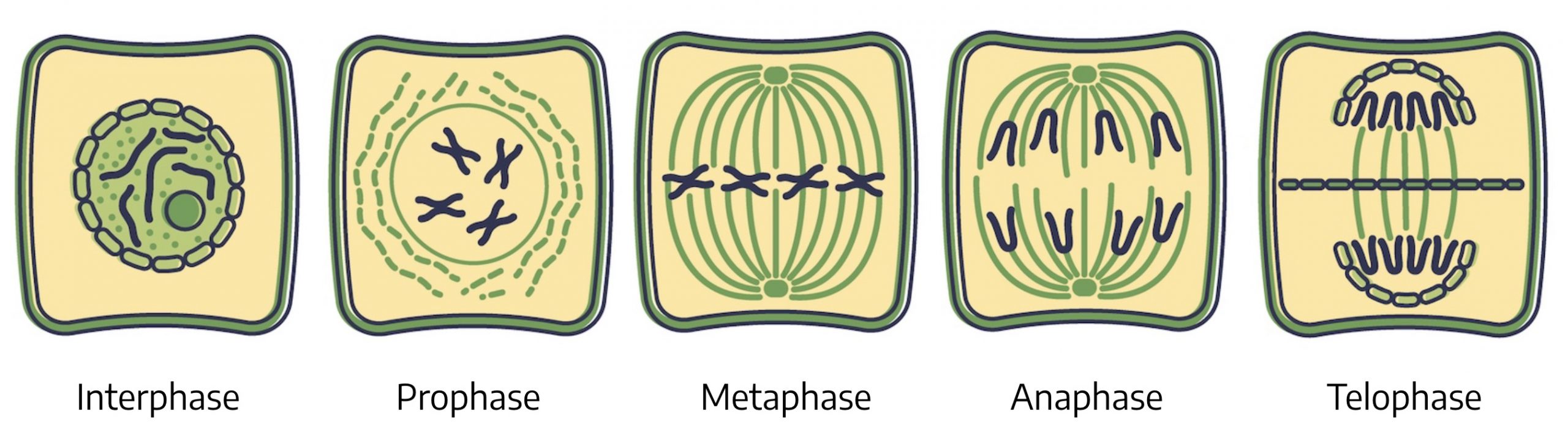
The micrographs below are onion (Allium cepa) root tip cells. Onion has 2n=16 chromosomes. Each of the cells has two sets of chromosomes where each set is made up of eight chromosomes. The micrographs are real examples of the illustrations above.
Interphase
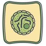 The lefthand frame of the illustration shows interphase cells. The deep red stained structures in the center of the onion cell micrograph are the chromosomes. They are corralled together within the nuclear membrane. Recall that during interphase the chromosomes are relaxed rather than highly condensed (that is, not extensively coiled or folded), and during the S phase of interphase each chromosome replicates. It makes sense that the chromosomes are relaxed because they can’t go through the replication process if they are tightly coiled, and because chromosomes only need to be coiled so that they can withstand movement and not break. They aren’t moving, just replicating, so being in a relaxed state is perfect. The two identical copies are called sister chromatids and they are held together at a site called the centromere. Note that sister chromatids are not the same as homologs. Homologs are corresponding chromosomes, one contributed through the sperm, the other through the egg. Sister chromatids are chromosomes that have replicated, are identical to each other, and are held together at centromeres. You can’t distinguish individual chromosomes in the picture because they are relaxed rather than tightly coiled and folded, making them so fine that they are difficult to see.
Prophase
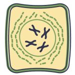 Prophase is the first stage of the M phase. In prophase the chromatin begins to coil and condense to form chromosomes. They are coiling because they are preparing to move around. You can begin to notice that each chromosome appears to have two strands (sister chromatids) and that these sister chromatids are attached to each other at a centromere. In prophase the nuclear membrane disappears and the chromosomes spread out to fill up much of the cell. During this phase, the spindle apparatus begins to appear. The spindles are microtubules associated with movement of the chromosomes during division.
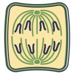
Metaphase
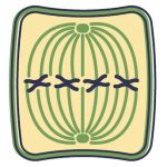 In metastage the spindle grows and forms attachments to the pairs of sister chromatids at the centromere that connects the sister chromatids. This point of attachment is called the kinetochore. The sister chromatids move to an imaginary equatorial plate (called the metaphase plate), which is formed along the midline of the cell between the poles. The sister chromatids are in their most condensed state at metaphase.
Anaphase
 The sister chromatids begin to separate at anaphase. When the sister chromatids separate, the centromeres divide so that one sister chromatid migrates to one pole, and the other migrates to the opposite pole. The chromatids that formed back in the S phase of interphase, when the chromosome replicated, now separate, and the spindle fibers shorten. With the sister chromatids separated, we can return to calling them chromosomes. Anaphase is the stage where the chromosomes carrying the DNA code are divided precisely so that each of the resulting cells has exactly the same chromosomes that were in the mother cell prior to division. One complete diploid complement of chromosomes (two sets) is delivered to each daughter cell.
Telophase
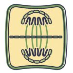 In telophase, the nuclear membrane forms around the chromosomes in each of the daughter cells, a cell plate forms between these cells, and cell walls separate the newly formed cells in a process called cytokinesis. The chromosomes decondense and again become relaxed chromatin. Telophase is the last stage of the M phase. After telophase and cytokinesis, the cells return to G1 of interphase.
- When do the sister chromatids separate from each other?
- Do the chromosomes replicate during mitosis or during interphase?
- Why are the chromosomes in their most condensed state during metaphase and retain this condensed state through chromatid migration in anaphase?
Review
Recall that the outcome of mitosis is two cells with DNA identical to that in the original cell. There are three keys to understanding how two cells are formed from one, both with the same DNA as the original cell:
- The DNA is completely replicated during the S stage of interphase. This replication results in twice as many sister chromatids as there were chromosomes, and once these sister chromatids separate and are evenly allocated to the two new sister cells, both sister cells have the diploid number of chromosomes, just like the original cell prior to division.
- As the cell prepares to divide, the DNA condenses. This packaging helps keep the very thin DNA helices from being broken, and keeps the DNA organized into a tight package so that the cell can keep track of it and move it around.
- The process is very organized. For instance, the sister chromatids all line up in the middle of the cell at metaphase, split at the centromere, and half the chromatids go to one side of the cell, half to the other. This orderly separation of the sister chromatids ensures that the right number of chromosomes is packaged into each of the new sister cells.
There are many sites online that illustrate mitosis, but particularly relevant here are ones that show micrographs of plant cells. You may discover that there are some details about the spindles and their apparent site of origin that differ between descriptions of mitosis in animal and plant cells; not everything online pertains to plants. Any mention of a structure called a “centriole” refers to animal cell mitosis, not plants (as plants don’t have centrioles).
John H. Wahlert and Mary Jean Holland, of Baruch College, authored this site showing stages of mitosis in onion. (You can ignore the stages of whitefish mitosis in the second half of the site unless you are interested in the differences between plant and animal mitosis.)