
6.1: Plant Cells and Tissues
- Page ID
- 93167

\( \newcommand{\vecs}[1]{\overset { \scriptstyle \rightharpoonup} {\mathbf{#1}} } \)
\( \newcommand{\vecd}[1]{\overset{-\!-\!\rightharpoonup}{\vphantom{a}\smash {#1}}} \)
\( \newcommand{\dsum}{\displaystyle\sum\limits} \)
\( \newcommand{\dint}{\displaystyle\int\limits} \)
\( \newcommand{\dlim}{\displaystyle\lim\limits} \)
\( \newcommand{\id}{\mathrm{id}}\) \( \newcommand{\Span}{\mathrm{span}}\)
( \newcommand{\kernel}{\mathrm{null}\,}\) \( \newcommand{\range}{\mathrm{range}\,}\)
\( \newcommand{\RealPart}{\mathrm{Re}}\) \( \newcommand{\ImaginaryPart}{\mathrm{Im}}\)
\( \newcommand{\Argument}{\mathrm{Arg}}\) \( \newcommand{\norm}[1]{\| #1 \|}\)
\( \newcommand{\inner}[2]{\langle #1, #2 \rangle}\)
\( \newcommand{\Span}{\mathrm{span}}\)
\( \newcommand{\id}{\mathrm{id}}\)
\( \newcommand{\Span}{\mathrm{span}}\)
\( \newcommand{\kernel}{\mathrm{null}\,}\)
\( \newcommand{\range}{\mathrm{range}\,}\)
\( \newcommand{\RealPart}{\mathrm{Re}}\)
\( \newcommand{\ImaginaryPart}{\mathrm{Im}}\)
\( \newcommand{\Argument}{\mathrm{Arg}}\)
\( \newcommand{\norm}[1]{\| #1 \|}\)
\( \newcommand{\inner}[2]{\langle #1, #2 \rangle}\)
\( \newcommand{\Span}{\mathrm{span}}\) \( \newcommand{\AA}{\unicode[.8,0]{x212B}}\)
\( \newcommand{\vectorA}[1]{\vec{#1}} % arrow\)
\( \newcommand{\vectorAt}[1]{\vec{\text{#1}}} % arrow\)
\( \newcommand{\vectorB}[1]{\overset { \scriptstyle \rightharpoonup} {\mathbf{#1}} } \)
\( \newcommand{\vectorC}[1]{\textbf{#1}} \)
\( \newcommand{\vectorD}[1]{\overrightarrow{#1}} \)
\( \newcommand{\vectorDt}[1]{\overrightarrow{\text{#1}}} \)
\( \newcommand{\vectE}[1]{\overset{-\!-\!\rightharpoonup}{\vphantom{a}\smash{\mathbf {#1}}}} \)
\( \newcommand{\vecs}[1]{\overset { \scriptstyle \rightharpoonup} {\mathbf{#1}} } \)
\(\newcommand{\longvect}{\overrightarrow}\)
\( \newcommand{\vecd}[1]{\overset{-\!-\!\rightharpoonup}{\vphantom{a}\smash {#1}}} \)
\(\newcommand{\avec}{\mathbf a}\) \(\newcommand{\bvec}{\mathbf b}\) \(\newcommand{\cvec}{\mathbf c}\) \(\newcommand{\dvec}{\mathbf d}\) \(\newcommand{\dtil}{\widetilde{\mathbf d}}\) \(\newcommand{\evec}{\mathbf e}\) \(\newcommand{\fvec}{\mathbf f}\) \(\newcommand{\nvec}{\mathbf n}\) \(\newcommand{\pvec}{\mathbf p}\) \(\newcommand{\qvec}{\mathbf q}\) \(\newcommand{\svec}{\mathbf s}\) \(\newcommand{\tvec}{\mathbf t}\) \(\newcommand{\uvec}{\mathbf u}\) \(\newcommand{\vvec}{\mathbf v}\) \(\newcommand{\wvec}{\mathbf w}\) \(\newcommand{\xvec}{\mathbf x}\) \(\newcommand{\yvec}{\mathbf y}\) \(\newcommand{\zvec}{\mathbf z}\) \(\newcommand{\rvec}{\mathbf r}\) \(\newcommand{\mvec}{\mathbf m}\) \(\newcommand{\zerovec}{\mathbf 0}\) \(\newcommand{\onevec}{\mathbf 1}\) \(\newcommand{\real}{\mathbb R}\) \(\newcommand{\twovec}[2]{\left[\begin{array}{r}#1 \\ #2 \end{array}\right]}\) \(\newcommand{\ctwovec}[2]{\left[\begin{array}{c}#1 \\ #2 \end{array}\right]}\) \(\newcommand{\threevec}[3]{\left[\begin{array}{r}#1 \\ #2 \\ #3 \end{array}\right]}\) \(\newcommand{\cthreevec}[3]{\left[\begin{array}{c}#1 \\ #2 \\ #3 \end{array}\right]}\) \(\newcommand{\fourvec}[4]{\left[\begin{array}{r}#1 \\ #2 \\ #3 \\ #4 \end{array}\right]}\) \(\newcommand{\cfourvec}[4]{\left[\begin{array}{c}#1 \\ #2 \\ #3 \\ #4 \end{array}\right]}\) \(\newcommand{\fivevec}[5]{\left[\begin{array}{r}#1 \\ #2 \\ #3 \\ #4 \\ #5 \\ \end{array}\right]}\) \(\newcommand{\cfivevec}[5]{\left[\begin{array}{c}#1 \\ #2 \\ #3 \\ #4 \\ #5 \\ \end{array}\right]}\) \(\newcommand{\mattwo}[4]{\left[\begin{array}{rr}#1 \amp #2 \\ #3 \amp #4 \\ \end{array}\right]}\) \(\newcommand{\laspan}[1]{\text{Span}\{#1\}}\) \(\newcommand{\bcal}{\cal B}\) \(\newcommand{\ccal}{\cal C}\) \(\newcommand{\scal}{\cal S}\) \(\newcommand{\wcal}{\cal W}\) \(\newcommand{\ecal}{\cal E}\) \(\newcommand{\coords}[2]{\left\{#1\right\}_{#2}}\) \(\newcommand{\gray}[1]{\color{gray}{#1}}\) \(\newcommand{\lgray}[1]{\color{lightgray}{#1}}\) \(\newcommand{\rank}{\operatorname{rank}}\) \(\newcommand{\row}{\text{Row}}\) \(\newcommand{\col}{\text{Col}}\) \(\renewcommand{\row}{\text{Row}}\) \(\newcommand{\nul}{\text{Nul}}\) \(\newcommand{\var}{\text{Var}}\) \(\newcommand{\corr}{\text{corr}}\) \(\newcommand{\len}[1]{\left|#1\right|}\) \(\newcommand{\bbar}{\overline{\bvec}}\) \(\newcommand{\bhat}{\widehat{\bvec}}\) \(\newcommand{\bperp}{\bvec^\perp}\) \(\newcommand{\xhat}{\widehat{\xvec}}\) \(\newcommand{\vhat}{\widehat{\vvec}}\) \(\newcommand{\uhat}{\widehat{\uvec}}\) \(\newcommand{\what}{\widehat{\wvec}}\) \(\newcommand{\Sighat}{\widehat{\Sigma}}\) \(\newcommand{\lt}{<}\) \(\newcommand{\gt}{>}\) \(\newcommand{\amp}{&}\) \(\definecolor{fillinmathshade}{gray}{0.9}\)By the end of this lesson you will be able to:
- Label the parts of a plant cell.
- List the types of tissues in a plant and describe where they are located and the specialized cells that make up each of these tissues.
- Summarize the key functions of those tissues.
Plant cell
Watch this video about plant cell parts (7:47)
The graphic below illustrates the key parts of the plant cell.

Cell wall
The outer covering of the cell, the cell wall is a rigid membrane that contains cellulose (a carbohydrate that is indigestible for humans). The cell wall protects the parts inside, and the cellulose molecules in the wall provide the support and rigidity needed to maintain the cell’s three-dimensional structure.
Cell membrane
The cell membrane is made up of layers of protein and lipid (fats and oils are examples of lipids). The cell membrane is semi-permeable — it allows select compounds in and out, but blocks other types of compounds. If the cell were like a bicycle tire, the cell wall would be the thick, protective outer tire tread and the cell membrane would be the inner tube.
Chloroplast
An organelle (“organelle” is the generic name for a plant organ) that contains chlorophyll. In the chloroplast, light energy is captured and the first steps are taken in the chemical pathway that converts the energy in light into forms of energy that the plant can transport and store, like sugar and starch. Chloroplasts are not evenly distributed throughout the plant but, as you might expect, are concentrated in parts of the plant that are exposed to and oriented toward the sun. A plant cell in the leaf blade will have many chloroplasts, while cells in the middle of the stem will have few or none.
Mitochondria
(Singular = mitochondrion)
The mitochondria is where stored sugars from photosynthesis are metabolized to produce forms of energy that the plant can use for growth. This metabolism is known as respiration and uses oxygen to convert sugars (and other carbohydrates) to energy and carbon dioxide. This is the cell’s power plant. All cells have numerous mitochondria.
Nucleus
An organelle that contains the chromosomes. Chromosomes contain the genetic material (deoxyribonucleic acid; DNA) that is carried within each cell and that directs which chemical reactions are turned on and off in the cell. Chromosomes are the hereditary material passed on to new cells and to subsequent generations. Each cell has one nucleus.
Vacuole
An organelle containing various fluids, ions, chemical energy, and waste products from the cell. The vacuole takes up much of the cell volume and gives shape to the cell.
Cytoplasm
The fluid inside the cell membrane in which the organelles and other plant cell parts are suspended.
Middle lamella
A material containing pectin that forms between cells and cements the cell wall of one cell to the cell wall of an adjacent cell. If bricks in a wall are like cells in a plant, the middle lamella in the plant is like the mortar between bricks in the wall.
Plant cells have other parts as well, but these are the key ones to know and understand now.
- What is the difference in function between the cell wall and the cell membrane?
- What is the mortar that holds cells together? If lettuce is grown in a soil with low calcium content, the outer edges of leaves can degenerate and die, causing tip burn. Could this involve the mortar that holds cells together?
- Where is light energy captured?
- What happens in the mitochondria, and what is the connection between that function in mitochondria and the function of chloroplasts?
Tissues
Watch this video about plant tissues (6:30)
A tissue is a group of cells that share a function. The cells within a tissue may differ from one another, but they all contribute to a particular function. We’re going to look at three types of tissues: dermal, cortex, and vascular.
Dermal tissue
Dermal tissues (derma is Greek for “skin”) are on the outside of the plant and provide protection for the plant cells they surround. The cells making up dermal tissues are tough so that they can protect against mechanical challenges to the plant, like abrasion. They have thick cell walls. In the shoot, the epidermis cells, which are the main cell type in dermal tissue, secrete a water-resistant substance called cutin (a waxy polymer), which coats the wall of the cell exposed to the environment. This coating helps limit the loss to the atmosphere of water that is inside the plant. Cutin is absent or greatly reduced in root tissue because roots need to reach out into the soil to absorb water and nutrients.


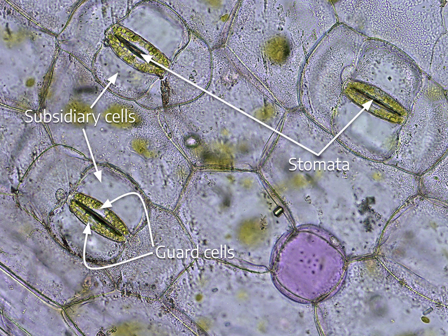
The epidermis is the outermost layer of cells in the plant. It is normally only one cell thick, but in some cases the epidermis can be a few cells thick. Epidermis cells typically have few if any chloroplasts. They are often called pavement cells because they are flat like tiles or puzzle pieces. Depending on the plant, the epidermis may have hairs, or trichomes, that extend out from the plant. Some of these trichomes are associated with glands that contain oils or other substances secreted by the plant.
The epidermis contains pairs of guard cells that will open to form stomata (Greek stoma = mouth; an opening in the leaf surface) through which gasses can move into and out of the deeper cell layers in the leaf. These guard cells are found most abundantly on the underside of leaves, but may also be on the upper leaf surface and on the stems.
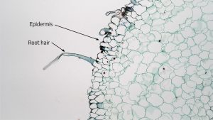
The root has dermal tissue as well. The predominant cell type, like in the shoot, is also epidermis, but as noted above there is no cutin covering the root epidermis because the root is underground and less prone to dehydration. There are no guard cells or trichomes, but there are root hairs . The root hair is a very small-diameter extension of the epidermis cell wall and cell membrane that extends out into the growth medium. Water and nutrients enter the plant through absorption into the root hairs.
- What unique feature of the epidermis is found in roots and not shoots?
- What is the function of the waxy cutin layer? Why don’t you find it on the root epidermis?
- Are stomata found in roots, shoots, or both? Why does this make sense?
- What’s the difference between a cell and a tissue?
Cortex or ground meristem tissue
The cortex (sometimes called “ground meristem“) tissue is found just inside the epidermis and extends toward the interior of the stem and root. Some types of plants also contain cortex tissue at the very center of the stem called the pith, but you won’t find pith in roots or in all plant stems. Cortex cells provide structural support for the stems. In leaves, this tissue just inside the epidermis is called the mesophyll (“middle of the leaf”). Mesophyll tissue is the site of most photosynthesis reactions in the leaf.
Three types of cells make up the cortex:
Parenchyma
- The most common type of cortex cell.
- Has thin cell walls (called a primary wall in the graphic below).
- The mature cell is alive.
- Has the ability to begin dividing to help heal wounds (by covering the wound with plant tissue called callus).
- Will also divide to initiate adventitious roots on stem cuttings.
- Site of many other functions, such as photosynthesis and storage of starch and other chemical compounds.
- Leaf mesophyll tissue is a type of parenchyma that is packed with chloroplasts.
Collenchyma
- A living cell at maturity.
- Cell walls are thicker than the thin parenchyma cell walls, which give collenchyma strength. However, these cells remain somewhat flexible compared to sclerenchyma, which you will read about next.
- The cells can connect together to form resilient strands, like the strands of a celery stalk. These strands provide support for young tissues.
- Because the cells are alive, they can respond to external stimuli. If the plant is regularly shaken by wind, for example, the collenchyma cells will respond by producing thicker cell walls for greater support of the plant stem so that it can remain upright.
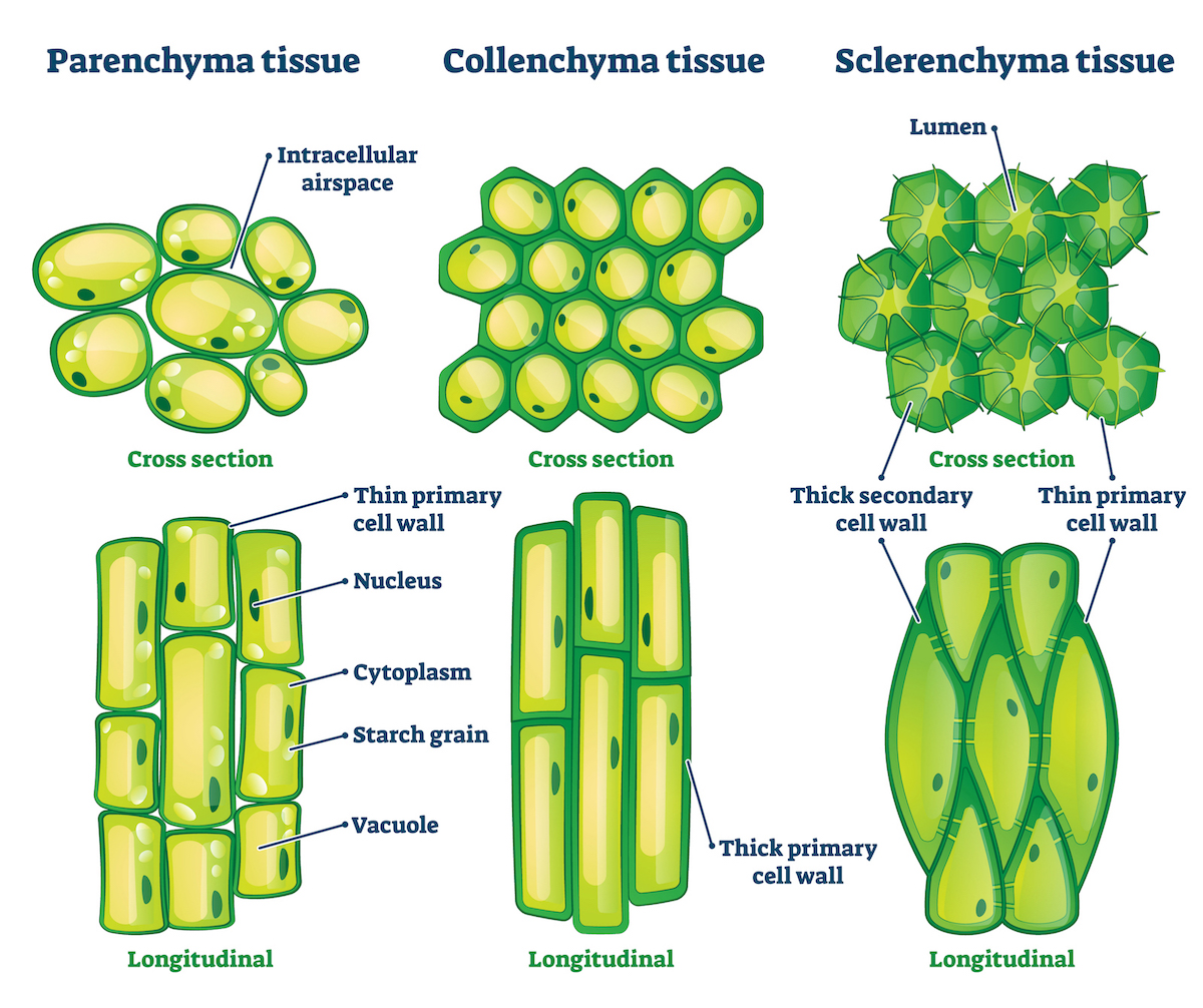
Sclerenchyma
- This type of cell has a primary and secondary cell wall. The primary cell wall, on the outside of the cell, is rich in cellulose, just like other plant cell walls. Once the cell has reached its final size, a secondary cell wall is deposited just inside the primary wall. The secondary wall has a high concentration of lignin that gives the cell rigidity. This rigid, lignified secondary cell wall is responsible for sclerenchyma’s hardness and strengthening properties. Sclerenchyma comes in two types:
- Fibers (see below) formed from long strands of sclerenchyma. These tough fibers give the plant rigidity. We extract these fibers from plants and use them in fabrics, carpets, and rope. Examples of plant fibers made up of sclerenchyma cells include jute, hemp, and flax (the fabric made of flax fibers is called linen). Cotton is not in this list; it is an epidermal fiber produced by the plant’s seed coats.
- Sclereids are cells with hard, tough cell walls. Sclereid cells can coalesce and cover other plant parts. For instance, they form the hard covering around the seeds (the endocarp) of stone fruits like cherries, the hard shell around walnuts, and the hard covering of coconut. Sclereids also make up the grit that crunches between your teeth when you eat a pear.
- Sclerenchyma cells are dead at maturity. They don’t thicken in response to external stimuli the way collenchyma can.
- Which cortex cells are alive and which are dead when mature?
- Which cells make up the tough fibers from which rope and fabrics can be made?
- Which cells divide to initiate adventitious roots?
- What tissue in the leaf corresponds to the cortex in the stem?
Vascular tissue
Vascular tissues form the plumbing system in the plant through which water, nutrients, sugars, and other compounds flow. These plumbing pipes and associated cells are bundled together in the plant in a structure called the vascular bundle. There are three main types of vascular tissue: xylem, phloem, and vascular cambium. Xylem and phloem are composed of different types of cells, listed below.
Xylem
- Moves water in the plant.
- The water flow is unidirectional. Water in xylem heads from root to stem to leaf and then out of the plant stomates through a process called transpiration.
- The part of the tree that we call “wood” is made up of xylem.
- These cells are dead at maturity, and they are hollow.
Xylem tissue is composed of four different types of cells:
Vessels
Elongated cells that connect end to end to form tubes. The cells are dead at maturity. The end walls of the vessels are perforated, so water can move freely through the holes and flow from cell to cell. Vessels have a relatively large diameter compared to other xylem cells and allow greater movement of water.
Tracheids
These cells are elongated and narrower than vessels, and connect by overlapping at their ends. These cells are also dead at maturity and contain pits through which water can move. Tracheids appear earlier in the paleontological record of plant evolutionary development than vessels and are thus considered “primitive” (not inferior, but appearing earlier in evolutionary time). Vessels are a subsequent evolutionary adaptation that allow for greater water flow because of their larger diameter.
Xylem fibers
Sclerenchyma cells lying near the vessels and tracheids, and thus part of the vascular bundle. They are strung together end to end like the vessels and tracheids, but unlike those water carriers they have no pits or perforations and instead have thick primary and secondary cell walls. They provide flexible support for the plant from within the vascular bundles.
Xylem parenchyma

In woody plants there are parenchyma cells around the vascular bundles that extend horizontally through the xylem (the woody part of the plant) and develop into rays moving laterally from the center to the exterior of the plant. Most of the vascular cell types are arranged in a linear fashion parallel to the long axis of the stem, but parenchyma rays are arranged laterally from the middle of the stem out toward the epidermis. They function to conduct water through the wood (xylem). Oak furniture for example, it will have a “grain” which is caused by the annual rings of xylem, and will have rays that, on edge, look like small pits in the wood. We will see this in later lectures when we deal more extensively with wood and secondary growth. As you can see in the photo to the right, some of the natural markings you see in an instrument’s wood are from parenchyma rays.
Phloem
- Moves some nutrients taken up by the roots to other parts of the plant.
- Moves sugars manufactured in leaves by photosynthesis, and other plant compounds such as plant hormones like auxin, to other parts of the plant.
- The flow in the phloem is multi-directional among leaf, stem, and root.
Phloem tissue also has four types of cells:
Sieve tube members
Elongated cells that join end to end to form tubes for passage of liquids. The end walls have pores. Unlike xylem cells, these cells are still alive. They have a thin cell membrane containing a layer of living protoplasm that hugs the wall of the cell.

Companion cells
Associated with sieve tube members. Contain a nucleus, may direct the metabolism of the sieve tube member, and are alive.
Phloem fibers (sclerenchyma cells)
Provide support, same as for xylem.
Phloem parenchyma cells
Adjoin the sieve tube cells, same as for xylem.
Vascular cambium
This third type of vascular tissue is a meristematic region (meaning that the cells can actively divide to form new growth) where new vascular tissues originate in plants with secondary growth, like trees. We will study secondary growth in Chapter 6.2.
- What substance flows in the xylem? Does it flow both directions or only up from the roots to the leaves?
- What are examples of substances that flow in the phloem? Do these flow both directions or only from roots to leaves?
- Which vascular cells are dead and which are alive at maturity?
- Look at a piece of wooden furniture near where you are sitting. What type of plant tissue and cell do you see? Look at the natural markings in the wood. What are those tissues and cells?