We see a massive amount of variation in the sizes and shapes of leaves. Similarly, the anatomical structure of leaves can vary considerably. Plant leaves may be specialised to maximise light utilisation, to minimise water loss, to facilitate C4 photosynthesis or CAM photosynthesis, to resist damage due to water stress, or to float on water.
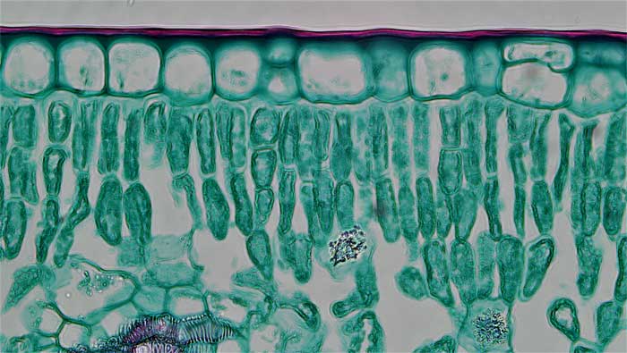
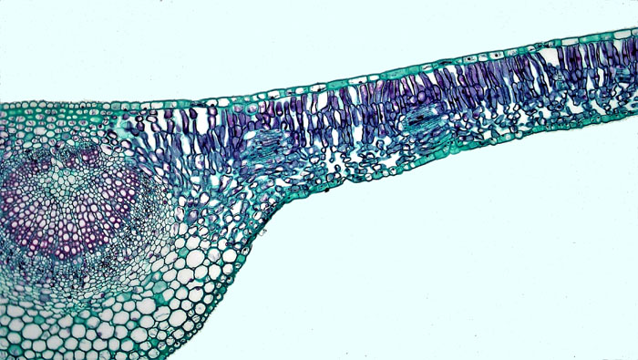
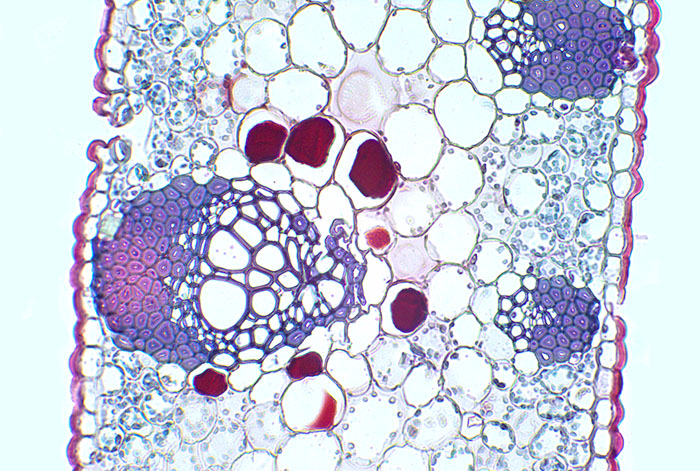
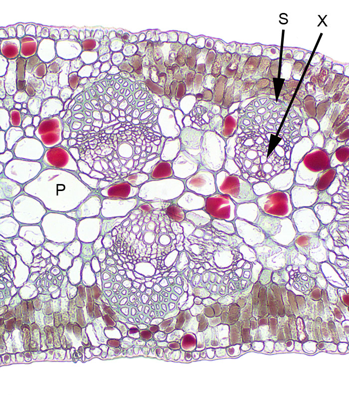
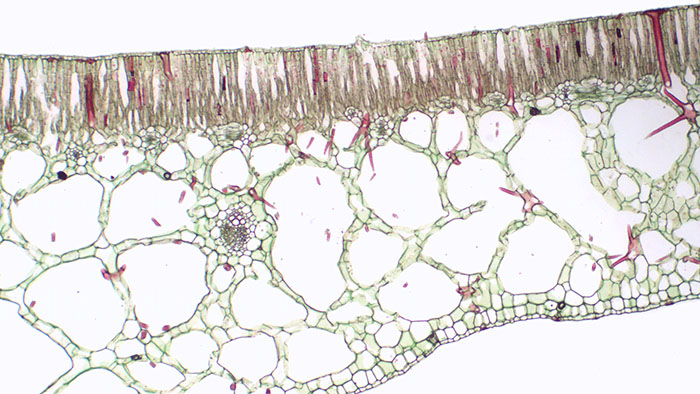
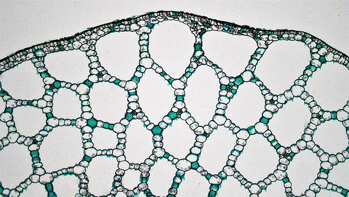
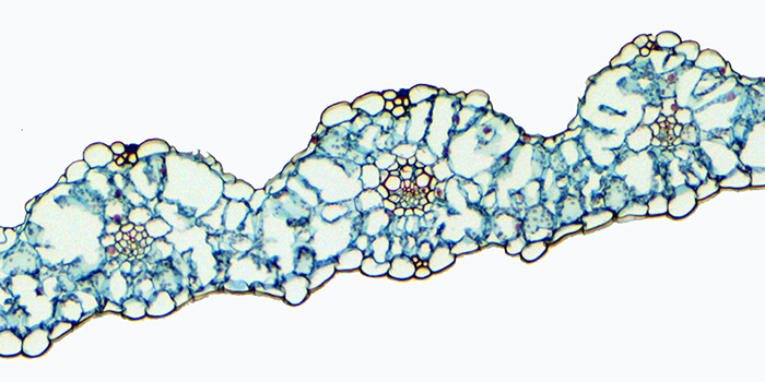
Figure 3.1. A typical leaf of a mesophyte plant with the cuticle above the dermal cells and then a later of closely packed palisade parenchyma cells. In the bottom third of the image, spongy parenchyma has large airspaces between the cells to maximise contact with carbon dioxide flowing in through the stomata. (Berkshire Community College Bioscience Image Library, public domain).Figure 3.2. Another example of a typical leaf of a mesophyte plant with the closely packed palisade parenchyma cells on the upper side of the leaf and the spongy parenchyma below. (Berkshire Community College Bioscience Image Library, public domain).Figure 3.3. An example of a leaf with some xerophyte features. Although it still has closely packed palisade parenchyma cells on the upper right surface and spongy parenchyma to the left, the cells are more closely packed and the cuticle is thicker. This is the leaf of a mangrove, Rhizophora. (Sean Bellairs, CC: attribute, share alike).Figure 3.4. An example of a leaf with clearer xerophyte features. Parenchyma tissues are not differentiated into palisade and spongy parenchyma. The cells are closely packed and as well as a thick cuticle there is substantial sclerenchyma (purple cells with very thick cell walls). The lignified cells have reddish-purple cell walls. This is the leaf of a drought adapted Myrtaceae, Callistemon. (Sean Bellairs, CC attribute, share alike).Figure 3.5. A drought adapted Acacia leaf with clear xerophyte features. Although parenchyma tissues are differentiated into palisade and storage parenchyma in the centre of the leaf (P), the cells are closely packed with few air spaces. The cuticle is thick. Lignified cells include sclerenchyma (S) for strength as well as xylem vessel cells (X). (Sean Bellairs, CC attribute, share alike).Figure 3.6. An example of a leaf with hydrophyte features. Parenchyma tissue below the palisade parenchyma has large air spaces between the cells. This tissue is termed aerenchyma and it allows the leaves to float. The cuticle is much thinner than on the other leaves. This is the leaf of a water lily, Nymphaea. (Sean Bellairs, CC attribute, share alike).Figure 3.7. Aerenchyma tissue in stem of the hydrophyte pondweed, Potamogeton. (Berkshire Community College Bioscience Image Library, public domain).
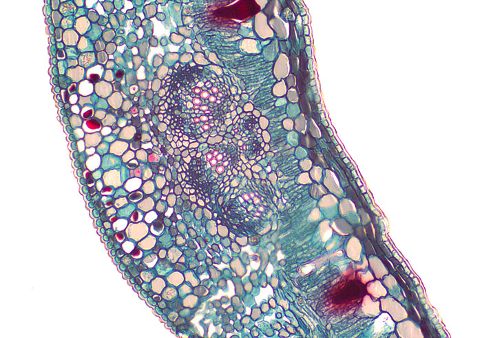
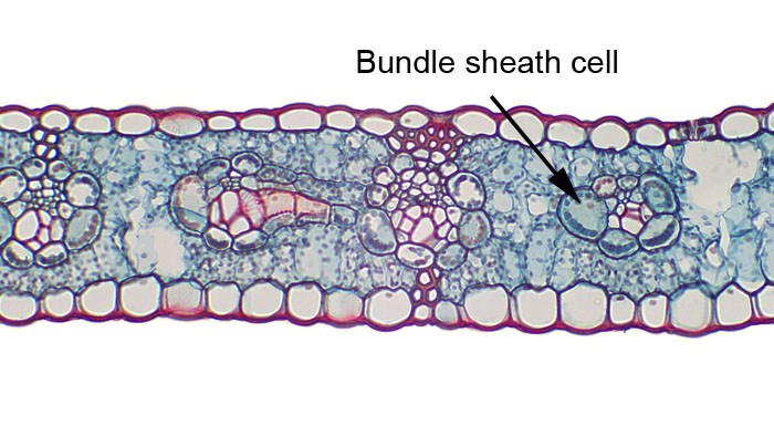
Figure 3.8. A leaf of a species with C3 leaf architecture (Triticum) (above) and of a leaf with C4 leaf architecture (Zea mays)(below). The C4 grass has a ring of parenchyma cells surrounding the veins, which are the bundle sheath cells. (Images by BlueRidgeKitties (CC attribute, share alike) above, and Sean Bellairs (CC attribute, share alike) below.)