21.4: Ferns (Class Polypodiopsida)
- Page ID
- 33892
\( \newcommand{\vecs}[1]{\overset { \scriptstyle \rightharpoonup} {\mathbf{#1}} } \)
\( \newcommand{\vecd}[1]{\overset{-\!-\!\rightharpoonup}{\vphantom{a}\smash {#1}}} \)
\( \newcommand{\id}{\mathrm{id}}\) \( \newcommand{\Span}{\mathrm{span}}\)
( \newcommand{\kernel}{\mathrm{null}\,}\) \( \newcommand{\range}{\mathrm{range}\,}\)
\( \newcommand{\RealPart}{\mathrm{Re}}\) \( \newcommand{\ImaginaryPart}{\mathrm{Im}}\)
\( \newcommand{\Argument}{\mathrm{Arg}}\) \( \newcommand{\norm}[1]{\| #1 \|}\)
\( \newcommand{\inner}[2]{\langle #1, #2 \rangle}\)
\( \newcommand{\Span}{\mathrm{span}}\)
\( \newcommand{\id}{\mathrm{id}}\)
\( \newcommand{\Span}{\mathrm{span}}\)
\( \newcommand{\kernel}{\mathrm{null}\,}\)
\( \newcommand{\range}{\mathrm{range}\,}\)
\( \newcommand{\RealPart}{\mathrm{Re}}\)
\( \newcommand{\ImaginaryPart}{\mathrm{Im}}\)
\( \newcommand{\Argument}{\mathrm{Arg}}\)
\( \newcommand{\norm}[1]{\| #1 \|}\)
\( \newcommand{\inner}[2]{\langle #1, #2 \rangle}\)
\( \newcommand{\Span}{\mathrm{span}}\) \( \newcommand{\AA}{\unicode[.8,0]{x212B}}\)
\( \newcommand{\vectorA}[1]{\vec{#1}} % arrow\)
\( \newcommand{\vectorAt}[1]{\vec{\text{#1}}} % arrow\)
\( \newcommand{\vectorB}[1]{\overset { \scriptstyle \rightharpoonup} {\mathbf{#1}} } \)
\( \newcommand{\vectorC}[1]{\textbf{#1}} \)
\( \newcommand{\vectorD}[1]{\overrightarrow{#1}} \)
\( \newcommand{\vectorDt}[1]{\overrightarrow{\text{#1}}} \)
\( \newcommand{\vectE}[1]{\overset{-\!-\!\rightharpoonup}{\vphantom{a}\smash{\mathbf {#1}}}} \)
\( \newcommand{\vecs}[1]{\overset { \scriptstyle \rightharpoonup} {\mathbf{#1}} } \)
\( \newcommand{\vecd}[1]{\overset{-\!-\!\rightharpoonup}{\vphantom{a}\smash {#1}}} \)
\(\newcommand{\avec}{\mathbf a}\) \(\newcommand{\bvec}{\mathbf b}\) \(\newcommand{\cvec}{\mathbf c}\) \(\newcommand{\dvec}{\mathbf d}\) \(\newcommand{\dtil}{\widetilde{\mathbf d}}\) \(\newcommand{\evec}{\mathbf e}\) \(\newcommand{\fvec}{\mathbf f}\) \(\newcommand{\nvec}{\mathbf n}\) \(\newcommand{\pvec}{\mathbf p}\) \(\newcommand{\qvec}{\mathbf q}\) \(\newcommand{\svec}{\mathbf s}\) \(\newcommand{\tvec}{\mathbf t}\) \(\newcommand{\uvec}{\mathbf u}\) \(\newcommand{\vvec}{\mathbf v}\) \(\newcommand{\wvec}{\mathbf w}\) \(\newcommand{\xvec}{\mathbf x}\) \(\newcommand{\yvec}{\mathbf y}\) \(\newcommand{\zvec}{\mathbf z}\) \(\newcommand{\rvec}{\mathbf r}\) \(\newcommand{\mvec}{\mathbf m}\) \(\newcommand{\zerovec}{\mathbf 0}\) \(\newcommand{\onevec}{\mathbf 1}\) \(\newcommand{\real}{\mathbb R}\) \(\newcommand{\twovec}[2]{\left[\begin{array}{r}#1 \\ #2 \end{array}\right]}\) \(\newcommand{\ctwovec}[2]{\left[\begin{array}{c}#1 \\ #2 \end{array}\right]}\) \(\newcommand{\threevec}[3]{\left[\begin{array}{r}#1 \\ #2 \\ #3 \end{array}\right]}\) \(\newcommand{\cthreevec}[3]{\left[\begin{array}{c}#1 \\ #2 \\ #3 \end{array}\right]}\) \(\newcommand{\fourvec}[4]{\left[\begin{array}{r}#1 \\ #2 \\ #3 \\ #4 \end{array}\right]}\) \(\newcommand{\cfourvec}[4]{\left[\begin{array}{c}#1 \\ #2 \\ #3 \\ #4 \end{array}\right]}\) \(\newcommand{\fivevec}[5]{\left[\begin{array}{r}#1 \\ #2 \\ #3 \\ #4 \\ #5 \\ \end{array}\right]}\) \(\newcommand{\cfivevec}[5]{\left[\begin{array}{c}#1 \\ #2 \\ #3 \\ #4 \\ #5 \\ \end{array}\right]}\) \(\newcommand{\mattwo}[4]{\left[\begin{array}{rr}#1 \amp #2 \\ #3 \amp #4 \\ \end{array}\right]}\) \(\newcommand{\laspan}[1]{\text{Span}\{#1\}}\) \(\newcommand{\bcal}{\cal B}\) \(\newcommand{\ccal}{\cal C}\) \(\newcommand{\scal}{\cal S}\) \(\newcommand{\wcal}{\cal W}\) \(\newcommand{\ecal}{\cal E}\) \(\newcommand{\coords}[2]{\left\{#1\right\}_{#2}}\) \(\newcommand{\gray}[1]{\color{gray}{#1}}\) \(\newcommand{\lgray}[1]{\color{lightgray}{#1}}\) \(\newcommand{\rank}{\operatorname{rank}}\) \(\newcommand{\row}{\text{Row}}\) \(\newcommand{\col}{\text{Col}}\) \(\renewcommand{\row}{\text{Row}}\) \(\newcommand{\nul}{\text{Nul}}\) \(\newcommand{\var}{\text{Var}}\) \(\newcommand{\corr}{\text{corr}}\) \(\newcommand{\len}[1]{\left|#1\right|}\) \(\newcommand{\bbar}{\overline{\bvec}}\) \(\newcommand{\bhat}{\widehat{\bvec}}\) \(\newcommand{\bperp}{\bvec^\perp}\) \(\newcommand{\xhat}{\widehat{\xvec}}\) \(\newcommand{\vhat}{\widehat{\vvec}}\) \(\newcommand{\uhat}{\widehat{\uvec}}\) \(\newcommand{\what}{\widehat{\wvec}}\) \(\newcommand{\Sighat}{\widehat{\Sigma}}\) \(\newcommand{\lt}{<}\) \(\newcommand{\gt}{>}\) \(\newcommand{\amp}{&}\) \(\definecolor{fillinmathshade}{gray}{0.9}\)- Megaphylls. Leaves have branching veins of vascular tissue.
- Rhizomes. Asexual propogation of the sporophyte through underground stems.
- Homosporous. Haploid spores grow into bisexual gametophytes that produce both antheridia and archegonia.
Horsetails (Subclass Equisetidae)
- Gametophyte morphology
- Reduced, thalloid. Bisexual gametophytes grow from homospores and produce both antheridia and archegonia.
- Sporophyte morphology
- Leaves are dark, papery and non-photosynthetic.
- Branches are photosynthetic and produced in whorls on the vegetative shoot.
- Sporangia produced in a terminal strobilus on the reproductive shoot.
- Shoots contain silica
- Spores are photosynthetic and have four hygroscopic arms called elaters
Horsetails are one of the most ancient lineages of plants and are relatively unchanged from the fossil record. If you look closely at the nodes of a green vegetative shoot, you will see that branches and leaves have not only switched roles, they have also switched places, with the photosynthetic branches emerging below the papery, non-photosynthetic leaves. The stems of horsetails are covered in silica, giving them the common name scouring rush, as they were formerly used to clean pots due to the abrasive nature of silicate granules. This is what gives the epidermis of the shoot its rough texture.
Observe a vegetative shoot of Equisetum. Draw what you see below and label a node, internode, branch, and leaf. Where would the rhizome be found?
You may notice that there are shoots without branches that appear more yellow than green. These are the reproductive shoots. Reproductive shoots of some Equisetum species (such as E. arvense) do not photosynthesize. Instead, they receive nutrients through the rhizome, which is connected to vegetative shoots. The function of the reproductive shoot is to produce a strobilus. In the strobilus, sporangia are produced on large, T-shaped structures called sporangiophores. Within the sporangia, photosynthetic homospores mature and develop projections called elaters that aid in spore dispersal via interaction with humidity in the air.
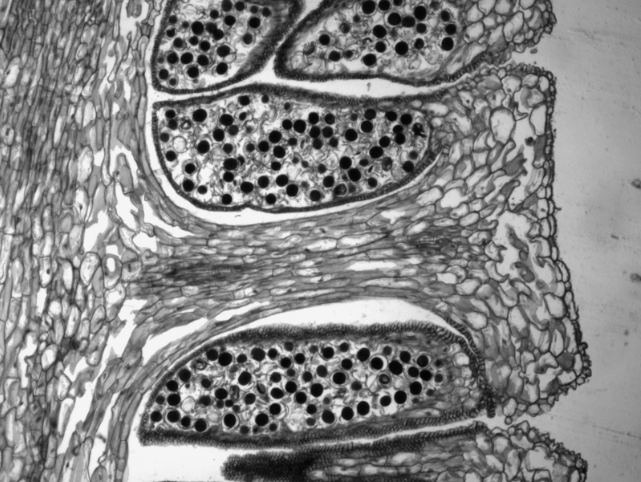
Obtain an Equisetum strobilus from a reproductive shoot. Tap the strobilus onto a dry slide to release mint green spores, do not cover with a coverslip. View this slide under the compound microscope and draw what you see below.
As you are looking through the ocular lenses of the microscope, add a drop of water to the slide near your field of view. What happens to the spores that are close to the droplet of water? What happens when they are touched by the water? Describe and draw what you see below.
Comparing Strobili
Lycophytes and horsetails produce spores in a cone-like structure called a strobilus. Obtain a prepared slide of a long section of the strobilus of each of the following: Equisetum, Lycopodium, and Selaginella.
|
Equisetum |
Lycopodium |
Selaginella |
|
|---|---|---|---|
|
Homosporous or heterosporous? |
|||
|
Sketch of the strobilus long section |
|||
|
Distinctive features? |
Ferns (Subclass Polypodiidae)
- Gametophyte morphology
- Reduced, thalloid, heart-shaped. Often referred to as a prothallus or prothallium.
- Sporophyte morphology
- Megaphylls often pinnately compound fronds, emerging as fiddleheads in the spring.
- Sporangia produced in clusters called sori (sorus, singular).
Fern Life Cycle
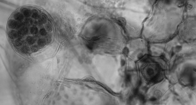
Observe a prepared slide of a fern gametophyte (sometimes referred to as a prothallus) under the compound microscope. Look for rhizoids, archegonia (each with a single egg), and antheridia containing many sperm. Label the bolded features in the life cycle diagram.
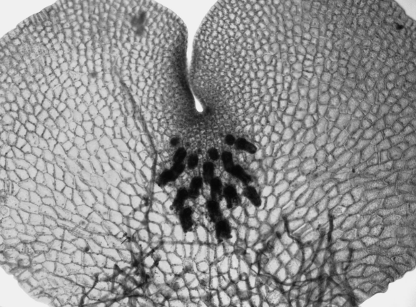
Archegonia and rhizoids are visible on the prothallium in the above figure.
Below: a side view of an archegonium
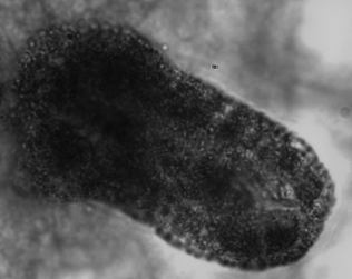
Antheridium with sperm (left) and an archegonium with an egg (right)
Note
One strategy ferns have evolved to avoid self-fertilization is to produce archegonia and antheridia at different times. Depending on the type of fern gametophyte you are looking at, you may need to view two different slides to see archegonia and antheridia.
Observe a fern fiddlehead, so named for its visual similarity to the scroll at the top of a violin. The fiddlehead uncoils in the spring by a process called circinate vernation (vernal meaning spring). Observe a mature fern frond. Locate the rachis, pinnae, and sori. Label the bolded structures in the life cycle diagram.
Obtain a prepared slide of a sorus. A sorus is a cluster of sporangia, often protected by an umbrella-like structure called the indusium as the spores mature. Each sporangium is lined by an inflated strip of cells called an annulus. When the spores have matured, the cells in the annulus begin to dry out, causing the cells to collapse and pull the sporangium open, releasing the spores. Label the bolded structures in the life cycle diagram.
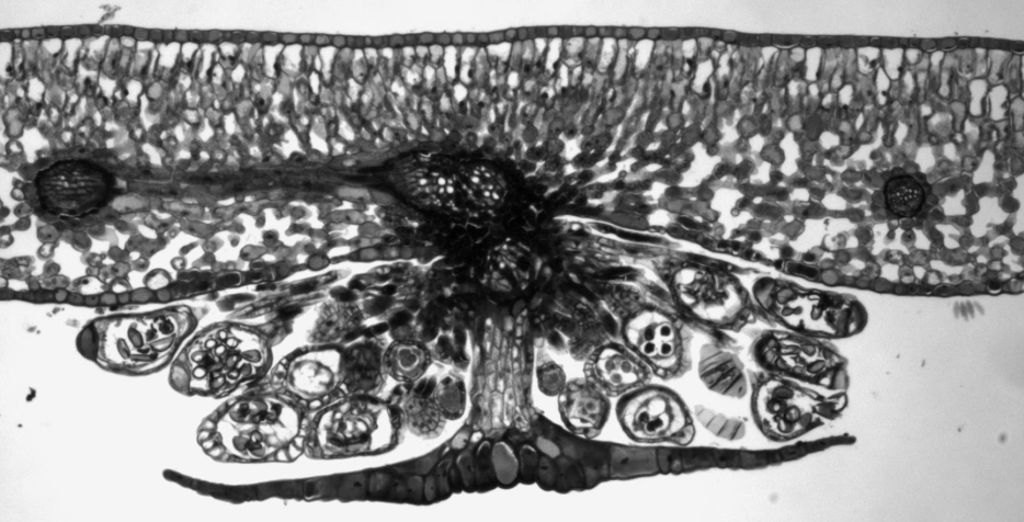
Based on the orientation of the palisade and spongy mesophylls in the leaf cross section above, is this sorus on the upper or lower surface of the leaf? Explain your reasoning.
Fern Life Cycle Diagram
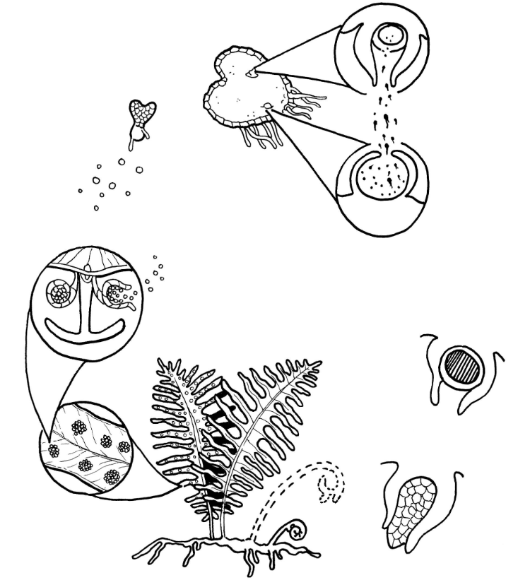
In the fern life cycle diagram, label the bolded structures described in the section above. Indicate where meiosis and fertilization occur and draw arrows to show where mitosis is happening. Choose different colors to represent haploid and diploid tissues.

