14.3: The Production of Genetic Diversity
- Page ID
- 29576
\( \newcommand{\vecs}[1]{\overset { \scriptstyle \rightharpoonup} {\mathbf{#1}} } \)
\( \newcommand{\vecd}[1]{\overset{-\!-\!\rightharpoonup}{\vphantom{a}\smash {#1}}} \)
\( \newcommand{\id}{\mathrm{id}}\) \( \newcommand{\Span}{\mathrm{span}}\)
( \newcommand{\kernel}{\mathrm{null}\,}\) \( \newcommand{\range}{\mathrm{range}\,}\)
\( \newcommand{\RealPart}{\mathrm{Re}}\) \( \newcommand{\ImaginaryPart}{\mathrm{Im}}\)
\( \newcommand{\Argument}{\mathrm{Arg}}\) \( \newcommand{\norm}[1]{\| #1 \|}\)
\( \newcommand{\inner}[2]{\langle #1, #2 \rangle}\)
\( \newcommand{\Span}{\mathrm{span}}\)
\( \newcommand{\id}{\mathrm{id}}\)
\( \newcommand{\Span}{\mathrm{span}}\)
\( \newcommand{\kernel}{\mathrm{null}\,}\)
\( \newcommand{\range}{\mathrm{range}\,}\)
\( \newcommand{\RealPart}{\mathrm{Re}}\)
\( \newcommand{\ImaginaryPart}{\mathrm{Im}}\)
\( \newcommand{\Argument}{\mathrm{Arg}}\)
\( \newcommand{\norm}[1]{\| #1 \|}\)
\( \newcommand{\inner}[2]{\langle #1, #2 \rangle}\)
\( \newcommand{\Span}{\mathrm{span}}\) \( \newcommand{\AA}{\unicode[.8,0]{x212B}}\)
\( \newcommand{\vectorA}[1]{\vec{#1}} % arrow\)
\( \newcommand{\vectorAt}[1]{\vec{\text{#1}}} % arrow\)
\( \newcommand{\vectorB}[1]{\overset { \scriptstyle \rightharpoonup} {\mathbf{#1}} } \)
\( \newcommand{\vectorC}[1]{\textbf{#1}} \)
\( \newcommand{\vectorD}[1]{\overrightarrow{#1}} \)
\( \newcommand{\vectorDt}[1]{\overrightarrow{\text{#1}}} \)
\( \newcommand{\vectE}[1]{\overset{-\!-\!\rightharpoonup}{\vphantom{a}\smash{\mathbf {#1}}}} \)
\( \newcommand{\vecs}[1]{\overset { \scriptstyle \rightharpoonup} {\mathbf{#1}} } \)
\( \newcommand{\vecd}[1]{\overset{-\!-\!\rightharpoonup}{\vphantom{a}\smash {#1}}} \)
\(\newcommand{\avec}{\mathbf a}\) \(\newcommand{\bvec}{\mathbf b}\) \(\newcommand{\cvec}{\mathbf c}\) \(\newcommand{\dvec}{\mathbf d}\) \(\newcommand{\dtil}{\widetilde{\mathbf d}}\) \(\newcommand{\evec}{\mathbf e}\) \(\newcommand{\fvec}{\mathbf f}\) \(\newcommand{\nvec}{\mathbf n}\) \(\newcommand{\pvec}{\mathbf p}\) \(\newcommand{\qvec}{\mathbf q}\) \(\newcommand{\svec}{\mathbf s}\) \(\newcommand{\tvec}{\mathbf t}\) \(\newcommand{\uvec}{\mathbf u}\) \(\newcommand{\vvec}{\mathbf v}\) \(\newcommand{\wvec}{\mathbf w}\) \(\newcommand{\xvec}{\mathbf x}\) \(\newcommand{\yvec}{\mathbf y}\) \(\newcommand{\zvec}{\mathbf z}\) \(\newcommand{\rvec}{\mathbf r}\) \(\newcommand{\mvec}{\mathbf m}\) \(\newcommand{\zerovec}{\mathbf 0}\) \(\newcommand{\onevec}{\mathbf 1}\) \(\newcommand{\real}{\mathbb R}\) \(\newcommand{\twovec}[2]{\left[\begin{array}{r}#1 \\ #2 \end{array}\right]}\) \(\newcommand{\ctwovec}[2]{\left[\begin{array}{c}#1 \\ #2 \end{array}\right]}\) \(\newcommand{\threevec}[3]{\left[\begin{array}{r}#1 \\ #2 \\ #3 \end{array}\right]}\) \(\newcommand{\cthreevec}[3]{\left[\begin{array}{c}#1 \\ #2 \\ #3 \end{array}\right]}\) \(\newcommand{\fourvec}[4]{\left[\begin{array}{r}#1 \\ #2 \\ #3 \\ #4 \end{array}\right]}\) \(\newcommand{\cfourvec}[4]{\left[\begin{array}{c}#1 \\ #2 \\ #3 \\ #4 \end{array}\right]}\) \(\newcommand{\fivevec}[5]{\left[\begin{array}{r}#1 \\ #2 \\ #3 \\ #4 \\ #5 \\ \end{array}\right]}\) \(\newcommand{\cfivevec}[5]{\left[\begin{array}{c}#1 \\ #2 \\ #3 \\ #4 \\ #5 \\ \end{array}\right]}\) \(\newcommand{\mattwo}[4]{\left[\begin{array}{rr}#1 \amp #2 \\ #3 \amp #4 \\ \end{array}\right]}\) \(\newcommand{\laspan}[1]{\text{Span}\{#1\}}\) \(\newcommand{\bcal}{\cal B}\) \(\newcommand{\ccal}{\cal C}\) \(\newcommand{\scal}{\cal S}\) \(\newcommand{\wcal}{\cal W}\) \(\newcommand{\ecal}{\cal E}\) \(\newcommand{\coords}[2]{\left\{#1\right\}_{#2}}\) \(\newcommand{\gray}[1]{\color{gray}{#1}}\) \(\newcommand{\lgray}[1]{\color{lightgray}{#1}}\) \(\newcommand{\rank}{\operatorname{rank}}\) \(\newcommand{\row}{\text{Row}}\) \(\newcommand{\col}{\text{Col}}\) \(\renewcommand{\row}{\text{Row}}\) \(\newcommand{\nul}{\text{Nul}}\) \(\newcommand{\var}{\text{Var}}\) \(\newcommand{\corr}{\text{corr}}\) \(\newcommand{\len}[1]{\left|#1\right|}\) \(\newcommand{\bbar}{\overline{\bvec}}\) \(\newcommand{\bhat}{\widehat{\bvec}}\) \(\newcommand{\bperp}{\bvec^\perp}\) \(\newcommand{\xhat}{\widehat{\xvec}}\) \(\newcommand{\vhat}{\widehat{\vvec}}\) \(\newcommand{\uhat}{\widehat{\uvec}}\) \(\newcommand{\what}{\widehat{\wvec}}\) \(\newcommand{\Sighat}{\widehat{\Sigma}}\) \(\newcommand{\lt}{<}\) \(\newcommand{\gt}{>}\) \(\newcommand{\amp}{&}\) \(\definecolor{fillinmathshade}{gray}{0.9}\)Mitosis is a type of cell division in which the original cell makes a duplicate copy. If this were the only form of cell division, the production of genetic diversity would be limited to mutations. Most lineages of organisms might not be able to adapt to the rapidly changing conditions of Earth’s many environments. This is not to say that asexually reproducing organisms cannot be successful. The most proliferate, widespread organisms on the planet, the Bacteria, are asexual and cannot undergo meiosis because they lack a nucleus. Most multicellular eukaryotic organisms (and some unicellular eukaryotes) have evolved to use sexual reproduction to create diversity through meiosis and random fertilization.
Meiosis = PMAT x 2
If a diploid cell undergoes meiosis, the product will be four genetically distinct haploid cells. In humans, this process is used to make eggs and sperm. Meiosis is almost like doing mitosis twice. This description will focus on the differences between the phases of meiosis and mitosis, rather than describing everything that occurs in each stage. For a refresher of the other events, see lab 4 (Multicellularity & Asexual Reproduction).
Meiosis I
- Prophase I: During prophase I, homologous chromosomes pair together. These chromosomes have the same genes in the same order but are derived from different parents. Due to their similarity, when they pair, they can overlap and trade segments of the chromosomal arms. The result is a mixture of genes from each parent on each chromosome. This process is called crossing over and it will happen differently in each cell that undergoes meiosis.
- Metaphase I: In metaphase I, homologous chromosomes line up across from each other on the metaphase plate. This is another source of variation, as they may align differently in each cell with a different combination of genes on either side of the metaphase plate. This is called independent assortment.
- Anaphase I: During anaphase in mitosis, sister chromatids are separated from each other. In meiosis, pairs of homologous chromosomes that are separated.
- Telophase I: Telophase I is similar to telophase in mitosis. The only difference is that the two daughter cells that emerge are genetically distinct from each other.
Meiosis II
Meiosis II is nearly identical to mitosis. It consists of prophase II, metaphase II, anaphase II, and telophase II. It is in anaphase II that sister chromatids are pulled apart. At the end of telophase II, four haploid cells have been produced, each genetically distinct.
Meiosis in Lilium Pollen
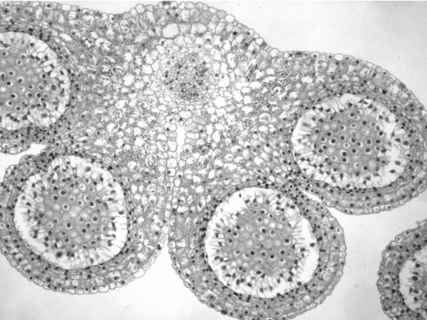
Anthers are one of the sexually reproducing structures in flowers, producing haploid pollen that will fertilize haploid eggs.

The above image shows a cross section through Lillium anthers. There are four circular areas, one of which is circled in the picture. These are the pollen sacs. It is inside these pollen sacs that meiosis occurs, so look within these areas for cells in different stages of meiosis.
Pollen divides synchronously, so most cells should be in approximately the same stage. However, meiosis is a continuous process, so some cells may be in anaphase while most are in metaphase. Additionally, dividing pollen cells remain attached, so you will be able to tell if you are in meiosis I or meiosis II by how many cell walls have formed within the dividing cell. In meiosis I, there should be one cell until cytokinesis occurs during telophase I, producing two joined cells. In meiosis II, there will be two cells stuck together until cytokinesis during telophase II, producing four cells.


The image on the left shows two cells in metaphase I (left and middle) and one cell in prophase I (upper right corner). The image on the right shows two “cells”, both in metaphase II and showing that each “cell” is now actually two cells stuck together.
Note the wall that travels down the center of each of the cells in the photo on the right. During which phase of meiosis would this wall have been formed?
Observe slides of Lilium anthers at different stages of cell division and find each phase of meiosis. Draw and label each phase below. Make sure to note where crossing over and independent assortment are happening.
Meiosis I:
Meiosis II:
At what point did the number of sets of chromosomes in each cell go from 2 sets to 1 set? Said another way, at what point did the ploidy change from diploid to haploid?


