
5.6: Meristems and Tissues in the Root
- Page ID
- 33234

The Root Apical Meristem (RAM, for short)
When you are viewing the onion root tip, you are viewing the beginning of the formation of tissues of the plant’s root system. This formation of tissues begins with the root apical meristem (RAM). Just below the tip of the root, there is a region of small, densely packed cells that are actively dividing. These cells make up the RAM. To the outside (below, in your slide) of the root tip, the RAM cells produce an ephemeral tissue called the root cap. Cells in the root cap protect the root apical meristem, secrete chemicals to attract beneficial bacteria and fungi, and secrete a mucilage that makes it easier for the root tip to penetrate the soil substrate. As the root tip grows, these cells are sloughed off into the environment and so the root cap must be continually produced by the RAM. To the inside (above, in your slide), the RAM produces cells that begin to form the three primary meristems. Remember, a meristem is a region of cells whose function is to produce more cells!
Based on the root tips you have looked at today, how might you identify meristematic regions in a plant organ under the microscope? What would you look for and why?
Primary Meristems and Their Primary Tissues
Primary meristems divide to form the primary tissues. The protoderm is the primary meristem that produces the epidermis. The procambium is the primary meristem that produces the vascular tissues (xylem, phloem, and any associated tissues). The ground meristem is the primary meristem that produces the ground tissue. Depending on the type of plant or the part of the plant, the ground tissue can be divided into distinct regions or not. The Zea mays root tip in the image below belongs to a monocot, so the ground tissue in this particular sample is divided into the pith (appearing as a column of tissue in the center of the root tip, bordered on either side by vascular tissue) and the cortex (located between the epidermis and the vascular tissue on either side of your slide).
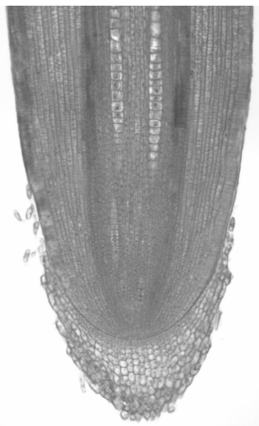
In the image above, label the RAM, protoderm, procambium, ground meristem, pith, cortex, and vascular tissue.
Where in the root above would you be likely to find cells in the G0 phase of the cell cycle?
Where would you be likely to find cells in G1, S-phase, or G2?