3.5: Using the Compound Microscope
- Page ID
- 33217
\( \newcommand{\vecs}[1]{\overset { \scriptstyle \rightharpoonup} {\mathbf{#1}} } \)
\( \newcommand{\vecd}[1]{\overset{-\!-\!\rightharpoonup}{\vphantom{a}\smash {#1}}} \)
\( \newcommand{\dsum}{\displaystyle\sum\limits} \)
\( \newcommand{\dint}{\displaystyle\int\limits} \)
\( \newcommand{\dlim}{\displaystyle\lim\limits} \)
\( \newcommand{\id}{\mathrm{id}}\) \( \newcommand{\Span}{\mathrm{span}}\)
( \newcommand{\kernel}{\mathrm{null}\,}\) \( \newcommand{\range}{\mathrm{range}\,}\)
\( \newcommand{\RealPart}{\mathrm{Re}}\) \( \newcommand{\ImaginaryPart}{\mathrm{Im}}\)
\( \newcommand{\Argument}{\mathrm{Arg}}\) \( \newcommand{\norm}[1]{\| #1 \|}\)
\( \newcommand{\inner}[2]{\langle #1, #2 \rangle}\)
\( \newcommand{\Span}{\mathrm{span}}\)
\( \newcommand{\id}{\mathrm{id}}\)
\( \newcommand{\Span}{\mathrm{span}}\)
\( \newcommand{\kernel}{\mathrm{null}\,}\)
\( \newcommand{\range}{\mathrm{range}\,}\)
\( \newcommand{\RealPart}{\mathrm{Re}}\)
\( \newcommand{\ImaginaryPart}{\mathrm{Im}}\)
\( \newcommand{\Argument}{\mathrm{Arg}}\)
\( \newcommand{\norm}[1]{\| #1 \|}\)
\( \newcommand{\inner}[2]{\langle #1, #2 \rangle}\)
\( \newcommand{\Span}{\mathrm{span}}\) \( \newcommand{\AA}{\unicode[.8,0]{x212B}}\)
\( \newcommand{\vectorA}[1]{\vec{#1}} % arrow\)
\( \newcommand{\vectorAt}[1]{\vec{\text{#1}}} % arrow\)
\( \newcommand{\vectorB}[1]{\overset { \scriptstyle \rightharpoonup} {\mathbf{#1}} } \)
\( \newcommand{\vectorC}[1]{\textbf{#1}} \)
\( \newcommand{\vectorD}[1]{\overrightarrow{#1}} \)
\( \newcommand{\vectorDt}[1]{\overrightarrow{\text{#1}}} \)
\( \newcommand{\vectE}[1]{\overset{-\!-\!\rightharpoonup}{\vphantom{a}\smash{\mathbf {#1}}}} \)
\( \newcommand{\vecs}[1]{\overset { \scriptstyle \rightharpoonup} {\mathbf{#1}} } \)
\(\newcommand{\longvect}{\overrightarrow}\)
\( \newcommand{\vecd}[1]{\overset{-\!-\!\rightharpoonup}{\vphantom{a}\smash {#1}}} \)
\(\newcommand{\avec}{\mathbf a}\) \(\newcommand{\bvec}{\mathbf b}\) \(\newcommand{\cvec}{\mathbf c}\) \(\newcommand{\dvec}{\mathbf d}\) \(\newcommand{\dtil}{\widetilde{\mathbf d}}\) \(\newcommand{\evec}{\mathbf e}\) \(\newcommand{\fvec}{\mathbf f}\) \(\newcommand{\nvec}{\mathbf n}\) \(\newcommand{\pvec}{\mathbf p}\) \(\newcommand{\qvec}{\mathbf q}\) \(\newcommand{\svec}{\mathbf s}\) \(\newcommand{\tvec}{\mathbf t}\) \(\newcommand{\uvec}{\mathbf u}\) \(\newcommand{\vvec}{\mathbf v}\) \(\newcommand{\wvec}{\mathbf w}\) \(\newcommand{\xvec}{\mathbf x}\) \(\newcommand{\yvec}{\mathbf y}\) \(\newcommand{\zvec}{\mathbf z}\) \(\newcommand{\rvec}{\mathbf r}\) \(\newcommand{\mvec}{\mathbf m}\) \(\newcommand{\zerovec}{\mathbf 0}\) \(\newcommand{\onevec}{\mathbf 1}\) \(\newcommand{\real}{\mathbb R}\) \(\newcommand{\twovec}[2]{\left[\begin{array}{r}#1 \\ #2 \end{array}\right]}\) \(\newcommand{\ctwovec}[2]{\left[\begin{array}{c}#1 \\ #2 \end{array}\right]}\) \(\newcommand{\threevec}[3]{\left[\begin{array}{r}#1 \\ #2 \\ #3 \end{array}\right]}\) \(\newcommand{\cthreevec}[3]{\left[\begin{array}{c}#1 \\ #2 \\ #3 \end{array}\right]}\) \(\newcommand{\fourvec}[4]{\left[\begin{array}{r}#1 \\ #2 \\ #3 \\ #4 \end{array}\right]}\) \(\newcommand{\cfourvec}[4]{\left[\begin{array}{c}#1 \\ #2 \\ #3 \\ #4 \end{array}\right]}\) \(\newcommand{\fivevec}[5]{\left[\begin{array}{r}#1 \\ #2 \\ #3 \\ #4 \\ #5 \\ \end{array}\right]}\) \(\newcommand{\cfivevec}[5]{\left[\begin{array}{c}#1 \\ #2 \\ #3 \\ #4 \\ #5 \\ \end{array}\right]}\) \(\newcommand{\mattwo}[4]{\left[\begin{array}{rr}#1 \amp #2 \\ #3 \amp #4 \\ \end{array}\right]}\) \(\newcommand{\laspan}[1]{\text{Span}\{#1\}}\) \(\newcommand{\bcal}{\cal B}\) \(\newcommand{\ccal}{\cal C}\) \(\newcommand{\scal}{\cal S}\) \(\newcommand{\wcal}{\cal W}\) \(\newcommand{\ecal}{\cal E}\) \(\newcommand{\coords}[2]{\left\{#1\right\}_{#2}}\) \(\newcommand{\gray}[1]{\color{gray}{#1}}\) \(\newcommand{\lgray}[1]{\color{lightgray}{#1}}\) \(\newcommand{\rank}{\operatorname{rank}}\) \(\newcommand{\row}{\text{Row}}\) \(\newcommand{\col}{\text{Col}}\) \(\renewcommand{\row}{\text{Row}}\) \(\newcommand{\nul}{\text{Nul}}\) \(\newcommand{\var}{\text{Var}}\) \(\newcommand{\corr}{\text{corr}}\) \(\newcommand{\len}[1]{\left|#1\right|}\) \(\newcommand{\bbar}{\overline{\bvec}}\) \(\newcommand{\bhat}{\widehat{\bvec}}\) \(\newcommand{\bperp}{\bvec^\perp}\) \(\newcommand{\xhat}{\widehat{\xvec}}\) \(\newcommand{\vhat}{\widehat{\vvec}}\) \(\newcommand{\uhat}{\widehat{\uvec}}\) \(\newcommand{\what}{\widehat{\wvec}}\) \(\newcommand{\Sighat}{\widehat{\Sigma}}\) \(\newcommand{\lt}{<}\) \(\newcommand{\gt}{>}\) \(\newcommand{\amp}{&}\) \(\definecolor{fillinmathshade}{gray}{0.9}\)Prepare a Wet Mount
Place a drop of water onto a glass slide. Obtain a tiny piece of the water fern, Azolla, and place it on the water droplet. You are looking for a cyanobacterium, Anabaena, that lives inside the leaves of the water fern. Using a razor blade, chop the sample into small pieces, much like you would mincing garlic, to release the bacteria. Hold a glass coverslip at an angle, touching the base of the coverslip to the water droplet on your slide. Slowly lower the coverslip over the drop of water until it is flat against the slide.
Observe your Specimen
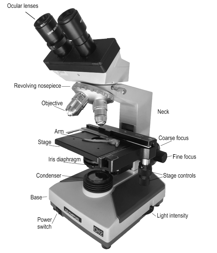
Place the slide you have prepared onto the microscope stage, holding it in place with the arm, and use the stage controls to place your specimen directly above the light source. Rotate the revolving nosepiece to the scanning objective (4x), adjust the light intensity (it should not hurt your eyes), and look through the ocular lenses (10x).
Checkpoint
Are there black crescents obstructing your field of view? If so, you need to adjust the ocular lenses to match your interpupillary distance (the distance between your eyes). As you look through the ocular lenses, adjust the distance between them until the image resolves into a single white circle.
Anatomy of a Compound Microscope

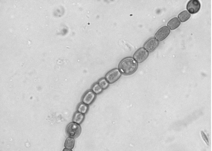
As you look through the ocular lenses, rotate the coarse focus knob away from you until your specimen comes into focus. You are viewing your specimen at 40x its actual size. What you are looking for are small strands of cells that look similar to a chain of tiny green beads. These are colonies of Anabaena that live inside the leaves of the water fern, fixing nitrogen in exchange for a safe place to live. The colonies will appear a slightly darker green than the pieces of the water fern cells.
Scan your specimen, using the stage controls to move the slide around, until you find a cluster of these dark green chains. When you find some, rotate the nosepiece to the low power objective (10x) and use the fine focus knob to adjust the focus on your specimen.
Note
If you lose your specimen when moving to a higher magnification, go back to the lower magnification and find it again.
Anabaena
When looking at the colonies of cyanobacteria, you should be able to see three different types of cells: vegetative cells, heterocysts, and akinetes.
Note
The image below shows only vegetative cells and heterocysts.

Vegetative cells are the “normal” smaller, darker green cells. These cells are doing photosynthesis and making sugars for the colony.
Heterocysts are larger, round, thick-walled cells that can appear yellow and have two polar bodies, one on each end where they attach to other cells in the colony.
Individual cyanobacteria that have converted into heterocysts are not photosynthesizing because photosynthesis produces oxygen. Instead, these cyanobacteria are fixing nitrogen -- the enzyme nitrogenase will not function in the presence of oxygen. This individual relies on the colony to make it food and, in return, it supplies the colony (and the landlord, Azolla) with nitrogen.
Akinetes are larger, football to nearly rectangular-shaped, thick-walled cells that tend to be more granular in appearance. These individuals still perform photosynthesis, but also function as a sort of failsafe. Akinetes store large amounts of lipids and carbohydrates so that they have enough energy to begin a new colony if conditions become too cold or too dry for survival. Their formation is triggered by these conditions (dry or cold), so you may not see them from a fresh water fern leaf, as this is a relatively stable, comfortable environment. If you left this slide out overnight and allowed the water to evaporate, you might see akinetes forming when you rehydrated it the following day.
Draw a colony of Anabaena, including all three cell types, labelling each with its name and function. Draw a cell of the water fern next to the colony for a size comparison.
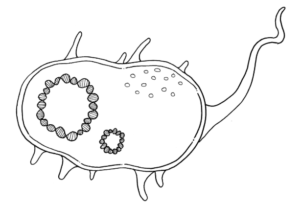
Cyanobacteria belong to Domain Bacteria, one of two large groups of organisms called prokaryotes. Prokaryotes are unicellular organisms that have relatively simple cellular makeup. The cell is surrounded by a structure called the cell wall that protects the organism from desiccation (drying out) and other external stressors. In bacteria, this cell wall contains a compound called peptidoglycan. Inside the cell wall is a semi-permeable membrane called the plasma membrane (also called the cell membrane), which controls what enters and exits the cytoplasm of the cell. Prokaryotes have no nucleus or organelles, instead bundling their circular DNA into a compact structure called a nucleoid.
In your drawing of the Anabaena above, label the cell wall, plasma membrane, and cytoplasm of one of the vegetative cells.
The diagram below is a generalized prokaryote. There is a large circle of DNA, the circular chromosome, and a small circle of DNA, a plasmid. On the exterior, there is a long projection called a flagellum which is used for movement. The smaller projections are called pili (pilus, singular) and are used for interacting with other cells. Label these structures in the diagram below, as well as the cell wall, cytoplasm, and ribosomes.


