2.5.2.1: Anthocerotophyta
- Page ID
- 37009

\( \newcommand{\vecs}[1]{\overset { \scriptstyle \rightharpoonup} {\mathbf{#1}} } \)
\( \newcommand{\vecd}[1]{\overset{-\!-\!\rightharpoonup}{\vphantom{a}\smash {#1}}} \)
\( \newcommand{\dsum}{\displaystyle\sum\limits} \)
\( \newcommand{\dint}{\displaystyle\int\limits} \)
\( \newcommand{\dlim}{\displaystyle\lim\limits} \)
\( \newcommand{\id}{\mathrm{id}}\) \( \newcommand{\Span}{\mathrm{span}}\)
( \newcommand{\kernel}{\mathrm{null}\,}\) \( \newcommand{\range}{\mathrm{range}\,}\)
\( \newcommand{\RealPart}{\mathrm{Re}}\) \( \newcommand{\ImaginaryPart}{\mathrm{Im}}\)
\( \newcommand{\Argument}{\mathrm{Arg}}\) \( \newcommand{\norm}[1]{\| #1 \|}\)
\( \newcommand{\inner}[2]{\langle #1, #2 \rangle}\)
\( \newcommand{\Span}{\mathrm{span}}\)
\( \newcommand{\id}{\mathrm{id}}\)
\( \newcommand{\Span}{\mathrm{span}}\)
\( \newcommand{\kernel}{\mathrm{null}\,}\)
\( \newcommand{\range}{\mathrm{range}\,}\)
\( \newcommand{\RealPart}{\mathrm{Re}}\)
\( \newcommand{\ImaginaryPart}{\mathrm{Im}}\)
\( \newcommand{\Argument}{\mathrm{Arg}}\)
\( \newcommand{\norm}[1]{\| #1 \|}\)
\( \newcommand{\inner}[2]{\langle #1, #2 \rangle}\)
\( \newcommand{\Span}{\mathrm{span}}\) \( \newcommand{\AA}{\unicode[.8,0]{x212B}}\)
\( \newcommand{\vectorA}[1]{\vec{#1}} % arrow\)
\( \newcommand{\vectorAt}[1]{\vec{\text{#1}}} % arrow\)
\( \newcommand{\vectorB}[1]{\overset { \scriptstyle \rightharpoonup} {\mathbf{#1}} } \)
\( \newcommand{\vectorC}[1]{\textbf{#1}} \)
\( \newcommand{\vectorD}[1]{\overrightarrow{#1}} \)
\( \newcommand{\vectorDt}[1]{\overrightarrow{\text{#1}}} \)
\( \newcommand{\vectE}[1]{\overset{-\!-\!\rightharpoonup}{\vphantom{a}\smash{\mathbf {#1}}}} \)
\( \newcommand{\vecs}[1]{\overset { \scriptstyle \rightharpoonup} {\mathbf{#1}} } \)
\(\newcommand{\longvect}{\overrightarrow}\)
\( \newcommand{\vecd}[1]{\overset{-\!-\!\rightharpoonup}{\vphantom{a}\smash {#1}}} \)
\(\newcommand{\avec}{\mathbf a}\) \(\newcommand{\bvec}{\mathbf b}\) \(\newcommand{\cvec}{\mathbf c}\) \(\newcommand{\dvec}{\mathbf d}\) \(\newcommand{\dtil}{\widetilde{\mathbf d}}\) \(\newcommand{\evec}{\mathbf e}\) \(\newcommand{\fvec}{\mathbf f}\) \(\newcommand{\nvec}{\mathbf n}\) \(\newcommand{\pvec}{\mathbf p}\) \(\newcommand{\qvec}{\mathbf q}\) \(\newcommand{\svec}{\mathbf s}\) \(\newcommand{\tvec}{\mathbf t}\) \(\newcommand{\uvec}{\mathbf u}\) \(\newcommand{\vvec}{\mathbf v}\) \(\newcommand{\wvec}{\mathbf w}\) \(\newcommand{\xvec}{\mathbf x}\) \(\newcommand{\yvec}{\mathbf y}\) \(\newcommand{\zvec}{\mathbf z}\) \(\newcommand{\rvec}{\mathbf r}\) \(\newcommand{\mvec}{\mathbf m}\) \(\newcommand{\zerovec}{\mathbf 0}\) \(\newcommand{\onevec}{\mathbf 1}\) \(\newcommand{\real}{\mathbb R}\) \(\newcommand{\twovec}[2]{\left[\begin{array}{r}#1 \\ #2 \end{array}\right]}\) \(\newcommand{\ctwovec}[2]{\left[\begin{array}{c}#1 \\ #2 \end{array}\right]}\) \(\newcommand{\threevec}[3]{\left[\begin{array}{r}#1 \\ #2 \\ #3 \end{array}\right]}\) \(\newcommand{\cthreevec}[3]{\left[\begin{array}{c}#1 \\ #2 \\ #3 \end{array}\right]}\) \(\newcommand{\fourvec}[4]{\left[\begin{array}{r}#1 \\ #2 \\ #3 \\ #4 \end{array}\right]}\) \(\newcommand{\cfourvec}[4]{\left[\begin{array}{c}#1 \\ #2 \\ #3 \\ #4 \end{array}\right]}\) \(\newcommand{\fivevec}[5]{\left[\begin{array}{r}#1 \\ #2 \\ #3 \\ #4 \\ #5 \\ \end{array}\right]}\) \(\newcommand{\cfivevec}[5]{\left[\begin{array}{c}#1 \\ #2 \\ #3 \\ #4 \\ #5 \\ \end{array}\right]}\) \(\newcommand{\mattwo}[4]{\left[\begin{array}{rr}#1 \amp #2 \\ #3 \amp #4 \\ \end{array}\right]}\) \(\newcommand{\laspan}[1]{\text{Span}\{#1\}}\) \(\newcommand{\bcal}{\cal B}\) \(\newcommand{\ccal}{\cal C}\) \(\newcommand{\scal}{\cal S}\) \(\newcommand{\wcal}{\cal W}\) \(\newcommand{\ecal}{\cal E}\) \(\newcommand{\coords}[2]{\left\{#1\right\}_{#2}}\) \(\newcommand{\gray}[1]{\color{gray}{#1}}\) \(\newcommand{\lgray}[1]{\color{lightgray}{#1}}\) \(\newcommand{\rank}{\operatorname{rank}}\) \(\newcommand{\row}{\text{Row}}\) \(\newcommand{\col}{\text{Col}}\) \(\renewcommand{\row}{\text{Row}}\) \(\newcommand{\nul}{\text{Nul}}\) \(\newcommand{\var}{\text{Var}}\) \(\newcommand{\corr}{\text{corr}}\) \(\newcommand{\len}[1]{\left|#1\right|}\) \(\newcommand{\bbar}{\overline{\bvec}}\) \(\newcommand{\bhat}{\widehat{\bvec}}\) \(\newcommand{\bperp}{\bvec^\perp}\) \(\newcommand{\xhat}{\widehat{\xvec}}\) \(\newcommand{\vhat}{\widehat{\vvec}}\) \(\newcommand{\uhat}{\widehat{\uvec}}\) \(\newcommand{\what}{\widehat{\wvec}}\) \(\newcommand{\Sighat}{\widehat{\Sigma}}\) \(\newcommand{\lt}{<}\) \(\newcommand{\gt}{>}\) \(\newcommand{\amp}{&}\) \(\definecolor{fillinmathshade}{gray}{0.9}\)- Use morphological traits and cellular components to distinguish between hornworts and other bryophytes.
- Identify structures and phases in the hornwort life cycle; know their ploidy.
- Label a hornwort sporophyte and describe its development.
Hornworts, Phylum Anthocerotophyta
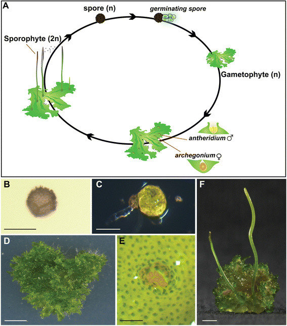
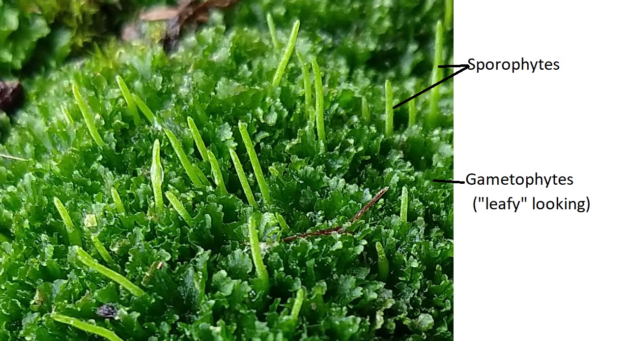
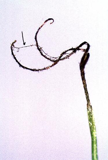
The name Anthocerotophyta means 'horn flower plant'. These strange plants, called the hornworts, get their name from the horn-like sporophytes they produce. Hornworts have a flattened thalloid gametophyte, out of which grow their long photosynthetic sporophytes (Figure \(\PageIndex{1}\)), composed of a sporangium with a columella and pseudoelaters. The sporophyte grows from a basal meristem, with the oldest tissues at the apex. When spores have matured, the sporangium dries and dehisces, twisting open to release spores. The long, multicellular pseudoelaters (true elaters are a single cell) within the sporangium assist in dispersing the spores. Hornworts are somewhat rare and quite small, and they prefer shady and wet places. They are often found growing on the muddy sides of waterways, from small ditches to larger creeks.
The presence of stomata on sporophytes and the ability of some hornwort sporophytes to branch (and sometimes even live independently from the gametophyte!) lend support for the hypothesis that hornworts are sister to vascular plants (tracheophytes). However, hornworts are discussed first in this chapter due to their strictly thalloid gametophyte morphology.
Gametophyte Morphology
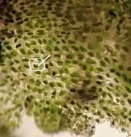
Hornwort gametophytes are exclusively thalloid, often with compartments of mutualistic cyanobacteria from the genus Nostoc. Cells within the gametophyte are monoplastidic (Figure \(\PageIndex{2}\)), containing one large chloroplast in each cell. Similar to the green algae, hornwort chloroplasts contain a pyrenoid where starch storage is concentrated. On the exterior of the thallus, simple pores allow for gas exchange. Unlike stomata, simple pores have no guard cells, meaning they are permanently open. Some hornwort species house colonies of Nostoc within the gametophyte thallus, providing access to nitrogen fixed by the cyanobacterial colony.
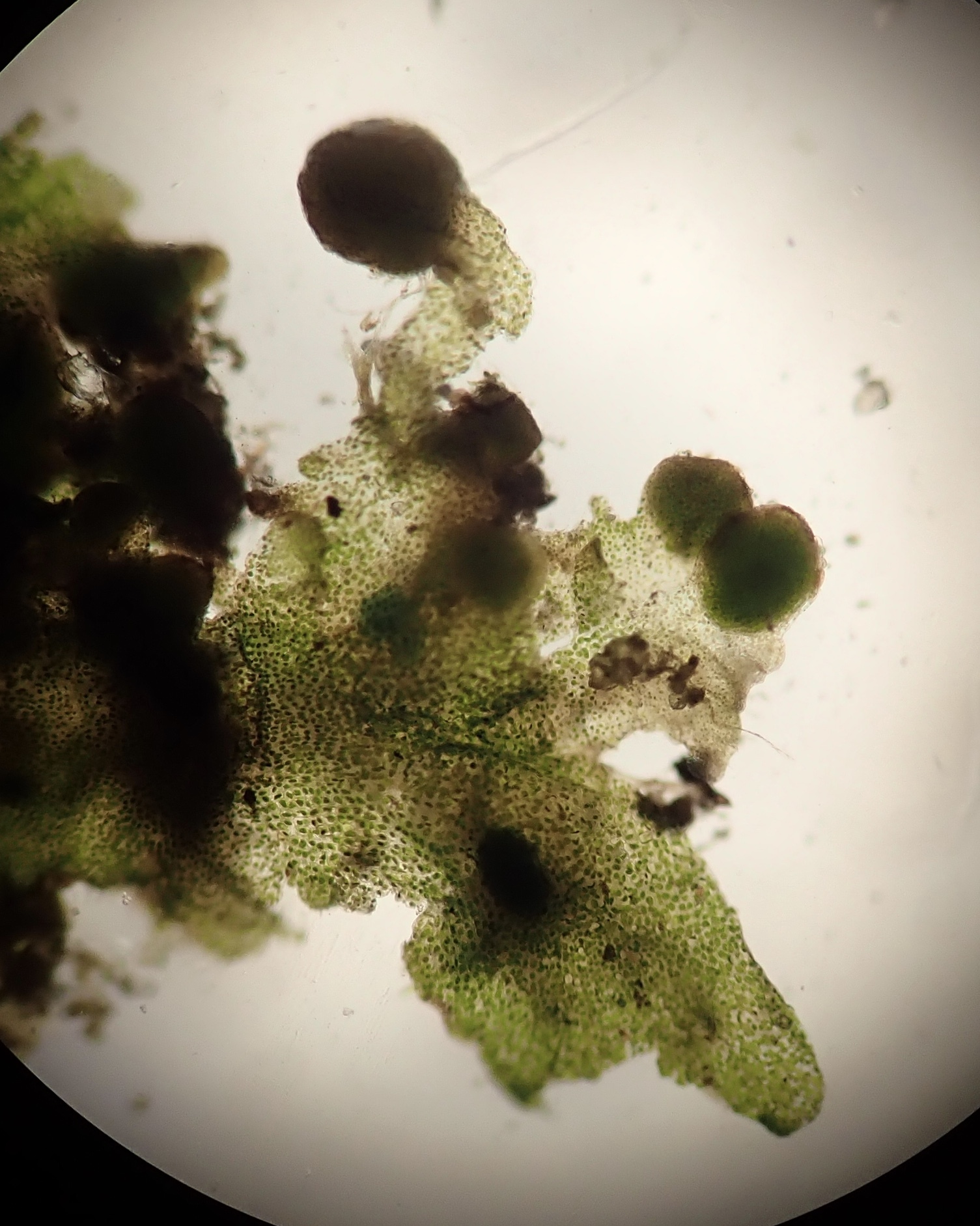
Sporophyte Morphology
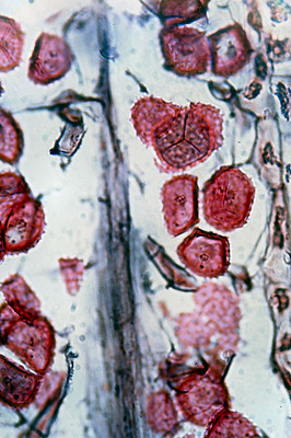
Hornwort sporophytes are comprised of a linear sporangium that lacks a seta. It grows from a basal meristem, meaning the cells at the apex are the oldest. When you see the sporangia mature, this becomes more obvious, as the tip dries out first and dehisces (Figure \(\PageIndex{3}\)) to release the spores. Unlike the gametophyte, the sporophyte possesses true stomata for gas exchange, though stomata have been lost in some lineages. Notably, hornwort sporophytes are photosynthetic and capable of transferring photosynthates to the gametophyte. In other bryophyte lineages, the sporophyte is almost entirely dependent on the gametophyte for nutrition.
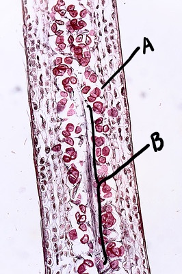
Within the sporgangium, there is a central columella (Figure \(\PageIndex{4}\)) and often pseudoelaters and/or sterile cells that aid in spore dispersal by twisting when dry.
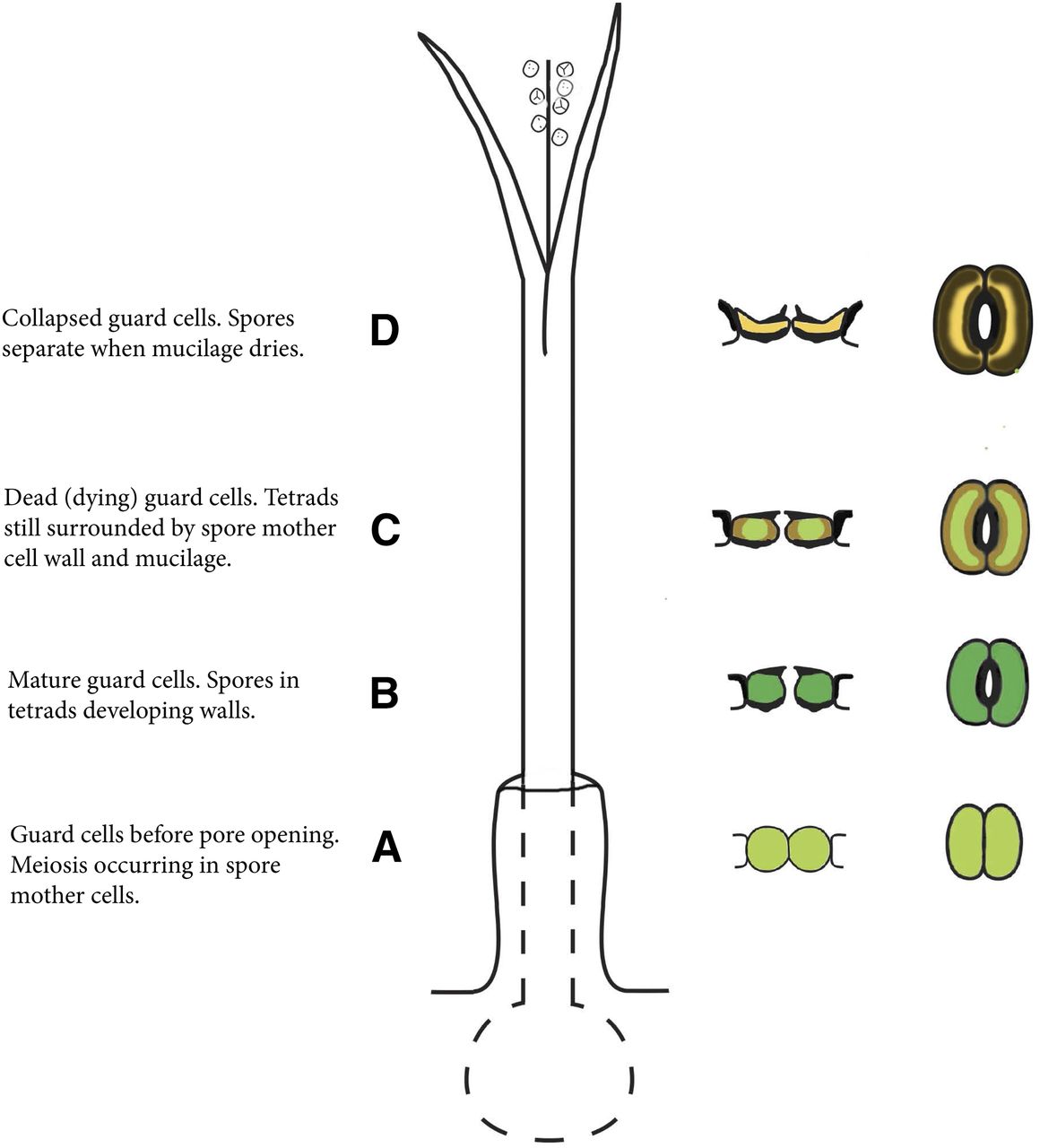
Stomata on the sporophyte may assist in spore dispersal by allowing the sporangium to dry, providing conditions necessary for dehiscence. Figure \(\PageIndex{5}\) walks through the stages of sporophyte maturation, showing the appearance of the stomata at each stage.
Life Cycle
The hornwort life cycle, like all plants, is alternation of generations. The multicellular haploid stage, the gametophyte, is the longer-lived and larger phase of this life cycle. Most hornwort gametophytes are monecious, meaning both types of gametangia (antheridia and archegonia) are produced on the same gametophyte. The life cycle shown in Figure \(\PageIndex{6}\) features a monoecious gametophyte. If a hornwort is dioecious, the life cycle would have antheridia and archegonia produced on separate gametophytes and the sporophyte would grow from the gametophyte producing archegonia.
As will be the case with all plants, archegonia produce one or more eggs and antheridia produce many sperm. These haploid gametes are produced by mitosis. The sperm have two flagella and must swim through water to reach the egg at the base of the archegonium. Fertilization (fusion of the egg and sperm) results in a diploid zygote. This zygote grows within the archegonium, nourished by the gametophyte. All land plants retain and nourish the zygote, called an embryo, and so are sometimes referred to as the embryophytes. Meiosis occurs within the sporangium (also called a capsule), producing haploid spores that are dispersed aerially.