
4.1.5: The Light-dependent Reactions
- Page ID
- 31990

Learning Objectives
- Relate wavelength, energy, and the type of electromagnetic radiation (and the color of visible light).
- Explain how plants absorb energy from sunlight.
- Detail the steps of the light-dependent interactions.
How can light be used to make food? When a person turns on a lamp, electrical energy becomes light energy. Like all other forms of kinetic energy, light can travel, change form, and be harnessed to do work. In the case of photosynthesis, light energy is converted into chemical energy, which photoautotrophs use to build carbohydrate molecules (Figure \(\PageIndex{1}\)). However, photoautotrophs only use a few specific components of sunlight.

What Is Light Energy?
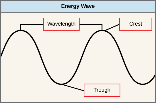
The sun emits an enormous amount of electromagnetic radiation (solar energy). Humans can see only a fraction of this energy, which portion is therefore referred to as “visible light”. The manner in which solar energy travels is described as waves. Scientists can determine the amount of energy of a wave by measuring its wavelength, the distance between consecutive points of a wave. A single wave is measured from two consecutive points, such as from crest to crest or from trough to trough (Figure \(\PageIndex{2}\)). The difference between wavelengths relates to the amount of energy carried by them.
Visible light constitutes only one of many types of electromagnetic radiation emitted from the sun and other stars. Scientists differentiate the various types of radiant energy from the sun within the electromagnetic spectrum. The electromagnetic spectrum is the range of all possible frequencies of radiation (Figure \(\PageIndex{3}\)). Several types of electromagnetic radiation originate from the sun, including X-rays and ultraviolet (UV) rays. The higher-energy waves can penetrate tissues and damage cells and DNA, explaining why both X-rays and UV rays can be harmful to living organisms.

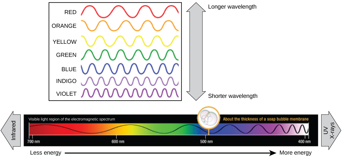
Each type of electromagnetic radiation travels at a particular wavelength. The longer the wavelength (or the more stretched out it appears in the diagram), the less energy is carried. Short, tight waves carry the most energy. This may seem illogical, but think of it in terms of a piece of moving a heavy rope. It takes little effort by a person to move a rope in long, wide waves. To make a rope move in short, tight waves, a person would need to apply significantly more energy.
Absorption of Light
Light energy initiates the process of photosynthesis when pigments absorb the light. Organic pigments, whether in the human retina or the chloroplast thylakoid, have a narrow range of energy levels that they can absorb. Energy levels lower than those represented by red light are insufficient to excite electrons in the retinal pigments. Energy levels higher than those in blue light will physically tear these molecules apart, called bleaching. So retinal pigments can only “see” (absorb) 700 nm to 400 nm light, which is therefore called visible light. For the same reasons, plants pigment molecules absorb only light in the wavelength range of 700 nm to 400 nm; plant physiologists refer to this range for plants as photosynthetically active radiation.
The visible light seen by humans as white light actually exists in a rainbow of colors. Certain objects, such as a prism or a drop of water, disperse white light to reveal the colors to the human eye. The visible light portion of the electromagnetic spectrum shows the rainbow of colors, with violet and blue having shorter wavelengths, and therefore higher energy. At the other end of the spectrum toward red, the wavelengths are longer and have lower energy (Figure \(\PageIndex{4}\)).
Understanding Pigments
Different kinds of pigments exist, and each has evolved to absorb only certain wavelengths (colors) of visible light. Pigments reflect or transmit the wavelengths they cannot absorb, making them appear in the corresponding color.
Chlorophylls and carotenoids are the two major classes of photosynthetic pigments found in plants and algae; each class has multiple types of pigment molecules. There are five major chlorophylls: a, b, c and d and a related molecule found in prokaryotes called bacteriochlorophyll. Chlorophyll a and chlorophyll b are found in plant chloroplasts and will be the focus of the following discussion.
With dozens of different forms, carotenoids are a much larger group of pigments. The carotenoids found in fruit—such as the red of tomato (lycopene), the yellow of corn seeds (zeaxanthin), or the orange of an orange peel (β-carotene)—are used as advertisements to attract seed dispersers. In photosynthesis, carotenoids function as photosynthetic pigments that are very efficient molecules for the disposal of excess energy. When a leaf is exposed to full sun, the light-dependent reactions are required to process an enormous amount of energy; if that energy is not handled properly, it can do significant damage. Therefore, many carotenoids reside in the thylakoid membrane, absorb excess energy, and safely dissipate that energy as heat.
Each type of pigment can be identified by the specific pattern of wavelengths it absorbs from visible light, which is the absorption spectrum. The graph in Figure \(\PageIndex{5}\) shows the absorption spectra for chlorophyll a, chlorophyll b, and a type of carotenoid pigment called β-carotene (which absorbs blue and green light). Notice how each pigment has a distinct set of peaks and troughs, revealing a highly specific pattern of absorption. Chlorophyll a absorbs wavelengths from either end of the visible spectrum (blue and red), but not green. Because green is reflected or transmitted, chlorophyll appears green. Carotenoids absorb in the short-wavelength blue region, and reflect the longer yellow, red, and orange wavelengths.

Many photosynthetic organisms have a mixture of pigments; using them, the organism can absorb energy from a wider range of wavelengths. Not all photosynthetic organisms have full access to sunlight. Some organisms grow underwater where light intensity and quality decrease and change with depth (Figure \(\PageIndex{6}\)). Other organisms grow in competition for light. Plants on the rainforest floor must be able to absorb any bit of light that comes through, because the taller trees absorb most of the sunlight and scatter the remaining solar radiation (Figure \(\PageIndex{7}\)).


When studying a photosynthetic organism, scientists can determine the types of pigments present by generating absorption spectra. An instrument called a spectrophotometer can differentiate which wavelengths of light a substance can absorb. Spectrophotometers measure transmitted light and compute from it the absorption. By extracting pigments from leaves and placing these samples into a spectrophotometer, scientists can identify which wavelengths of light an organism can absorb. Additional methods for the identification of plant pigments include various types of chromatography that separate the pigments by their relative affinities to solid and mobile phases.
How Light-Dependent Reactions Work
The overall function of light-dependent reactions is to convert solar energy into chemical energy in the form of NADPH and ATP. This chemical energy supports the light-independent reactions and fuels the assembly of sugar molecules. In the light-dependent reactions protein complexes and pigment molecules work together to produce NADPH and ATP (Figure \(\PageIndex{8}\) and Video \(\PageIndex{1}\)).
Video \(\PageIndex{1}\): Here is an animation of the light-dependent reactions.
Photosystems Absorb Light Energy
The actual step that converts light energy into chemical energy takes place in a multiprotein complex called a photosystem, two types of which are found embedded in the thylakoid membrane, photosystem II (PSII) and photosystem I (PSI) (Figure \(\PageIndex{9}\)). The two complexes differ on the basis of what they oxidize (that is, the source of the low-energy electron supply) and what they reduce (the place to which they deliver their energized electrons).
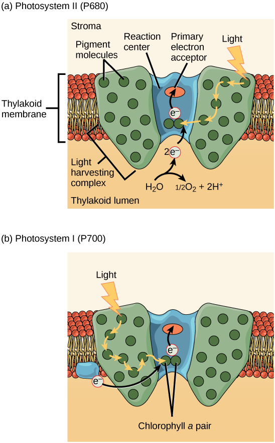
Both photosystems have the same basic structure; a number of antenna proteins to which the chlorophyll molecules are bound surround the reaction center, where the photochemistry takes place. Each photosystem is serviced antenna complex, which passes energy from sunlight to the reaction center. The antenna complex consists of multiple antenna proteins that contain a mixture of 300–400 chlorophyll a and b molecules as well as other pigments like carotenoids. (Technically, photosystems consist of the reaction center and the antennae complex, and a photosytem plus light-harvesting complexes comprises a photosystem complex, but in Figure \(\PageIndex{9}\), the light-harvesting complex is labeled as the antennae complex.) The absorption of a single photon, or distinct quantity or “packet” of light, by any of the chlorophylls pushes that molecule into an excited state. In short, the light energy has now been captured by biological molecules but is not stored in any useful form yet. The energy is transferred from chlorophyll to chlorophyll until eventually (after about a millionth of a second), it is delivered to the reaction center. Up to this point, only energy has been transferred between molecules, not electrons.
The reaction center contains a pair of chlorophyll a molecules with a special property. Those two chlorophylls can undergo oxidation upon excitation; they can actually give up an electron. It is at this step in the reaction center, this step in photosynthesis, that light energy is converted into an excited electron.
Electrons Move Down the Electron Transport Chain, Providing Energy to Pump Protons into the Thylakoid Lumen
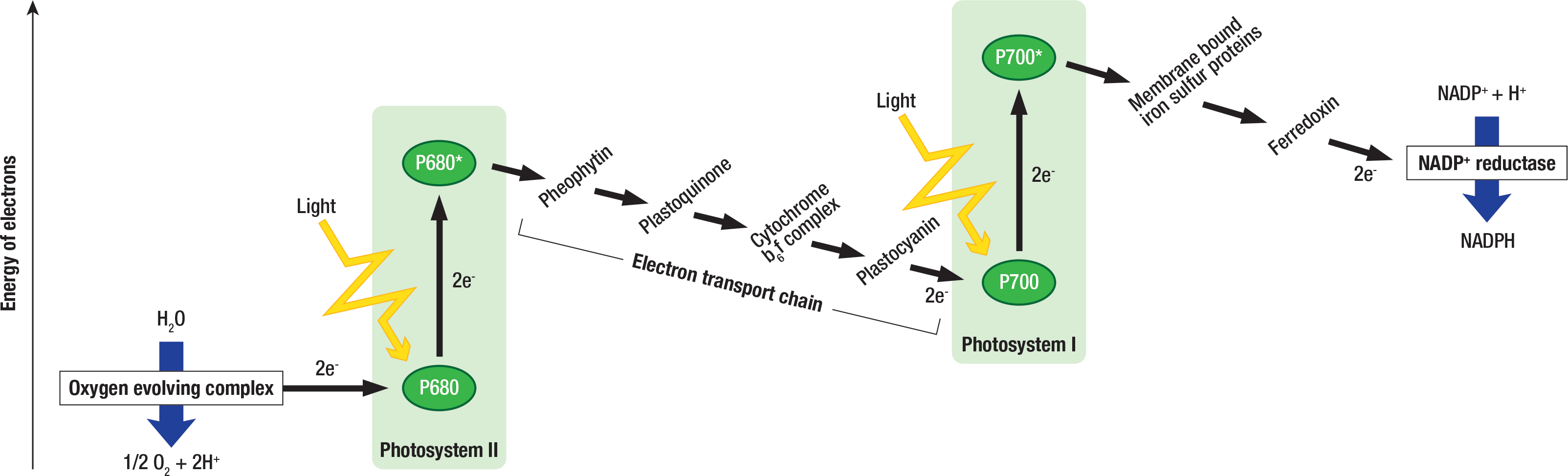
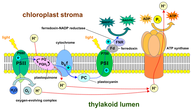
The reaction center of PSII (called P680) delivers its high-energy electrons, one at the time, to the primary electron acceptor, and through the electron transport chain (Pq to cytochrome b6f complex to plastocyanine) to PSI (Figure \(\PageIndex{10}\)). The cytochrome b6f complex, an enzyme composed of two protein complexes, transfers the electrons from the carrier molecule plastoquinone (Pq) to the protein plastocyanin (Pc), ultimately facilitating the transfer of electrons from PSII to PSI. During this process, cytochrome b6f pumps protons from the stroma into the thylakoid lumen using the energy from electrons moving down the electron transport chain.

Photophosphorylation
The synthesis of ATP in the light-dependent reactions is called photophosphorylation. The buildup of protons inside the thylakoid lumen creates a concentration gradient. The passive movement (facilitated diffusion) of protons from high concentration (in the thylakoid lumen) to low concentration (in the stroma) is harnessed to create ATP. The protons build up energy at high concentrations because they all have the same electrical charge, repelling each other.
To release this energy, protons will rush through any opening, similar to water jetting through a hole in a dam. In the thylakoid, that opening is a passage through a specialized protein channel called the ATP synthase. The energy released by the proton stream allows ATP synthase to attach a third phosphate group to ADP, which forms a molecule of ATP (Figures \(\PageIndex{10-11}\)). The flow of protons through ATP synthase is called chemiosmosis because the ions move from an area of high to an area of low concentration through a semi-permeable structure.
Water Photolysis
P680’s missing electron is replaced by extracting a low-energy electron from water; thus, water is split (water photolysis) and PSII is re-reduced. Splitting one H2O molecule releases two electrons, two protons, and one atom of oxygen (Figures \(\PageIndex{10-11}\)). Those protons, in addition to the ones transported by cytrochrome b6f, accumulate in the thylakoid lumen and fuel photophosphorylation. Splitting two molecules is required to form one molecule of diatomic O2 gas. About 10 percent of the oxygen is used by mitochondria in the leaf to support aerobic cellular respiration, the process of breaking down glucose to generate ATP. The remainder escapes to the atmosphere where it is used by aerobic organisms to support respiration.
Reduction of NADP+
Because the electrons have lost energy prior to their arrival at PSI, they must be re-energized by PSI, hence, another photon is absorbed by the PSI antenna. That energy is relayed to the PSI reaction center (called P700). P700 is oxidized and sends high-energy electrons to the electron carrier NADP+ to form NADPH. Thus, PSII captures the energy to create proton gradients to make ATP, and PSI captures the energy to reduce NADP+ into NADPH. The two photosystems work in concert, in part, to guarantee that the correct proportions of NADPH and ATP needed for the light-independent reactions are generated. Other mechanisms exist to fine tune that ratio to exactly match the chloroplast’s constantly changing energy needs.
Attribution
Curated and authored by Melissa Ha using 8.2 The Light-Dependent Reactions of Photosynthesis from Biology 2e by OpenStax (licensed CC-BY). Access for free at openstax.org.