
2.2.1.3.1: Cyanobacteria
- Page ID
- 43211

Learning Objectives
- Explain the role of Cyanobacteria in changing Earth's atmosphere
- Describe a few mutualistic relationships formed with Cyanobacteria
- Distinguish between different Cyanobacterial cell types and describe their functions
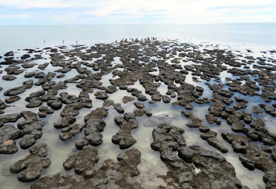
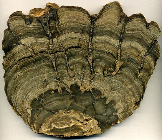
Stromatolites
The earliest fossils are interpreted to be unicellular organisms similar to modern day cyanobacteria, sometimes referred to as blue green algae. The most widely accepted of these fossils dates back to 3.4 billion years ago from the Strelley Pool Formation in Western Australia. These particular fossils are called stromatolites and are composed of alternating layers of fossilized cells and calcium carbonate (Figure \(\PageIndex{1}\)). We can use evidence from modern day stromatolite formation in Western Australia to infer that these fossilized cells were doing a process called photosynthesis, using dissolved CO2 in the water to form sugar molecules. This causes calcium to precipitate out of the seawater, forming hardened layers of calcium carbonate on top of the colony of organisms. Because they need access to light to continue photosynthesizing, living cells begin forming a new layer on top of the calcium carbonate. This process continues, making a ringed pattern as the formation grows, much like we see in trees and corals.
Cyanobacteria perform oxygenic photosynthesis. The ancestors of modern day cyanobacteria are responsible for the initial production of our oxygen-rich atmosphere. In addition to these initial inputs, cyanobacteria are the origin of chloroplasts in all eukaryotic phototrophs, including plants. In an event called primary endosymbiosis, a cyanobacterium was engulfed by a heterotrophic eukaryote. Instead of being digested, the prokaryote lived within the larger cell. Over time, genes were exchanged between the prokaryote and the eukaryote, eventually resulting in the first chloroplasts.
Modern Cyanobacteria
Cyanobacteria can be found free-living, often in colonies, or living in symbiotic relationships with other organisms, such as fungi and plants.
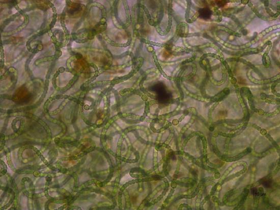
Free-Living
Cyanobacteria can be found in a vast diversity of places, from floating in the ocean to living in cryptobiotic crusts in the desert. Nostoc is a type of cyanobacteria that can often be found living in gelatinous colonies in wet, terrestrial environments. The colony secretes a mucilaginous sheath that provides a protective barrier and allows for the exchange of materials between cells in the colony.
.jpg?revision=1&size=bestfit&width=633&height=420)
.jpg?revision=1&size=bestfit&width=536&height=402)
.jpg?revision=1)
Mutualists
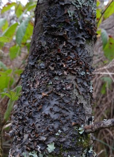
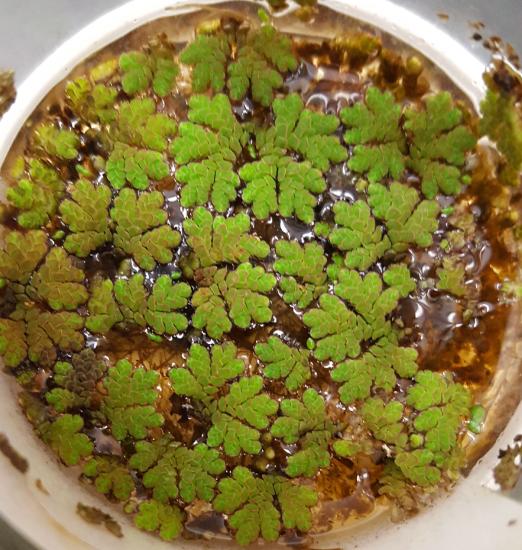
Many cyanobacteria that you'll see in botany will be in mutualistic relationships. Anabaena is a colonial cyanobacterium that lives within the water fern Azolla, fixing nitrogen in the fern's relatively nutrient-poor aquatic environment. Nostoc is another colonial cyanobacterium capable of fixing nitrogen. It can be found free-living in gelatinous colonies shown above or, as you are likely to see it in your botany course, in compartments of a hornwort thallus. Cyanobacteria can also be found in a mutualistic relationship with fungi in cyanolichens.
.jpeg?revision=1&size=bestfit&width=598&height=448)
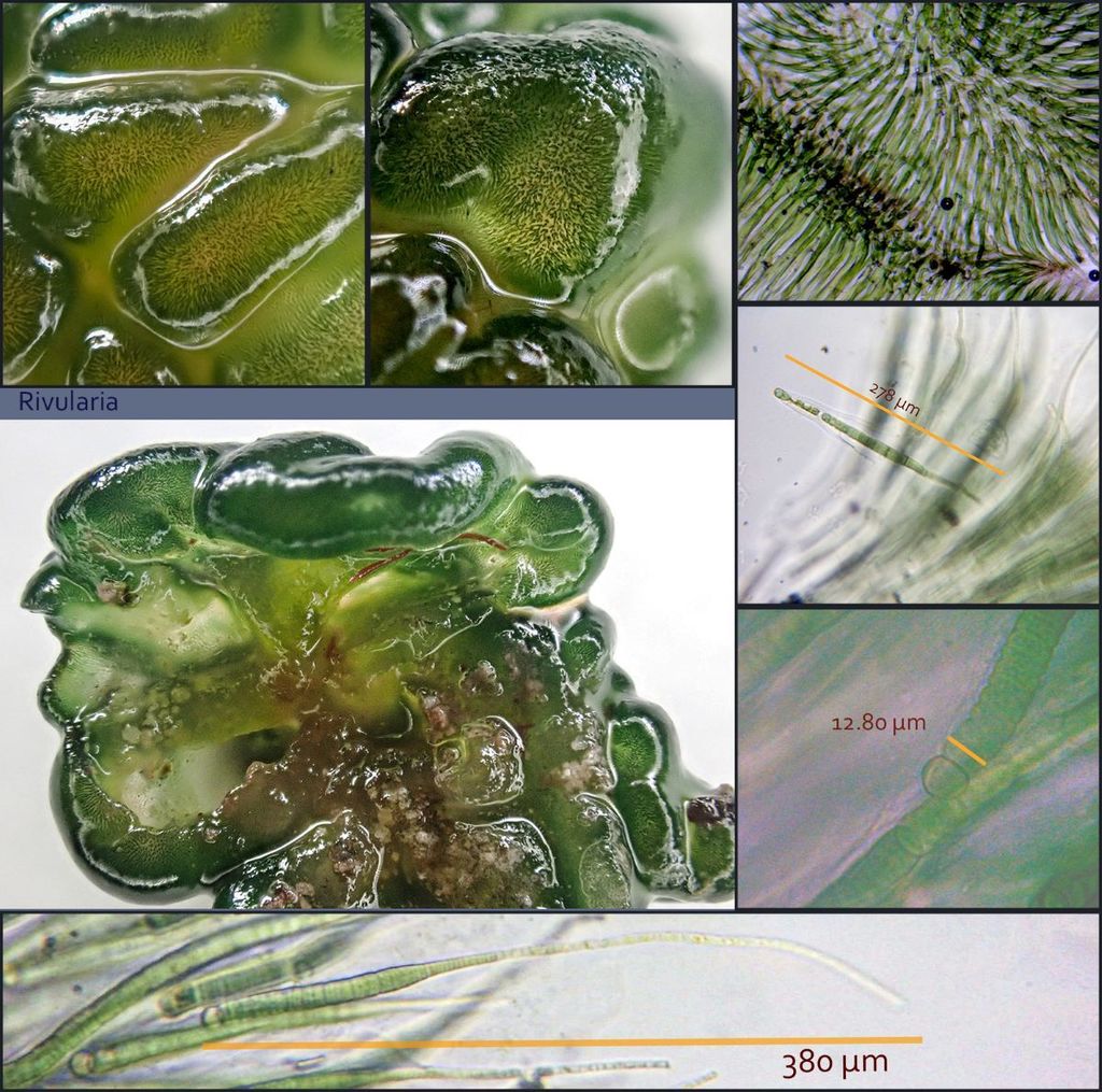
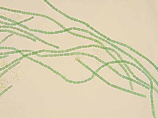
Anabaena
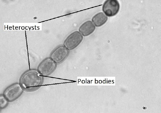
If you were to chop up a sample of Azolla and look at it under the microscope, you'd see what looked like strings of green beads. Each bead is an individual cyanobacterium of the genus Anabaena. However, even though each one is an individual, some cells will specialize to provide a service for the colony, as a whole.
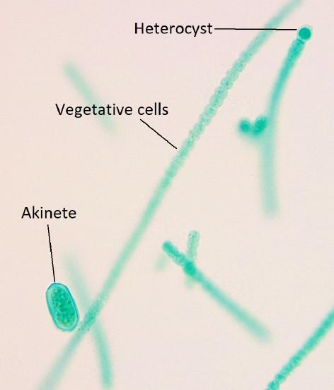
- Heterocysts are thick-walled, chlorophyll-free cells that are fixing atmospheric nitrogen into bioavailable forms using the enzyme nitrogenase. Heterocysts cannot do photosynthesis, as that process produces oxygen and nitrogenase cannot function in the presence of oxygen.
- Akinetes are individuals that still perform photosynthesis, but also function as a sort of failsafe. Akinetes store large amounts of lipids and carbohydrates so that they have enough energy to begin a new colony if conditions become too cold or too dry for survival. Their formation is triggered by these conditions (dry or cold), so you may not see them from a fresh water fern leaf, as this is a relatively stable, comfortable environment.
Summary
Cyanobacteria are a group of bacteria that perform oxygenic photosynthesis. The ancestors of modern cyanobacteria were responsible for the initial input of large amounts of oxygen into Earth's atmosphere. Evidence of these early cyanobacteria can be found in fossilized structures called stromatolites, which are still formed in some regions of the world. Cyanobacteria can be found free-living or as mutualists within the tissues of other organisms. Colonies of individuals are often encased within a protective mucilage. Within a colony, individual cells might specialize to fix nitrogen (heterocysts) or to survive cold and/or dry conditions (akinetes).
Attribution
Content by Maria Morrow, CC BY-NC