The fruiting body of a basidiomycete is called a basidiocarp. These structures produce haploid spores by meiosis and come in an incredible variety of shapes and sizes.
General Mushroom Anatomy
Though only a subset of basidiocarps look this way, they are the model for how we describe "mushrooms". In mycology, this type of basidiocarp is called "agaricoid" or "agaric" because it is the general form we see in the genus Agaricus. A more complex version of the agaric mushroom is seen in the genus Amanita, which is shown below.
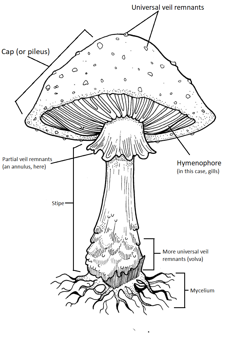
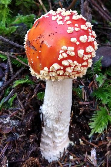
Figure \(\PageIndex{1}\): The anatomy of an Amanita muscaria basidiocarp. The cap (also called the pileus) protects the spore producing region, the hymenophore. The diagram shows a hymenophore composed of gills. However, you might see mushrooms with pores, teeth, or other types of spore-producing surfaces. The partial veil covers the hymenophore while the mushroom develops. When the cap extends as the mushroom grows, the partial veil is pulled and can either end up as an annulus or attached to the edges of the cap. Not all mushrooms have a partial veil and those that do often look quite different from this one! The universal veil (only present in a few mushrooms) covers the entire mushroom when it is young. As the mushroom expands, the universal veil is also pulled apart. Here, the cap has tiny fragments of the universal veil (warts) and the rest is at the base of the stipe (forming a volva). The stipe is the "stem" or "stalk" of the mushroom. Artwork by Nikki Harris CC-BY-NC.
Veils
Partial Veils
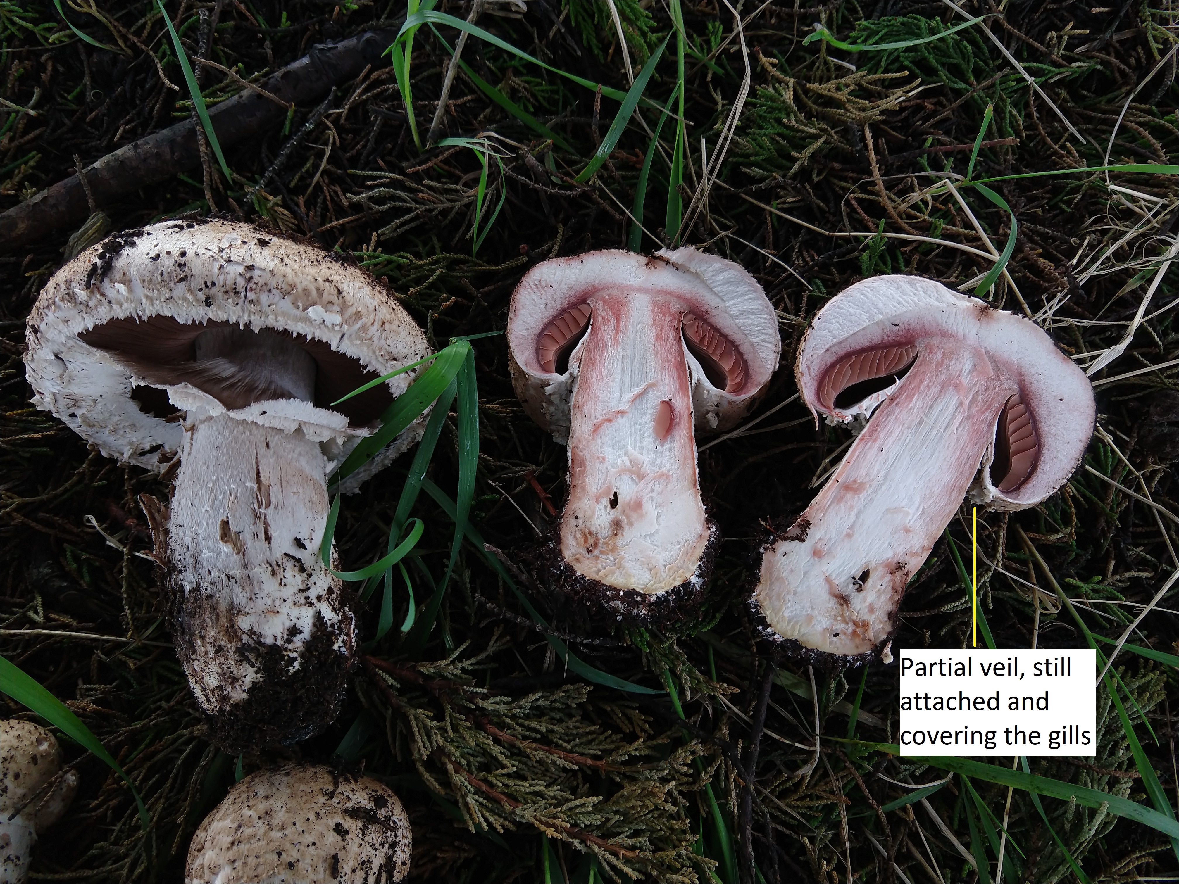
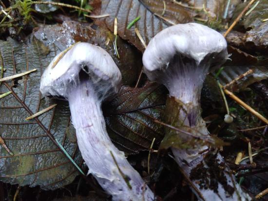
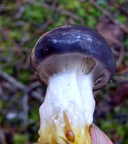
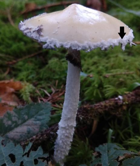
Figure \(\PageIndex{2}\): Veils can be difficult to conceptualize until you see them in person. The image above shows a few developmental stages of an Agaricus basidiocarp. In the fruiting body on the right (which has been cut in half), you can see the partial veil is still covering the gills. In the older fruiting body on the left, you can see the partial veil pulling away from the edges of the cap. This will end up as an annulus, as shown in the diagram of mushroom anatomy above. Photo by Maria Morrow, CC-BY-NC.Figure \(\PageIndex{3}\): Partial veils can also take on a variety of forms. In the image on the left (Cortinarius), the partial veil is cobwebby instead of thick and sheet-like. In the image on the right (Gomphidius), the partial veil is made of slime! What you can also see in the image on the right are arthropods (likely springtails) on the gills. The partial veil protects the spores from predators like these, until it begins to open. Photos by Maria Morrow, CC-BY-NC.Figure \(\PageIndex{4}\): Partial veils can also end up on the margin of the cap, instead of the stipe. In the image above, you can see the cottony partial veil (black arrow) of Stropharia ambigua dangling from the margin (edge) of the cap. Photo by Maria Morrow, CC-BY-NC.
Universal Veils
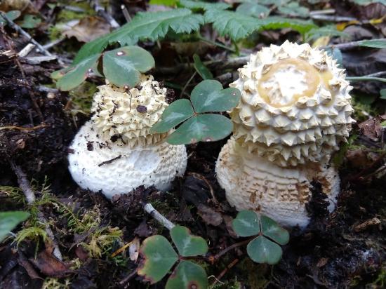
Figure \(\PageIndex{5}\): In the image on the left, you can see two Amanita buttons with a universal veil. As the veil dries out and the mushroom expands, it begins to crack into peaks. In the image on the right, an Amanita in a later stage of development shows similar peak-shaped universal veil remnants on the cap, but they are much farther apart. This is because the cap is expanding, much like a balloon, and the universal veil is staying the same size. Photos by Maria Morrow, CC-BY-NC.
Variety of Hymenophores
Gills (lamellate)
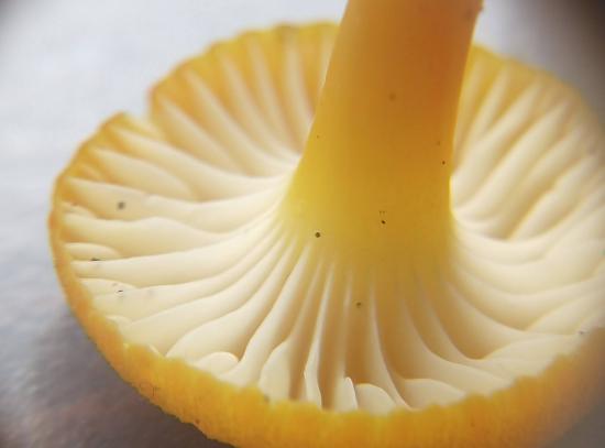
Figure \(\PageIndex{6}\): The underside of a gilled mushroom. The sheet-like membranes that travel between the cap and the stipe are called gills or lamellae. The spores are produced from basidia that cover the gills. Photo by Maria Morrow, CC-BY-NC.
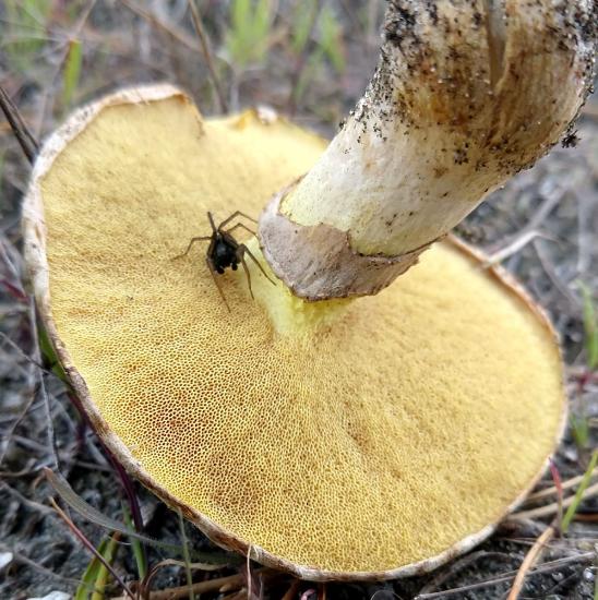
Pores (poroid)
Figure \(\PageIndex{7}\): The underside of three mushrooms with pores. The pores of the Suillus umbonatus in the image on the far left are large and easily distinguished. The spores are produced on the internal surfaces of the pores, which are tube-like in structure. In the center, another Suillus species has smaller pores, but they can still be distinguished with the naked eye. In the Picipes badius on the right, the pores are so small that it almost looks smooth. Photos by Maria Morrow, CC-BY-NC.
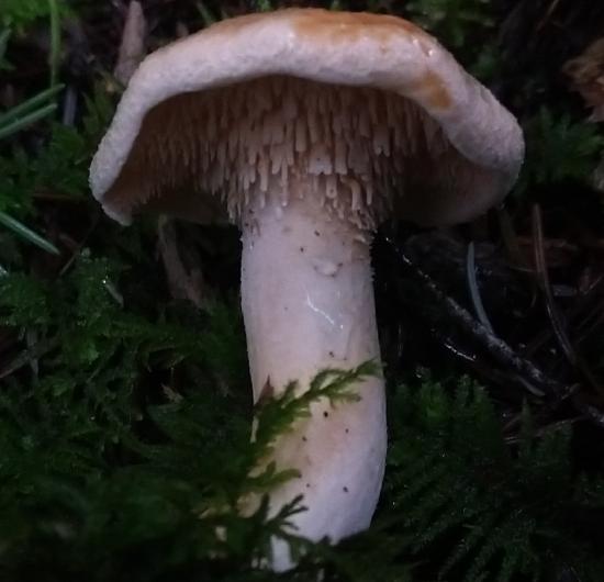
Teeth (dentate)
Figure \(\PageIndex{8}\): The underside of two mushrooms with teeth. Teeth are like a pore that has been turned inside-out. Each stalactite-like tooth is covered by spore-producing basidia. The Hydnellum on the left has much smaller teeth than the Hydnum on the right. Photos by Maria Morrow, CC-BY-NC.
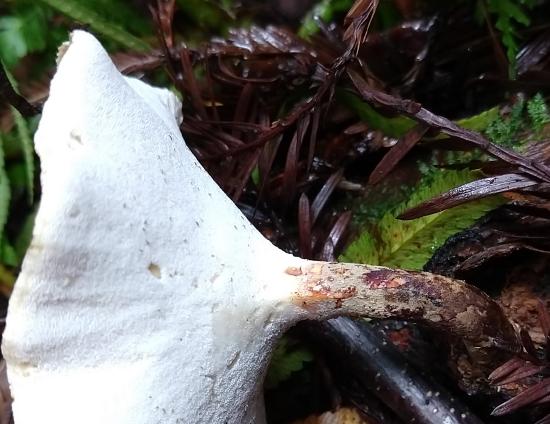
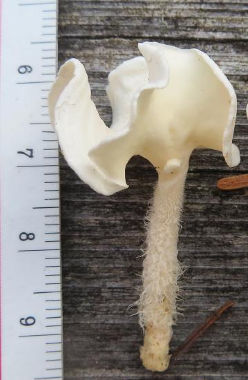
Smooth
Figure \(\PageIndex{8}\): This Stereopsis humphreyi has a hairy stipe and a saddle-shaped cap. On the underside of the cap, where you might expect to find gills or pores, the surface is completely smooth. However, spores are still formed here! Photo by Kilasiak, CC-BY-NC.
Other Basidiocarps
There are a vast diversity of basidiocarp forms out there. Below are a few examples of the more common ones you might run into.
Puffballs
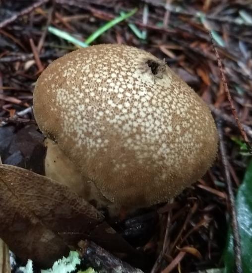
Figure \(\PageIndex{9}\): Lycoperdon is a puffball-producing group. The spores are produced in a mass within the puffball. As it matures, it dries and a pore in the top opens. When the puffball is disturbed, such as by feet or raindrops, it puffs out spores, much like a bellows. Photo by Maria Morrow, CC-BY-NC.
Clubs & Corals
Figure \(\PageIndex{10}\): On the left are the club-shaped fruiting bodies of Clavariadelphus occidentale. On the right, is the branched, antler-like fruiting body. The branching-forms are generally referred to as corals. Spores can be produced on both clubs and corals on any exterior surface. Photos by Maria Morrow, CC-BY-NC.
Brackets and Shelves
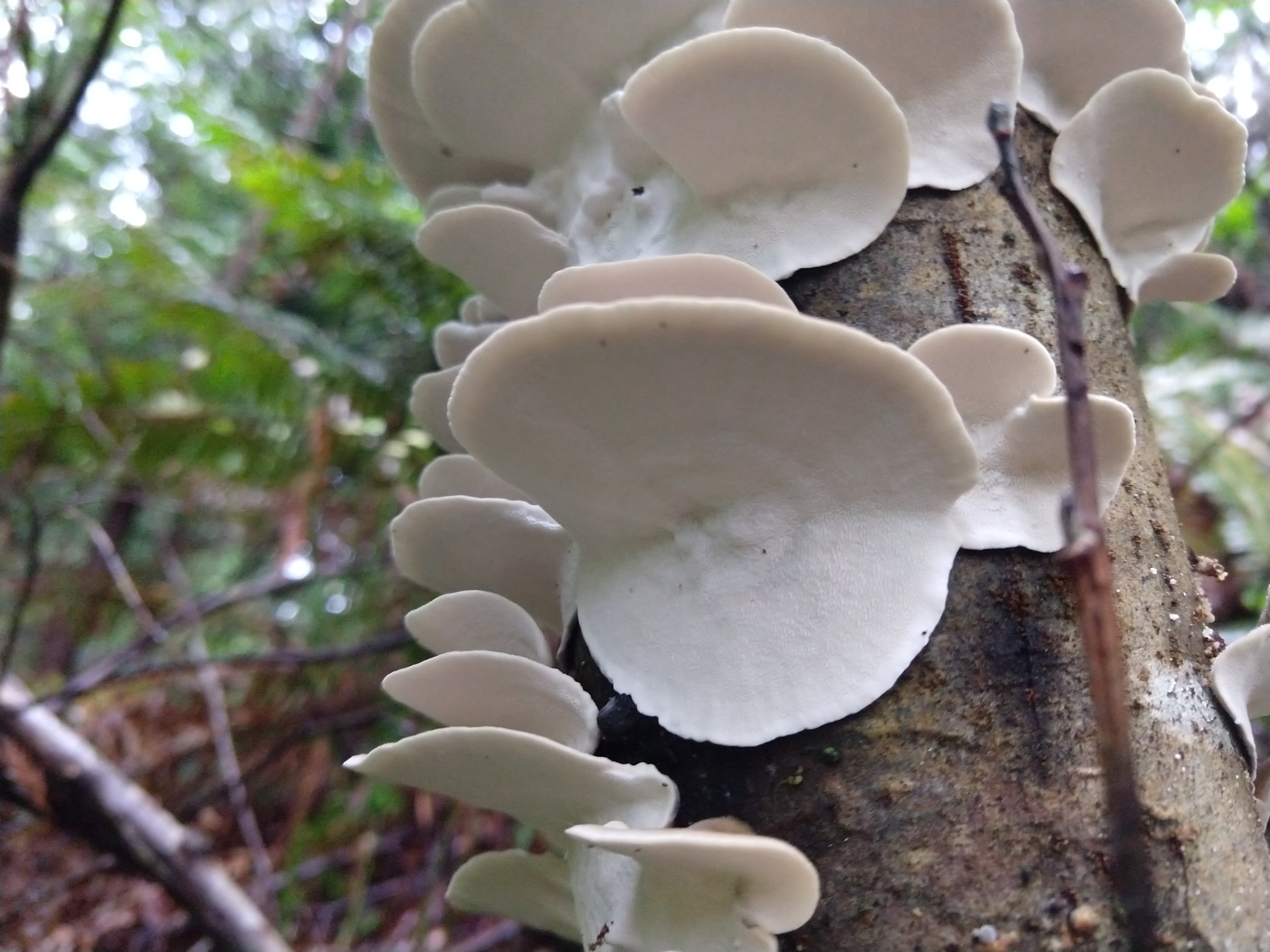
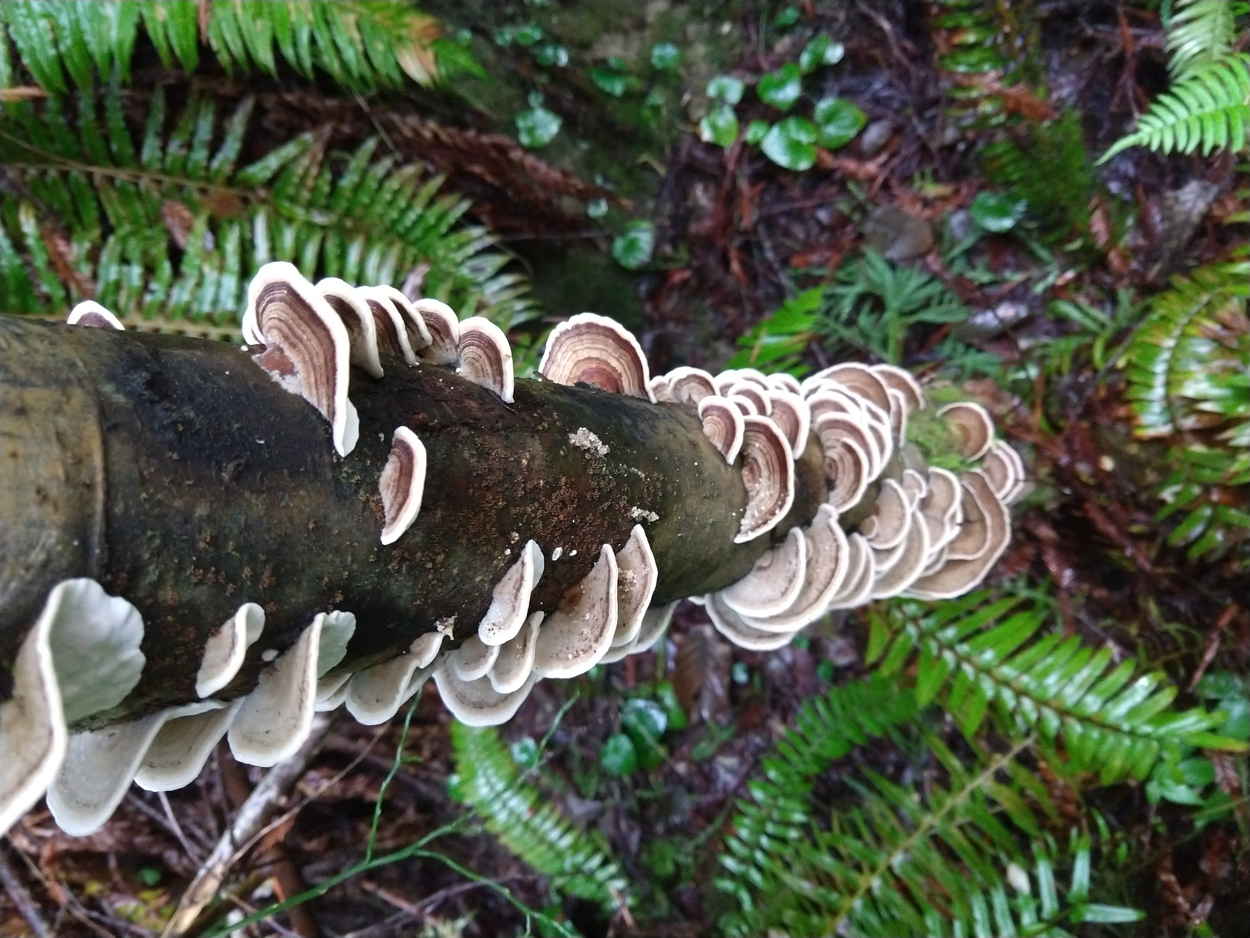
Figure \(\PageIndex{11}\): Brackets formed by Trametes versicolor. These brackets have a hymenophore composed of tiny pores that cover the underside. The pileus (top) on these brackets has many concentric zones of brown stipes. Brackets tend to be relatively small and flimsy. Photos by Maria Morrow, CC-BY-NC.
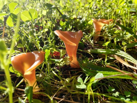
Jellies
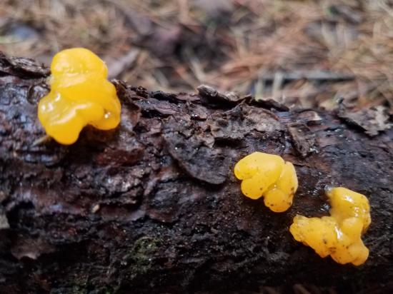
Figure \(\PageIndex{12}\): Two different types of jelly fungi. Jelly fungi can occur in a variety of forms. The gelatinous blobs on the left are produced by Dacrymyces. The vase-shaped jellies on the right are found in Guepinia helvelloides. Spores are produced on the exterior surface of jelly fruiting bodies. Photos by Maria Morrow, CC-BY-NC.
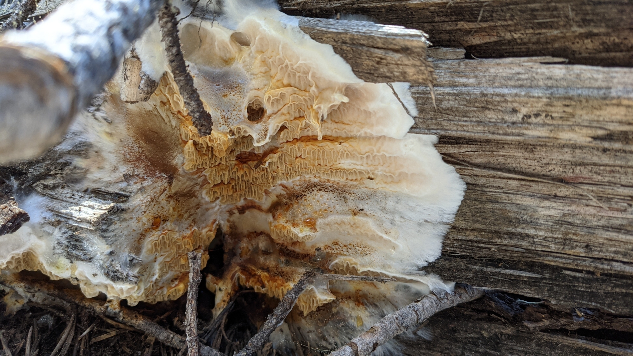
Crusts
Figure \(\PageIndex{13}\): Some fungi form fruiting bodies that spread flat across the surface of what they are eating, like this Serpula himantioides fruiting on a log. The spores are produced on the exposed surface. This type of fruiting body is referred to as a crust. Photo by Maria Morrow, CC-BY-NC.