Lab 1: The Microscopic World
- Page ID
- 23954

\( \newcommand{\vecs}[1]{\overset { \scriptstyle \rightharpoonup} {\mathbf{#1}} } \)
\( \newcommand{\vecd}[1]{\overset{-\!-\!\rightharpoonup}{\vphantom{a}\smash {#1}}} \)
\( \newcommand{\dsum}{\displaystyle\sum\limits} \)
\( \newcommand{\dint}{\displaystyle\int\limits} \)
\( \newcommand{\dlim}{\displaystyle\lim\limits} \)
\( \newcommand{\id}{\mathrm{id}}\) \( \newcommand{\Span}{\mathrm{span}}\)
( \newcommand{\kernel}{\mathrm{null}\,}\) \( \newcommand{\range}{\mathrm{range}\,}\)
\( \newcommand{\RealPart}{\mathrm{Re}}\) \( \newcommand{\ImaginaryPart}{\mathrm{Im}}\)
\( \newcommand{\Argument}{\mathrm{Arg}}\) \( \newcommand{\norm}[1]{\| #1 \|}\)
\( \newcommand{\inner}[2]{\langle #1, #2 \rangle}\)
\( \newcommand{\Span}{\mathrm{span}}\)
\( \newcommand{\id}{\mathrm{id}}\)
\( \newcommand{\Span}{\mathrm{span}}\)
\( \newcommand{\kernel}{\mathrm{null}\,}\)
\( \newcommand{\range}{\mathrm{range}\,}\)
\( \newcommand{\RealPart}{\mathrm{Re}}\)
\( \newcommand{\ImaginaryPart}{\mathrm{Im}}\)
\( \newcommand{\Argument}{\mathrm{Arg}}\)
\( \newcommand{\norm}[1]{\| #1 \|}\)
\( \newcommand{\inner}[2]{\langle #1, #2 \rangle}\)
\( \newcommand{\Span}{\mathrm{span}}\) \( \newcommand{\AA}{\unicode[.8,0]{x212B}}\)
\( \newcommand{\vectorA}[1]{\vec{#1}} % arrow\)
\( \newcommand{\vectorAt}[1]{\vec{\text{#1}}} % arrow\)
\( \newcommand{\vectorB}[1]{\overset { \scriptstyle \rightharpoonup} {\mathbf{#1}} } \)
\( \newcommand{\vectorC}[1]{\textbf{#1}} \)
\( \newcommand{\vectorD}[1]{\overrightarrow{#1}} \)
\( \newcommand{\vectorDt}[1]{\overrightarrow{\text{#1}}} \)
\( \newcommand{\vectE}[1]{\overset{-\!-\!\rightharpoonup}{\vphantom{a}\smash{\mathbf {#1}}}} \)
\( \newcommand{\vecs}[1]{\overset { \scriptstyle \rightharpoonup} {\mathbf{#1}} } \)
\(\newcommand{\longvect}{\overrightarrow}\)
\( \newcommand{\vecd}[1]{\overset{-\!-\!\rightharpoonup}{\vphantom{a}\smash {#1}}} \)
\(\newcommand{\avec}{\mathbf a}\) \(\newcommand{\bvec}{\mathbf b}\) \(\newcommand{\cvec}{\mathbf c}\) \(\newcommand{\dvec}{\mathbf d}\) \(\newcommand{\dtil}{\widetilde{\mathbf d}}\) \(\newcommand{\evec}{\mathbf e}\) \(\newcommand{\fvec}{\mathbf f}\) \(\newcommand{\nvec}{\mathbf n}\) \(\newcommand{\pvec}{\mathbf p}\) \(\newcommand{\qvec}{\mathbf q}\) \(\newcommand{\svec}{\mathbf s}\) \(\newcommand{\tvec}{\mathbf t}\) \(\newcommand{\uvec}{\mathbf u}\) \(\newcommand{\vvec}{\mathbf v}\) \(\newcommand{\wvec}{\mathbf w}\) \(\newcommand{\xvec}{\mathbf x}\) \(\newcommand{\yvec}{\mathbf y}\) \(\newcommand{\zvec}{\mathbf z}\) \(\newcommand{\rvec}{\mathbf r}\) \(\newcommand{\mvec}{\mathbf m}\) \(\newcommand{\zerovec}{\mathbf 0}\) \(\newcommand{\onevec}{\mathbf 1}\) \(\newcommand{\real}{\mathbb R}\) \(\newcommand{\twovec}[2]{\left[\begin{array}{r}#1 \\ #2 \end{array}\right]}\) \(\newcommand{\ctwovec}[2]{\left[\begin{array}{c}#1 \\ #2 \end{array}\right]}\) \(\newcommand{\threevec}[3]{\left[\begin{array}{r}#1 \\ #2 \\ #3 \end{array}\right]}\) \(\newcommand{\cthreevec}[3]{\left[\begin{array}{c}#1 \\ #2 \\ #3 \end{array}\right]}\) \(\newcommand{\fourvec}[4]{\left[\begin{array}{r}#1 \\ #2 \\ #3 \\ #4 \end{array}\right]}\) \(\newcommand{\cfourvec}[4]{\left[\begin{array}{c}#1 \\ #2 \\ #3 \\ #4 \end{array}\right]}\) \(\newcommand{\fivevec}[5]{\left[\begin{array}{r}#1 \\ #2 \\ #3 \\ #4 \\ #5 \\ \end{array}\right]}\) \(\newcommand{\cfivevec}[5]{\left[\begin{array}{c}#1 \\ #2 \\ #3 \\ #4 \\ #5 \\ \end{array}\right]}\) \(\newcommand{\mattwo}[4]{\left[\begin{array}{rr}#1 \amp #2 \\ #3 \amp #4 \\ \end{array}\right]}\) \(\newcommand{\laspan}[1]{\text{Span}\{#1\}}\) \(\newcommand{\bcal}{\cal B}\) \(\newcommand{\ccal}{\cal C}\) \(\newcommand{\scal}{\cal S}\) \(\newcommand{\wcal}{\cal W}\) \(\newcommand{\ecal}{\cal E}\) \(\newcommand{\coords}[2]{\left\{#1\right\}_{#2}}\) \(\newcommand{\gray}[1]{\color{gray}{#1}}\) \(\newcommand{\lgray}[1]{\color{lightgray}{#1}}\) \(\newcommand{\rank}{\operatorname{rank}}\) \(\newcommand{\row}{\text{Row}}\) \(\newcommand{\col}{\text{Col}}\) \(\renewcommand{\row}{\text{Row}}\) \(\newcommand{\nul}{\text{Nul}}\) \(\newcommand{\var}{\text{Var}}\) \(\newcommand{\corr}{\text{corr}}\) \(\newcommand{\len}[1]{\left|#1\right|}\) \(\newcommand{\bbar}{\overline{\bvec}}\) \(\newcommand{\bhat}{\widehat{\bvec}}\) \(\newcommand{\bperp}{\bvec^\perp}\) \(\newcommand{\xhat}{\widehat{\xvec}}\) \(\newcommand{\vhat}{\widehat{\vvec}}\) \(\newcommand{\uhat}{\widehat{\uvec}}\) \(\newcommand{\what}{\widehat{\wvec}}\) \(\newcommand{\Sighat}{\widehat{\Sigma}}\) \(\newcommand{\lt}{<}\) \(\newcommand{\gt}{>}\) \(\newcommand{\amp}{&}\) \(\definecolor{fillinmathshade}{gray}{0.9}\)ACTIVITY 1: Getting to Know Your Microscope
1. Individually, get the combination from the instructor for your microscope drawer.
2. Open combination drawer and take out the microscope.
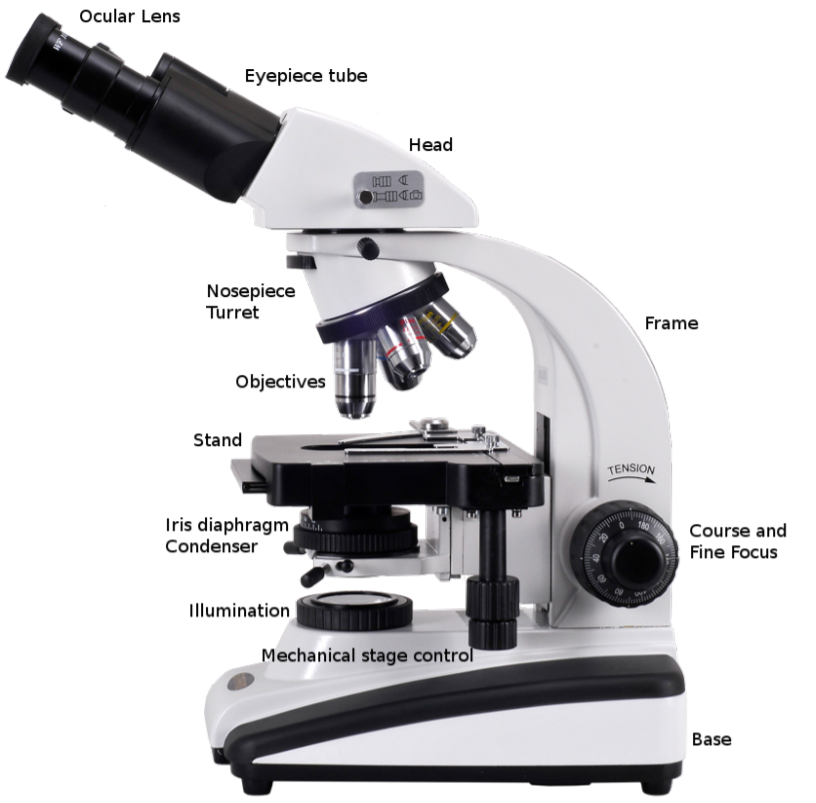
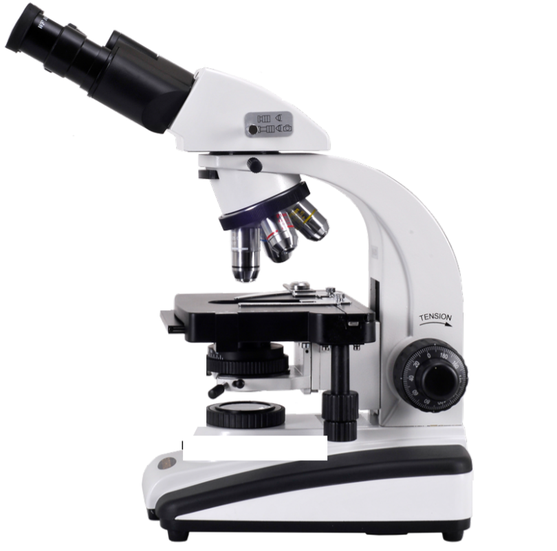
3. Label all the parts of the microscope with the provided post-its using the image below or the laboratory manual.
Note
The image below does not match your microscope perfectly, you will be responsible for knowing the parts of your microscope on the lab practical.
Practice worksheet (this is not an assignment)
Can you label all the parts of the microscope?
Complete the following:
The magnification is = __________ multiplied by _____________
| Ocular | Objective | Total Magnification |
ACTIVITY 2: How to Use Your Microscope
Note
Microscope magnification: enlarging an object’s appearance
The magnification is = (power of the oculars) multiplied by (power of the objectives)
Our microscopes have 10X oculars and 4X, 10X, 40X and 100X objectives
Never carry a microscope with just one hand!
FOCUSING:
1. Use one of the pre-made, gram-stained, bacterial slides.
2. Make sure the condenser is all the way up and the iris diaphragm is all the way open, letting the maximum amount of light to contact your slide.
3. ALWAYS start at 4X, stage lowered, focus with coarse focus knob first.
4. Once in focus move to 10X and focus using the fine focus knob.
5. Once in focus move to 40X and focus using the fine focus knob.
6. Move objectives half-way between 40X and 100X, add 1 drop of oil.
7. MAKE SURE THE 40X DOES NOT TOUCH THE OIL!
8. Move to 100x and SLOWLY focus with the fine focus knob If you do not see your image clearly, DO NOT go back to the 40X and try to refocus.

CLEANING A MICROSCOPE:
1. Lower stage.
2. Remove slide, turn the power off. 
3. Wipe oil from all surfaces and 100X with lens paper.
4. With the second piece of lens paper, moistened with alcohol, wipe all surfaces. Never use Kimwipes to clean microscope.
5. Wipe surfaces with a new dry piece of lens paper.
6. Return to the lowest lens (4x).
Once you are ready, invite the instructor over for your first skill tests.
SKILL TEST #1 (2.5 PTS): FOCUSING MICROScope
Under the supervision of the instructor or lab technician, bring your slide into focus under 100X. You can use your notes, books, etc. but you CAN NOT get help from other students.
SKILL TEST #2 (2.5 pts): Cleaning your microscope
Under the supervision of the instructor or lab technician, clean your microscope. You can use your notes, books, etc. but you CAN NOT get help from other students.
PLEASE READ THE FOLLOWING BEFORE PROCEEDING: 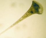
Stentors are a genus of single-celled eukaryotic organisms with cilia. They are usually horn-shaped are among the biggest known unicellular organisms. They reproduce asexually through binary fission and found in most freshwater lakes and streams.
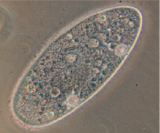
Paramecia are a genus of single-celled eukaryotic organisms with cilia. They are usually oval shaped and were some of the first microorganisms studied under a microscope. They reproduce asexually through binary fission and found in freshwater, brackish, and marine environments.
Euglenas are a genus of single-celled eukaryotic organisms with flagella. They are cylindrical with bright green chloroplasts inside. They are found in 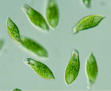 fresh and salt waters. Even though they have the ability to photosynthesize (like plants) they can also feed on other organisms (like animals).
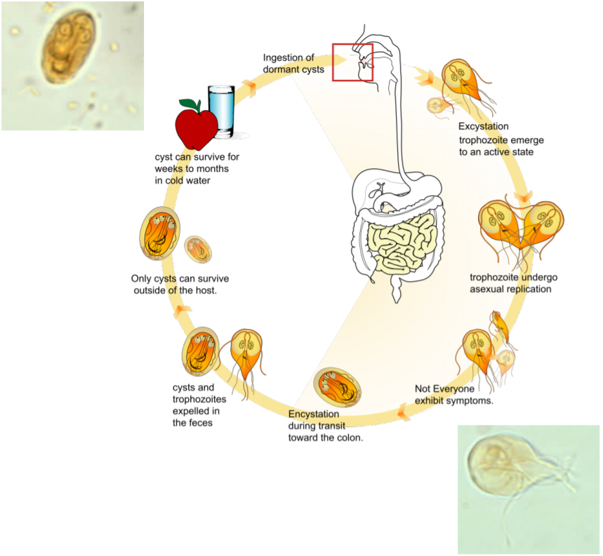
Giardia lamblia is a single-celled eukaryotic parasitic organism. The flagellated form of the parasite colonizes and reproduces in the small intestine, causing giardiasis. Giardia trophozoites absorb their nutrients from the lumen of the small intestine and are anaerobes (do not use oxygen). Human infection occurs via ingestion of water contaminated with the dormant cyst form of the parasite or through fecal-oral contamination. The cyst form is very hardy and can survive for weeks to months in cold water, and are resistant to chlorination. Giardia infects humans but is also one of the most common parasites infecting cats, dogs, cattle, sheep, and birds. The life cycle begins with a cyst being excreted in the feces of an infected individual. A distinguishing characteristic of the cyst is four nuclei. Once ingested by a host, the trophozoite emerges from the cyst. This is the active stage of the parasite capable of feeding and movement. After the feeding stage, the trophozoite undergoes asexual replication through longitudinal binary fission. The resulting trophozoites and cysts then pass through the digestive system in the feces. While the trophozoites may be found in the feces, only the cysts are capable of surviving outside of the host.
AFTER you have completed your skill tests, examine each of the following slides and sketch the organism you observe below.
1. Stentor
2. Paramecium
3. Euglena
4. Giardia cyst and trophozoite
ACTIVITY 3 (with your lab partner):
1. Label 5 TSA plates.
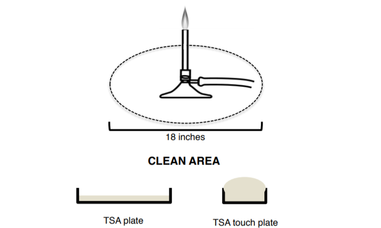
2. Place plate #1 within 6 inches of Bunsen burner open for 2 min.
3. Place plate #2 FAR away from Bunsen burner open for 2 min.
4. Place plate #3 FAR away from Bunsen burner open for 10 min.
5. Place plate #4 FAR away from Bunsen burner open for 30 min.
6. Take plate #5 and use a wet swab (use the sterile water to wet) to swab something (your partner, outside, your nose, be creative).
7. Take one touch plate and touch one place (face, walls, floor, shoes, bathrooms… be creative).
8. Put all your plates in the 37°C incubator, make sure your plates are agar side up.
Trypticase soy agar (TSA)
Trypticase soy agar (TSA) is a growth medium for the culturing of bacteria. It is one of the most common general-purpose media used in microbiology labs. TSA provides enough nutrients to allow for a wide variety of microorganisms to grow and is used for storage (4°C), enumeration (counting), isolation of pure cultures, or just general culturing. TSA contains enzymatic digests of casein and soybean meal, which provides amino acids and other nitrogenous substances, making it a nutritious medium for a variety of organisms. Glucose is the energy source. Sodium chloride maintains the osmotic equilibrium, while dipotassium phosphate acts as a buffer to maintain pH. Agar is used as a gelling agent.
Sometimes the medium is supplemented with blood to facilitate the growth of more fastidious bacteria or antimicrobial agents (antibiotics) to permit the selection of various microbial groups. TSA is frequently the base media of other agar plate types. For example, blood agar plates (BAP) are made by enriching TSA plates with defibrinated sheep blood, while chocolate agar is made through additional cooking of BAP.
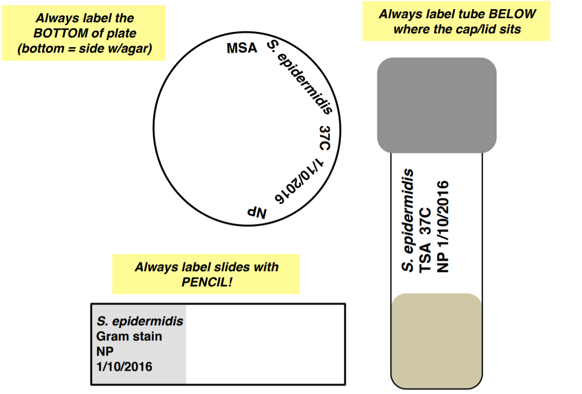
HOW TO LABEL SAMPLES:
In microbiology, it is important to properly label the media plates, broths, or slants PRIOR to inoculation with different microorganisms to avoid contamination.
Label your samples with a SHARPIE with the following information:
1. Your name or initials or group name
2. Date
3. Medium type
4. Organism name or abbreviation
5. Temperature that plate/tube will be incubated at
6. Miscellaneous info (antibiotic, time point, etc.)