
Tissues
- Page ID
- 23980

\( \newcommand{\vecs}[1]{\overset { \scriptstyle \rightharpoonup} {\mathbf{#1}} } \)
\( \newcommand{\vecd}[1]{\overset{-\!-\!\rightharpoonup}{\vphantom{a}\smash {#1}}} \)
\( \newcommand{\dsum}{\displaystyle\sum\limits} \)
\( \newcommand{\dint}{\displaystyle\int\limits} \)
\( \newcommand{\dlim}{\displaystyle\lim\limits} \)
\( \newcommand{\id}{\mathrm{id}}\) \( \newcommand{\Span}{\mathrm{span}}\)
( \newcommand{\kernel}{\mathrm{null}\,}\) \( \newcommand{\range}{\mathrm{range}\,}\)
\( \newcommand{\RealPart}{\mathrm{Re}}\) \( \newcommand{\ImaginaryPart}{\mathrm{Im}}\)
\( \newcommand{\Argument}{\mathrm{Arg}}\) \( \newcommand{\norm}[1]{\| #1 \|}\)
\( \newcommand{\inner}[2]{\langle #1, #2 \rangle}\)
\( \newcommand{\Span}{\mathrm{span}}\)
\( \newcommand{\id}{\mathrm{id}}\)
\( \newcommand{\Span}{\mathrm{span}}\)
\( \newcommand{\kernel}{\mathrm{null}\,}\)
\( \newcommand{\range}{\mathrm{range}\,}\)
\( \newcommand{\RealPart}{\mathrm{Re}}\)
\( \newcommand{\ImaginaryPart}{\mathrm{Im}}\)
\( \newcommand{\Argument}{\mathrm{Arg}}\)
\( \newcommand{\norm}[1]{\| #1 \|}\)
\( \newcommand{\inner}[2]{\langle #1, #2 \rangle}\)
\( \newcommand{\Span}{\mathrm{span}}\) \( \newcommand{\AA}{\unicode[.8,0]{x212B}}\)
\( \newcommand{\vectorA}[1]{\vec{#1}} % arrow\)
\( \newcommand{\vectorAt}[1]{\vec{\text{#1}}} % arrow\)
\( \newcommand{\vectorB}[1]{\overset { \scriptstyle \rightharpoonup} {\mathbf{#1}} } \)
\( \newcommand{\vectorC}[1]{\textbf{#1}} \)
\( \newcommand{\vectorD}[1]{\overrightarrow{#1}} \)
\( \newcommand{\vectorDt}[1]{\overrightarrow{\text{#1}}} \)
\( \newcommand{\vectE}[1]{\overset{-\!-\!\rightharpoonup}{\vphantom{a}\smash{\mathbf {#1}}}} \)
\( \newcommand{\vecs}[1]{\overset { \scriptstyle \rightharpoonup} {\mathbf{#1}} } \)
\(\newcommand{\longvect}{\overrightarrow}\)
\( \newcommand{\vecd}[1]{\overset{-\!-\!\rightharpoonup}{\vphantom{a}\smash {#1}}} \)
\(\newcommand{\avec}{\mathbf a}\) \(\newcommand{\bvec}{\mathbf b}\) \(\newcommand{\cvec}{\mathbf c}\) \(\newcommand{\dvec}{\mathbf d}\) \(\newcommand{\dtil}{\widetilde{\mathbf d}}\) \(\newcommand{\evec}{\mathbf e}\) \(\newcommand{\fvec}{\mathbf f}\) \(\newcommand{\nvec}{\mathbf n}\) \(\newcommand{\pvec}{\mathbf p}\) \(\newcommand{\qvec}{\mathbf q}\) \(\newcommand{\svec}{\mathbf s}\) \(\newcommand{\tvec}{\mathbf t}\) \(\newcommand{\uvec}{\mathbf u}\) \(\newcommand{\vvec}{\mathbf v}\) \(\newcommand{\wvec}{\mathbf w}\) \(\newcommand{\xvec}{\mathbf x}\) \(\newcommand{\yvec}{\mathbf y}\) \(\newcommand{\zvec}{\mathbf z}\) \(\newcommand{\rvec}{\mathbf r}\) \(\newcommand{\mvec}{\mathbf m}\) \(\newcommand{\zerovec}{\mathbf 0}\) \(\newcommand{\onevec}{\mathbf 1}\) \(\newcommand{\real}{\mathbb R}\) \(\newcommand{\twovec}[2]{\left[\begin{array}{r}#1 \\ #2 \end{array}\right]}\) \(\newcommand{\ctwovec}[2]{\left[\begin{array}{c}#1 \\ #2 \end{array}\right]}\) \(\newcommand{\threevec}[3]{\left[\begin{array}{r}#1 \\ #2 \\ #3 \end{array}\right]}\) \(\newcommand{\cthreevec}[3]{\left[\begin{array}{c}#1 \\ #2 \\ #3 \end{array}\right]}\) \(\newcommand{\fourvec}[4]{\left[\begin{array}{r}#1 \\ #2 \\ #3 \\ #4 \end{array}\right]}\) \(\newcommand{\cfourvec}[4]{\left[\begin{array}{c}#1 \\ #2 \\ #3 \\ #4 \end{array}\right]}\) \(\newcommand{\fivevec}[5]{\left[\begin{array}{r}#1 \\ #2 \\ #3 \\ #4 \\ #5 \\ \end{array}\right]}\) \(\newcommand{\cfivevec}[5]{\left[\begin{array}{c}#1 \\ #2 \\ #3 \\ #4 \\ #5 \\ \end{array}\right]}\) \(\newcommand{\mattwo}[4]{\left[\begin{array}{rr}#1 \amp #2 \\ #3 \amp #4 \\ \end{array}\right]}\) \(\newcommand{\laspan}[1]{\text{Span}\{#1\}}\) \(\newcommand{\bcal}{\cal B}\) \(\newcommand{\ccal}{\cal C}\) \(\newcommand{\scal}{\cal S}\) \(\newcommand{\wcal}{\cal W}\) \(\newcommand{\ecal}{\cal E}\) \(\newcommand{\coords}[2]{\left\{#1\right\}_{#2}}\) \(\newcommand{\gray}[1]{\color{gray}{#1}}\) \(\newcommand{\lgray}[1]{\color{lightgray}{#1}}\) \(\newcommand{\rank}{\operatorname{rank}}\) \(\newcommand{\row}{\text{Row}}\) \(\newcommand{\col}{\text{Col}}\) \(\renewcommand{\row}{\text{Row}}\) \(\newcommand{\nul}{\text{Nul}}\) \(\newcommand{\var}{\text{Var}}\) \(\newcommand{\corr}{\text{corr}}\) \(\newcommand{\len}[1]{\left|#1\right|}\) \(\newcommand{\bbar}{\overline{\bvec}}\) \(\newcommand{\bhat}{\widehat{\bvec}}\) \(\newcommand{\bperp}{\bvec^\perp}\) \(\newcommand{\xhat}{\widehat{\xvec}}\) \(\newcommand{\vhat}{\widehat{\vvec}}\) \(\newcommand{\uhat}{\widehat{\uvec}}\) \(\newcommand{\what}{\widehat{\wvec}}\) \(\newcommand{\Sighat}{\widehat{\Sigma}}\) \(\newcommand{\lt}{<}\) \(\newcommand{\gt}{>}\) \(\newcommand{\amp}{&}\) \(\definecolor{fillinmathshade}{gray}{0.9}\)1. Description of Tissues.
The structure of living organisms is organized in a hierarchical, step-wise fashion. Small units make up larger units, that contribute to still larger units. For the purpose of this tutorial, the levels of organization from atom to the organism will be discussed.
Hierarchy of Organization of Life
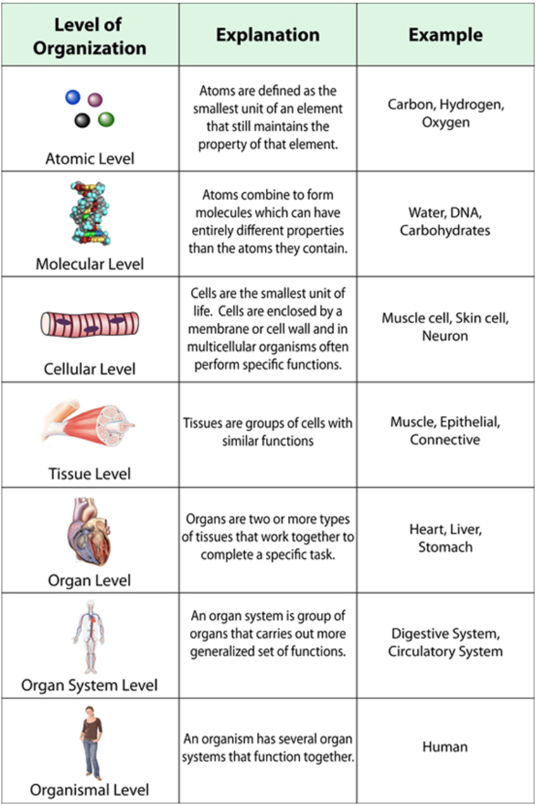
Figure \(\PageIndex{1}\). (CC BY-NC-SA)
4 Tissue Types
Although our body parts are diverse in both structure and function, they are all constructed from four basic tissue types:
1. Epithelial tissue: Covers the body, lines the cavities of the body and composes the glands.
2. Connective tissue: Connects and supports the structures of the body, providing structural support and binding organs together.
3. Muscular tissue: Has the unique capability to contract or shorten, provides movement and heat for the body.
4. Nervous tissue: Composed of specialized cells that respond to the environment by detecting, processing and coordinating information.
Epithelial Tissue
Epithelial tissue is composed of tightly connected cells arranged in one or more layers. Epithelial tissues covers the whole surface of the body (it's your skin!) as well as lining all cavities and forming glands. Epithelial tissues as many functions including protection, sensation, diffusion, secretion, absorption and excretion.
There are 8 different epithelial tissues types and they can be divided into two groups, depending on the number of layers in the tissue. Epithelial tissue that is only one cell thick is called simple epithelium. If the tissue is two or more cells thick it is known as stratified epithelium. The shape of the topmost layer of cells determines the name of stratified epithelium.
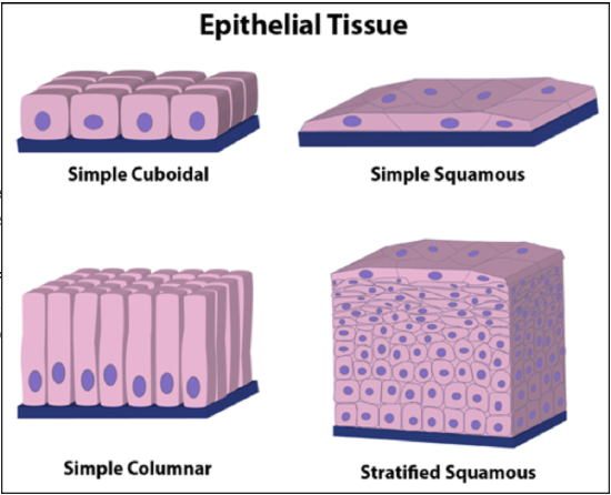
Figure \(\PageIndex{2}\). (CC BY-NC-SA)
Epithelial tissue, regardless of the type, is separated from the underlying tissue by a thin sheet of connective tissue, known as the basement membrane (pictured in blue in the image on the right.) The basement membrane provides structure and support for the epithelium and binds it to underlying structures.
Listed below are descriptions of the 8 different epithelial types. See the image on the right for a few examples of what these tissue types look like.
1. Simple squamous: A single layer of squamous cells that are thin, flat cells that resemble fried eggs. They form the lining of cavities such as the mouth, blood vessels and lungs.
2. Simple cuboidal: One layer of cuboidal cells which are plump cells that are roughly square or cube-like in shape. These cells are found in glands, duct and portions of the kidney tubules.
3. Simple columnar: A single layer of tall, skinny cells (column shaped) that are found in places like the lining of the intestine and gallbladder.
4. Pseudostratified columnar: This type of epithelium appears to be composed of layers of cells, but is in fact composed of just a single layer of cells, as each cell touches the basement membrane. Pseudostratified columnar cells line the nasal cavity, bronchi and trachea.
5. Stratified squamous: Many layers of cells are present, the topmost layer is made up of squamous cells. Stratified squamous cells are often described as looking like "piles of tiles." This is the type of epithelium that makes up the skin surface and lining of the mouth, through and esophagus.
6. Stratified columnar: Many layers of cells, the topmost layer is made up of columnar cells. Epithelium of this type is found in the mammary ducts and epididymus.
7. Transitional: Multiple layers of cells, but surface cells change from rounded to flat to permit expansion when needed. Transitional epithelium is found in the urinary bladder, renal pelvis and ureters.
8. Glandular: Columnar and cuboidal cells often become specialized as gland cells which are capable of secreting substances such as enzymes, hormones, mucus, sweat and saliva. Examples include the salivary, sweat and adrenal glands.
The most diverse and abundant of tissue, connective tissue holds cells together and supports the body. Connective tissue is made up of cells suspended in a noncellular matrix. The matrix (also known as ground substance) is secreted by the connective tissue cells and determines the characteristics of the connective tissue. It is the consistency of the matrix that determines the function of the connective tissue. The matrix can be liquid, gel-like or solid, all depending on the type of connective tissue.
Fibroblast cells are responsible for synthesizing protein fibers for the matrix. Collagen fibers are strong, elastic fibers are flexible and reticular fibers form a supportive framework for organs and basement membranes.
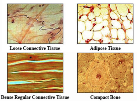
Figure \(\PageIndex{3}\). (CC BY-NC-SA)
Types of connective tissue include:
1. Loose connective tissue: Thin and soft, this tissue contains many collagen and elastic fibers in a jell-like matrix. The cells in loose connective tissue are not close together. This tissue functions in binding the skin to underlying structures.
2. Dense connective tissue: This tissue consists of two categories, dense irregular connective tissue and dense regular connective tissue which differ on the arrangement of the fibrous elements of the extracellular matrix. Dense irregular connective tissuecontains collage and elastic fibers which are found running in all different directions and planes. The dermis of the skin is composed of dense irregular connective tissue. Dense regular connective tissue has extracellular fibers that all run in the same direction and plane. Muscle tendons are a type of dense regular connective tissue.
3. Elastic connective tissue: Made up of freely branching elastic fibers with fibroblasts in the spaces between the fibers, this tissue allows the kind of stretch that is found in the walls of arteries.
4. Blood: Considered a fluid connective tissues because the matrix of blood is not a solid. The fluid matrix is called plasma, and formed elements of this tissue include white blood cells, red blood cells and platelets.
5. Cartilage: This connective tissue is relatively solid and is a non-vascularized tissue (does not have a blood supply). There are three types of cartilage: hyaline cartilage, elastic cartilage and fibrocartilage. Hyaline cartilage is the most common type of cartilage, contains many collagen fibers and is found in many places including the nose, betwween the ribs and the sternum and in the rings of the trachea. Elastic cartilage has many elastic fibers in the matrix and support the shape of hte ears and forms part of the larynx. Fibrocartilage is tough and contains many collagen fibers and is responsible for cushioning the knee joint and for forming the disks between the vertebrae.
6. Bone: A hard, mineralized tissue found in the skeleton. The bone matrix contains many collagen fibers as well as inorganic mineral salts, calcium carbonate and calcium phosphate, all features that make it a very rigid structure. Bone cells, called osteocytes, secrete the osteoid substance that eventuallys hardens around the cells to form an ossified matrix. The osteon forms the basic unit of compact bone.
7. Adipose tissue: Commonly known as fat, this tissue is related loose connective tissue. Adipose tissue contains fat cells which are specialized for lipid storage. In addition to storing energy, this tissue also cushions and protects the organs.
Muscular Tissue
Muscle tissue is characterized by the ability to contract when stimulated. When muscle cells contract, they get shorter, generating force and often movement. Muscle cells contain filaments of two proteins: actin and myosin. When these two filaments slide past each other, the muscle contracts.
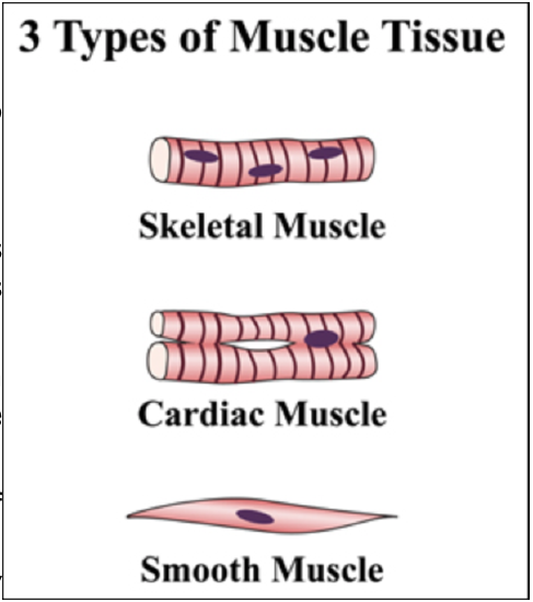
Figure \(\PageIndex{4}\). (CC BY-NC-SA)
There are three different types of muscle tissue:
1. Skeletal muscle: This tissue is composed of long, multinucleate cells with visible striations. Skeletal muscle allows movement by being attached to bones in the body. Because skeletal muscle contraction is consciously controlled, it is known as a voluntary muscle.
2. Smooth muscle: Composed of short, cylindrical cells that taper at the ends, smooth muscles are commonly involved in involuntary motions. Involuntary muscle contractions are not consciously controlled and occurs in places like the digestive tract an in the walls of blood vessels.
3. Cardiac (heart) muscle: Cardiac muscle tissue contains short, branched, striated cells, with one nucleus at the center of each cell. Cardiac muscle cells within a fiber are joined to their neighbors by intercalated discs. These specialized communication junctions facilitate the heart beat by transmitting the signal to contract. Cardiac muscles are also involuntary muscles.
Nervous Tissue
Nervous tissue responds to changes in the environment and conducts impulses to various organs in the body to respond to these changes. Nervous tissue is found in the brain, spinal cord and peripheral nerves that branch throughout the body.
Nervous tissue contains two types of cells:
1. Neuroglia: These cells do not send or receive electrical impulses, but instead function to support neurons. They have important functions such as providing physical support, providing nutrients, removing debris and providing electrical insulation.
2. Neurons: These are the cells that carry electrical impulses. There are three main types of neurons, which are classified by their function. Sensory neurons conduct impulses from the sensory organs (eyes, nose, ears, etc) to the central nervous system (brain and spinal cord). Motor neurons are responsible for conducting impulses from the central nervous system to the effector organs (muscles and glands). Finally, interneurons are those neurons that connect sensory neurons to motor neurons.l
Important Structures of a Motor Neuron
1. Cell body: Enclosed by a plasma membrane and central nucleus, the cell body is responsible for producing all of the proteins for the dendrites, axons and synaptic terminals. The nucleus of the neuron is located in the cell body.
2. Dendrites: These structures branch out from the cell body in a tree-like fashion. Dendrites function in receiving signals from other nerve cells.
3. Axon: The axon is the main conducting unit of the neuron and is capable of conveying electrical signals over long and short distances. The axon transmits impulses from the cell body to other cells. Individual neurons may have many dendrites, but each neuron has only one axon.
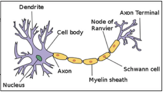
Figure \(\PageIndex{5}\). (CC BY-NC-SA)

Tissues Tutorial by Dr. Katherine Harris is licensed under a Creative Commons Attribution-NonCommercial-ShareAlike 3.0 Unported License.
This tutorial was funded by the Title V-STEM Grant #P031S090007.