
1.9: Mitosis and Meiosis
- Page ID
- 24112

\( \newcommand{\vecs}[1]{\overset { \scriptstyle \rightharpoonup} {\mathbf{#1}} } \)
\( \newcommand{\vecd}[1]{\overset{-\!-\!\rightharpoonup}{\vphantom{a}\smash {#1}}} \)
\( \newcommand{\dsum}{\displaystyle\sum\limits} \)
\( \newcommand{\dint}{\displaystyle\int\limits} \)
\( \newcommand{\dlim}{\displaystyle\lim\limits} \)
\( \newcommand{\id}{\mathrm{id}}\) \( \newcommand{\Span}{\mathrm{span}}\)
( \newcommand{\kernel}{\mathrm{null}\,}\) \( \newcommand{\range}{\mathrm{range}\,}\)
\( \newcommand{\RealPart}{\mathrm{Re}}\) \( \newcommand{\ImaginaryPart}{\mathrm{Im}}\)
\( \newcommand{\Argument}{\mathrm{Arg}}\) \( \newcommand{\norm}[1]{\| #1 \|}\)
\( \newcommand{\inner}[2]{\langle #1, #2 \rangle}\)
\( \newcommand{\Span}{\mathrm{span}}\)
\( \newcommand{\id}{\mathrm{id}}\)
\( \newcommand{\Span}{\mathrm{span}}\)
\( \newcommand{\kernel}{\mathrm{null}\,}\)
\( \newcommand{\range}{\mathrm{range}\,}\)
\( \newcommand{\RealPart}{\mathrm{Re}}\)
\( \newcommand{\ImaginaryPart}{\mathrm{Im}}\)
\( \newcommand{\Argument}{\mathrm{Arg}}\)
\( \newcommand{\norm}[1]{\| #1 \|}\)
\( \newcommand{\inner}[2]{\langle #1, #2 \rangle}\)
\( \newcommand{\Span}{\mathrm{span}}\) \( \newcommand{\AA}{\unicode[.8,0]{x212B}}\)
\( \newcommand{\vectorA}[1]{\vec{#1}} % arrow\)
\( \newcommand{\vectorAt}[1]{\vec{\text{#1}}} % arrow\)
\( \newcommand{\vectorB}[1]{\overset { \scriptstyle \rightharpoonup} {\mathbf{#1}} } \)
\( \newcommand{\vectorC}[1]{\textbf{#1}} \)
\( \newcommand{\vectorD}[1]{\overrightarrow{#1}} \)
\( \newcommand{\vectorDt}[1]{\overrightarrow{\text{#1}}} \)
\( \newcommand{\vectE}[1]{\overset{-\!-\!\rightharpoonup}{\vphantom{a}\smash{\mathbf {#1}}}} \)
\( \newcommand{\vecs}[1]{\overset { \scriptstyle \rightharpoonup} {\mathbf{#1}} } \)
\(\newcommand{\longvect}{\overrightarrow}\)
\( \newcommand{\vecd}[1]{\overset{-\!-\!\rightharpoonup}{\vphantom{a}\smash {#1}}} \)
\(\newcommand{\avec}{\mathbf a}\) \(\newcommand{\bvec}{\mathbf b}\) \(\newcommand{\cvec}{\mathbf c}\) \(\newcommand{\dvec}{\mathbf d}\) \(\newcommand{\dtil}{\widetilde{\mathbf d}}\) \(\newcommand{\evec}{\mathbf e}\) \(\newcommand{\fvec}{\mathbf f}\) \(\newcommand{\nvec}{\mathbf n}\) \(\newcommand{\pvec}{\mathbf p}\) \(\newcommand{\qvec}{\mathbf q}\) \(\newcommand{\svec}{\mathbf s}\) \(\newcommand{\tvec}{\mathbf t}\) \(\newcommand{\uvec}{\mathbf u}\) \(\newcommand{\vvec}{\mathbf v}\) \(\newcommand{\wvec}{\mathbf w}\) \(\newcommand{\xvec}{\mathbf x}\) \(\newcommand{\yvec}{\mathbf y}\) \(\newcommand{\zvec}{\mathbf z}\) \(\newcommand{\rvec}{\mathbf r}\) \(\newcommand{\mvec}{\mathbf m}\) \(\newcommand{\zerovec}{\mathbf 0}\) \(\newcommand{\onevec}{\mathbf 1}\) \(\newcommand{\real}{\mathbb R}\) \(\newcommand{\twovec}[2]{\left[\begin{array}{r}#1 \\ #2 \end{array}\right]}\) \(\newcommand{\ctwovec}[2]{\left[\begin{array}{c}#1 \\ #2 \end{array}\right]}\) \(\newcommand{\threevec}[3]{\left[\begin{array}{r}#1 \\ #2 \\ #3 \end{array}\right]}\) \(\newcommand{\cthreevec}[3]{\left[\begin{array}{c}#1 \\ #2 \\ #3 \end{array}\right]}\) \(\newcommand{\fourvec}[4]{\left[\begin{array}{r}#1 \\ #2 \\ #3 \\ #4 \end{array}\right]}\) \(\newcommand{\cfourvec}[4]{\left[\begin{array}{c}#1 \\ #2 \\ #3 \\ #4 \end{array}\right]}\) \(\newcommand{\fivevec}[5]{\left[\begin{array}{r}#1 \\ #2 \\ #3 \\ #4 \\ #5 \\ \end{array}\right]}\) \(\newcommand{\cfivevec}[5]{\left[\begin{array}{c}#1 \\ #2 \\ #3 \\ #4 \\ #5 \\ \end{array}\right]}\) \(\newcommand{\mattwo}[4]{\left[\begin{array}{rr}#1 \amp #2 \\ #3 \amp #4 \\ \end{array}\right]}\) \(\newcommand{\laspan}[1]{\text{Span}\{#1\}}\) \(\newcommand{\bcal}{\cal B}\) \(\newcommand{\ccal}{\cal C}\) \(\newcommand{\scal}{\cal S}\) \(\newcommand{\wcal}{\cal W}\) \(\newcommand{\ecal}{\cal E}\) \(\newcommand{\coords}[2]{\left\{#1\right\}_{#2}}\) \(\newcommand{\gray}[1]{\color{gray}{#1}}\) \(\newcommand{\lgray}[1]{\color{lightgray}{#1}}\) \(\newcommand{\rank}{\operatorname{rank}}\) \(\newcommand{\row}{\text{Row}}\) \(\newcommand{\col}{\text{Col}}\) \(\renewcommand{\row}{\text{Row}}\) \(\newcommand{\nul}{\text{Nul}}\) \(\newcommand{\var}{\text{Var}}\) \(\newcommand{\corr}{\text{corr}}\) \(\newcommand{\len}[1]{\left|#1\right|}\) \(\newcommand{\bbar}{\overline{\bvec}}\) \(\newcommand{\bhat}{\widehat{\bvec}}\) \(\newcommand{\bperp}{\bvec^\perp}\) \(\newcommand{\xhat}{\widehat{\xvec}}\) \(\newcommand{\vhat}{\widehat{\vvec}}\) \(\newcommand{\uhat}{\widehat{\uvec}}\) \(\newcommand{\what}{\widehat{\wvec}}\) \(\newcommand{\Sighat}{\widehat{\Sigma}}\) \(\newcommand{\lt}{<}\) \(\newcommand{\gt}{>}\) \(\newcommand{\amp}{&}\) \(\definecolor{fillinmathshade}{gray}{0.9}\)By Dr. Leah Howell, Dalton State College
Introduction:
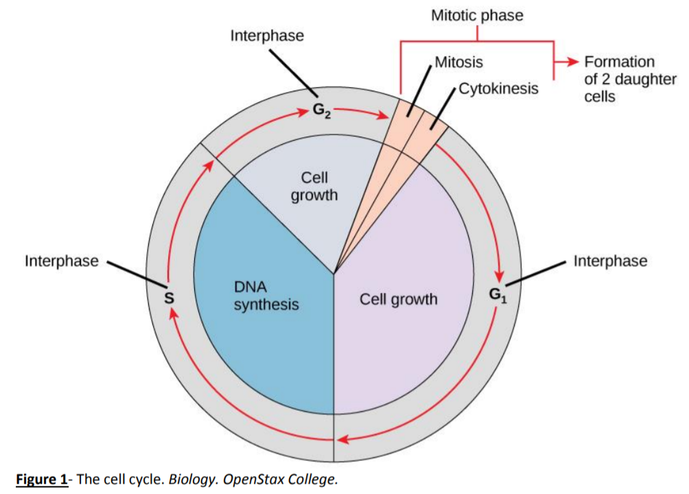
All cells come from preexisting cells and eukaryotic cells must undergo mitosis in order to form new cells. The replication of a cell is part of the overall cell cycle (Figure 1) which is composed of interphase and M phase (mitotic phase). M phase, which consists of mitosis and cytokinesis, is the portion of the cell cycle where the cell divides, reproducing itself. Mitosis is the division of the nucleus and its contents. In mitosis, DNA which has been copied in the S phase of interphase is separated into two individual copies. Each copy will end up in its own cell at the end of M phase. Mitosis has several steps: prophase, prometaphase, metaphase, anaphase, and telophase (Figure 2). The spindle fibers, which are formed by the cell as mitosis progresses, are used to attach to chromosomes, align them down the middle of the cell, and pull chromosomes apart into their identical individual chromatids which will end up in separate cells. As mitosis is nearing its end and the cell is in telophase, the cytoplasm also divides so that both new cells will have their own fluid, organelles, etc. This division of the cytoplasm is called cytokinesis. Mitosis and cytokinesis can be viewed under a microscope.

Exercise 1: Mitosis of Onion Root Tip
(Adapted from Cell Biology Laboratory Manual Online Dr. William H. Heidcamp, Biology Department, Gustavus Adolphus College, St. Peter, MN 56082 -- cellab@gac.edu)
Materials:
- Prepared slide of onion (allium) root tip
- Microscope
Procedure:
- Obtain a slide of allium root tip for observation of the stages of mitosis in a plant cell.
- Examine the slide under a microscope.
- Draw and label all stages of mitosis below. Figure 3 can be used to help with this.
Interphase Prophase Metaphase
Anaphase Telophase and Cytokinesis

Exercise 2: Mitosis of Whitefish Blastula
(Adapted from Cell Biology Laboratory Manual Online Dr. William H. Heidcamp, Biology Department, Gustavus Adolphus College, St. Peter, MN 56082 -- cellab@gac.edu)
Materials:
- Prepared slide of whitefish blastula
- Microscope
Procedure:
- Obtain a slide of a whitefish blastula for observation of the stages of mitosis in an animal cell. Since early embryogenesis involves rapid cellular division, the whitefish blastula has long served as a model of mitotic division in animals. It also has the advantage of demonstrating clear spindle formation in the cytoplasm.
- Examine the slide under a microscope.
- Draw and label all stages of mitosis below.
Interphase Prophase Metaphase
Anaphase Telophase and Cytokinesis
Exercise 3: Simulating Mitosis Using Beads
Materials:
- Baggy full of beads and strings
Procedure:
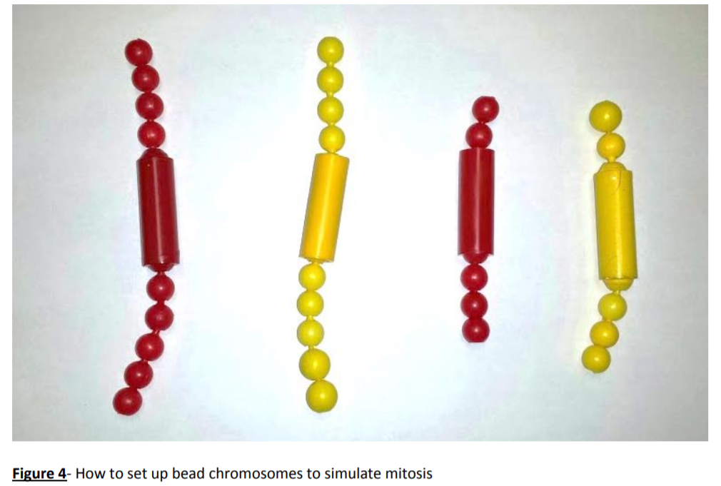
- Organize beads into “chromosomes” as shown in Figure 4.
- Simulate the steps of interphase (specifically S phase) and then M phase using the beads. Hint: The chromosomes in Figure 4 have not been through S phase yet, so you will eventually need more beads than shown in Figure 4. The strings in the bag are used to simulate spindle fibers.
Exercise 4: Nondisjunction Events During Meiosis
Failure of chromosomes to separate during mitosis or meiosis will result in an incorrect number of chromosomes in daughter cells. This occurrence is known as nondisjunction, and it is often triggered by a lapse during a mitotic checkpoint. Should nondisjunction occur during meiosis, the resulting egg or sperm cell will have an incorrect number of chromosomes; if this sex cell is then fertilized, the fetus will have a chromosomal abnormality. The term given for having an incorrect number of chromosomes is aneuploidy. A common type of aneuploidy is trisomy, which is when there are 3 copies of a particular chromosome instead of 2. Several common chromosomal abnormalities are listed in the table below. The most common trisomy that a human can survive is Down syndrome, which occurs at chromosome 21.
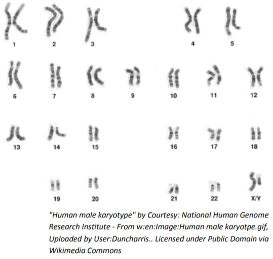
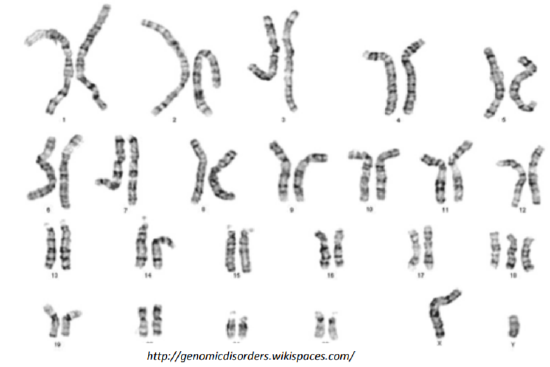
To diagnose a chromosomal abnormality, doctors use a map of the chromosomes known as a karyotype. Each chromosome pair is laid out side-by-side so it is relatively easy to determine if there are any irregularities. Referring to the karyotype below, it is clear that each chromosome pair is present and of relatively equal length. Note that last chromosome pair (23) is labeled X/Y; these chromosomes are the only 2 that do not exactly match.
| Chromosome Pair Affected | Type | Diagnosis |
| 13 | Trisomy | Patau Syndrome |
| 18 | Trisomy | Edward Syndrome |
| 21 | Trisomy | Down Syndrome |
| 23 (XO) | Monosomy | Turner Syndrome |
| 23 (XXY) | Trisomy | Klinefelter Syndrome |
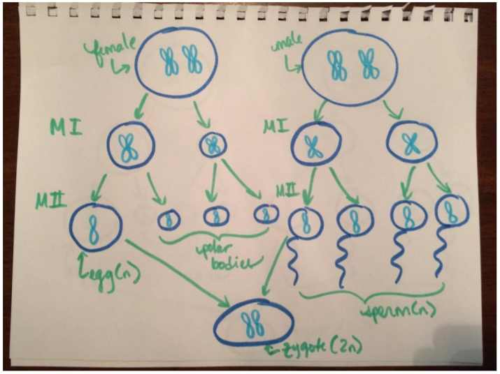
In order for any aneuploidy to occur, there must be an error during meiosis I or II. In the image at right, meiosis occurs without error and the resulting gametes are haploid, leading to a diploid zygote.
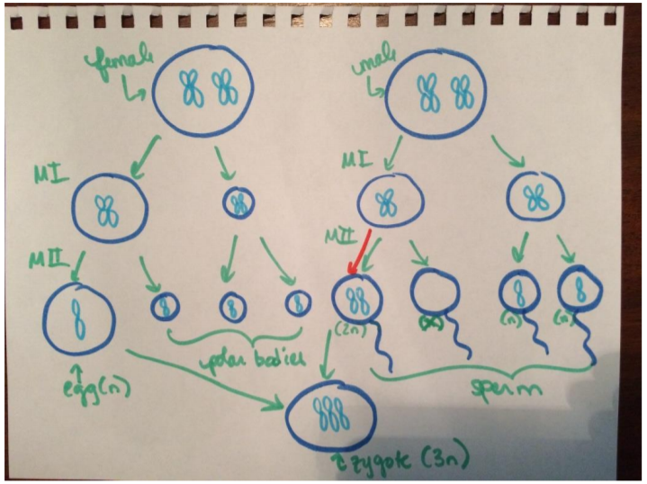
In the next image, a nondisjunction event occurs during meiosis II, resulting in trisomy in the zygote.
Questions:
1. Most nerve cells in the adult human central nervous system, as well as heart muscle cells, do not divide. In contrast, cells lining the inside of the small intestine divide frequently. Discuss this difference in terms of why damage to the nervous system and heart muscle cells (think stroke or heart attack) is so dangerous. What do you think might happen to tissues such as the intestinal lining if a disorder blocked mitotic cell division in all cells of the body?
2. How do mitosis and cytokinesis differ?
3. Ultimately, is it the paternal or maternal gamete that determines sex? Explain.
4. In what way are the 23 pairs of human chromosomes “matched” pairs of chromosomes?
5. In order to make a karyotype, cell division is arrested at a point when the chromosomes have condensed and the nuclear envelope has disappeared, but before the sister chromatids separate. Which stage of the cell cycle would be a good point to perform a karyotype?
6. Imagine you are an obstetrician and are performing early genetic testing on a 10-week old fetus. Below is the resulting karyotype. What can you tell about the fetus?
7. Look up the prognosis for any chromosomal abnormalities you may have detected. What can the parents expect?
8. Use the space below to draw out meiotic divisions that could result in trisomy, assuming that the error occurred during meiosis I.