
15.3: Spinal Nerves
- Page ID
- 53740

Spinal Nerves

Above: Position and numbering of the cervical spinal nerves.
There are 31 pairs (right and left) of spinal nerves:
- 8 pairs of cervical spinal nerves
- 12 pairs of thoracic spinal nerves
- 5 pairs of lumbar spinal nerves
- 5 pairs of sacral spinal nerves
- 1 pair of coccygeal spinal nerves
All spinal nerves, except for the first pair and last six pairs, exit the vertebral column through intervertebral foramina. The first pair of spinal nerves (C1 spinal nerves) exit the vertebral column between the occipital bone and atlas (C1).
The ventral and dorsal rami of sacral nerves 1- 4 exit the vertebral column through the anterior and posterior sacral foramina respectively, and the 5th pair of sacral nerves and the one pair coccygeal nerves exits the vertebral column through the sacral hiatus

Above: Branches of spinal nerves. Blue represents sensory neurons and red represents motor neurons.
A plexus is a network of nerves. The three biggest nerve plexuses in the body are the cervical plexus (paired, right and left) which serves the head and neck, the brachial plexus (paired, right and left) which serves the upper limb, and the lumbosacral plexus (paired, right and left) which serves the pelvis, genitalia, and lower limb.

Above: Diagram numbering each spinal nerve (those with "n." after are the spinal nerves) and the nerve plexuses.
Cervical Plexus
The cervical plexus is a plexus of the ventral rami of the first four cervical spinal nerves (C1 through C4). They are located laterally to the transverse processes between prevertebral muscles from the medial side and vertebral from lateral side. There is anastomosis (cross-connection) with accessory nerve, hypoglossal nerve and sympathetic trunk.
It is located in the neck, deep to sternocleidomastoid. Nerves formed from the cervical plexus innervate the back of the head, as well as some neck muscles. The branches of the cervical plexus emerge from the posterior triangle at the nerve point, a point which lies midway on the posterior border of the sternocleidomastoid. Also from the dorsal ramus of C2 greater occipital nerve arises.

Above: Nerves of the cervical plexus shown with some of cranial nerves (C.N. IX glossopharyngeal nerve, C.N. X vagus nerve, and XII hypoglossal nerve).
The cervical plexus has two types of branches: cutaneous and muscular.
|
Nerve Name |
Nerves Involved |
Innervation |
|---|---|---|
|
Cutaneous branch |
||
|
greater auricular nerve |
ventral rami C2 and C3 spinal nerves |
Skin near the outer ear and external acoustic meatus (ear canal) |
|
lesser occipital nerve |
ventral rami of C2 spinal nerve |
Skin and the scalp posterosuperior to the outer ear |
|
supraclavicular nerves (lateral, intermediate, and medial) |
ventral rami C3 and C4 spinal nerves |
Skin below the clavicle |
|
transverse cervical nerve |
ventral rami C2 and C3 spinal nerves |
Anterior region of the neck |
|
Muscular branch |
||
|
ansa cervicalis nerve |
A loop formed from ventral rami of C1, C2, and C3 spinal nerves |
thyrohyoid, sternothyroid, sternohyoid, and omohyoid muscles |
|
phrenic nerve |
ventral rami of C3 through C5 spinal nerves |
Diaphragm, pericardium |
|
segmental branches |
ventral rami of C1 through C4 spinal nerves |
Anterior and middle scalenes muscles |
Brachial Plexus

Above: (Left) Diagram and (right) cadaver image of the right brachial plexus, anterior view

Above: Nerves of the upper limb (left) anterior left upper limb and (right) posterior right upper limb.
The brachial plexus is divided into roots, trunks, divisions, cords, and branches:
- roots: ventral roots of spinal nerves C5, C6, C7, C8, and T1
- trunks: superior, middle, and inferior trunks
- divisions: each of the superior, middle, and inferior trunks have an anterior division and a posterior division (6 divisions total)
- cords:
- lateral cord: the anterior divisions from the superior and middle trunks combine to form the lateral cord
- posterior cord: the posterior divisions from the superior, middle, and inferior trunks combine to form the posterior cord
- medial cord: comprised of the anterior division of the inferior trunk
- branches: musculocutaneous nerve, axillary nerve, radial nerve, median nerve, and ulnar nerve

Above: Diagram of the structure of the brachial plexus.
The terminal branches of the brachial plexus are the nerves that will innervate the muscles of the upper limb. Some muscles are listed under the nerve that innervates them.
|
Nerve Name |
Nerves Involved |
Cutaneous Innervation |
Muscular Innervation |
|---|---|---|---|
|
axillary nerve |
ventral rami of C5 and C6 spinal nerves |
skin of the shoulder |
deltoid and teres minor muscles |
|
dorsal scapular nerve |
ventral rami of C5 spinal nerve |
-- |
levator scapulae, rhomboid major, and rhomboid minor muscles |
|
long thoracic nerve |
ventral rami of C5 through C7 spinal nerve |
-- |
serratus anterior muscles |
|
median nerve |
ventral rami of C5 through T1 spinal nerves |
some skin of the hand |
anterior forearm flexor, palm, and digit muscles |
|
musculocutaneous nerve |
ventral rami of C5 through C7 spinal nerves |
some skin of the forearm |
biceps brachii, brachialis, and coracobrachialis muscles |
|
pectoral nerve |
ventral rami of C5 through T1 spinal nerves |
-- |
pectoralis major and pectoralis minor muscles |
|
radial nerve |
ventral rami of C5 through T1 spinal nerves |
posterolateral skin of the upper limb |
triceps brachii, brachioradialis, and posterior forearm muscles |
|
subscapular nerve |
ventral rami of C5 and C6 spinal nerves |
-- |
subscapularis and teres major muscles |
|
suprascapular nerve |
ventral rami of C5 and C6 spinal nerves |
-- |
supraspinatus and infraspinatus muscles |
|
ulnar nerve |
ventral rami of C8 and T1 spinal nerves |
some skin of the hand |
anterior forearm flexor and hand muscles |
Intercostal Nerves
Intercostal nerves are not part of a plexus; they are simply ventral rami from thoracic spinal nerves that travel directly to the region they innervate. Intercostal nerves are found between ribs and between the external and internal intercostal muscles (ventral rami of spinal nerves T1 through T11). Notice how there are only 11 intercostal nerves, but there are 12 pair of ribs. This is because for a nerve to be an intercostal nerve, it must be between two ribs. The 12th thoracic spinal nerve is not between two ribs; it is below the last rib, which is why this nerve is called the subcostal nerve.
Lumbosacral Plexus
The lumbosacral plexus involves ventral rami from spinal nerves T12 through S4.
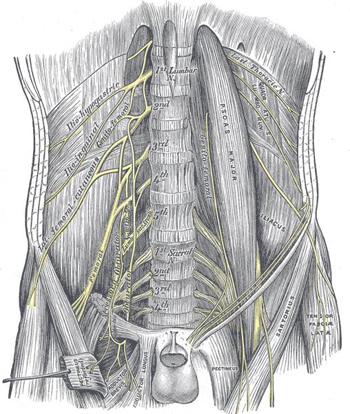
Above: Nerves of the lumbosacral plexus, anterior view.
|
Nerve Name |
Nerves Involved |
Cutaneous Innervation |
Muscular Innervation |
|---|---|---|---|
|
common fibular nerve (superficial and deep branches) |
ventral rami of L4 through S2 spinal nerves |
skin of the anterolateral leg and foot |
tibialis anterior, extensor digitorum longus, fibularis longus and fibularis brevis muscles |
|
femoral nerve |
ventral rami of L2 through L4 spinal nerves |
skin of the medial lower limb and anterior thigh |
iliacus, sartorius, pectineus, rectus femoris, vastus lateralis, vastus medialis, and vastus intermedius muscles |
|
genitofemoral nerve |
ventral rami of L1 and L2 spinal nerves |
skin of external genitalia |
cremasteric muscles (males only) |
|
iliohypogastric nerve |
ventral rami of L1 spinal nerves |
skin of the hip and inferior abdomen |
internal oblique and transversus abdominis muscles |
|
ilioinguinal nerve |
ventral rami of L1 spinal nerves |
skin of the medial thigh and external genitalia |
internal oblique and Transversus abdominis muscles |
|
inferior gluteal nerve |
ventral rami of L5 through S2 spinal nerves |
-- |
gluteus maximus muscles |
|
lateral femoral cutaneous nerve |
ventral rami of L2 and L3 spinal nerves |
skin of the anterolateral thigh and knee |
-- |
|
obturator nerve |
ventral rami of L2 through L |
skin of thigh (medial) |
gracilis, adductor magnus, adductor longus, and adductor brevis muscles |
|
posterior femoral cutaneous nerve |
ventral rami of S1 through S3 spinal nerves |
skin of the posterior lower limb |
-- |
|
pudendal nerve |
ventral rami of S2 through S |
skin of external genitalia |
external anal sphincter, external urethral sphincter, and pelvic muscles |
|
sciatic nerve (branches into tibial and common fibular nerves) |
ventral rami of L4 through S3 spinal nerves |
skin of the leg and foot |
semitendinosus, semimembranosus, and biceps femoris muscles |
|
superior gluteal nerve |
ventral rami of L4 through S1 spinal nerves |
-- |
gluteus medius, gluteus minimus, and tensor fasciae latae muscles |
|
tibial nerve |
ventral rami of L4 through S3 spinal nerves |
skin of the sole of the foot and posterior leg |
flexor digitorum longus, gastrocnemius, and soleus muscles |

Above: Arrangement of nerves in the right side of the lumbosacral plexus, anterior view.

Above: Nerves of the lower limb (left) anterior right lower limb and (right) posterior right lower limb.
Clinical Application: Epidural (Childbirth)
An epidural is a medical procedure commonly done to relieve pain during labor and delivery. In this procedure, a needle is inserted into the vertebral canal without puncturing the dura mater. With the tip of the needle/catheter above the dura mater (epi = above; dural= of the dura mater), anesthetics are injected into the epidural space. The anesthetics then pass through the meninges and block neuronal impulses, thus inhibiting the transmission of sensation (in this case, pain) to the brain. Just like in a lumbar puncture, the epidural needle can be inserted between L3/L4 vertebrae, but because the pain impulses from the uterus reach the spinal cord at levels between T10-L1, epidurals are more commonly placed more superiorly. A common location for epidural placement is T7, which is easily identifiable at about the level of the inferior border of the scapula. When the drugs are injected, gravity can pull the drugs downward, blocking everything below the epidural location. Therefore, administering an epidural above the level of T10 can relieve labor pain from uterine contractions as well as pain from the pelvic floor transmitted by the pudendal nerve (from S2, S3, and S4 spinal nerves).
Attributions
- "Anatomy 204L: Laboratory Manual (Second Edition)" by Ethan Snow, University of North Dakota is licensed under CC BY-NC 4.0
- "Anatomy and Physiology I Lab" by Victoria Vidal is licensed under CC BY 4.0
- "Cervical Spine Anterior View.png" by DrJanaOfficial is licensed under CC BY-SA 4.0
- "Cervical Spine Oblique View.png" by DrJanaOfficial is licensed under CC BY-SA 4.0
- "Gray's Anatomy plates" by Henry Vandyke Carte is in the Public Domain
- "Spinal nerve-es.svg" by Tristanb and Mysid is licensed under CC BY-SA 3.0