
14.5: Laboratory Activities and Assignment
- Page ID
- 53729

\( \newcommand{\vecs}[1]{\overset { \scriptstyle \rightharpoonup} {\mathbf{#1}} } \)
\( \newcommand{\vecd}[1]{\overset{-\!-\!\rightharpoonup}{\vphantom{a}\smash {#1}}} \)
\( \newcommand{\id}{\mathrm{id}}\) \( \newcommand{\Span}{\mathrm{span}}\)
( \newcommand{\kernel}{\mathrm{null}\,}\) \( \newcommand{\range}{\mathrm{range}\,}\)
\( \newcommand{\RealPart}{\mathrm{Re}}\) \( \newcommand{\ImaginaryPart}{\mathrm{Im}}\)
\( \newcommand{\Argument}{\mathrm{Arg}}\) \( \newcommand{\norm}[1]{\| #1 \|}\)
\( \newcommand{\inner}[2]{\langle #1, #2 \rangle}\)
\( \newcommand{\Span}{\mathrm{span}}\)
\( \newcommand{\id}{\mathrm{id}}\)
\( \newcommand{\Span}{\mathrm{span}}\)
\( \newcommand{\kernel}{\mathrm{null}\,}\)
\( \newcommand{\range}{\mathrm{range}\,}\)
\( \newcommand{\RealPart}{\mathrm{Re}}\)
\( \newcommand{\ImaginaryPart}{\mathrm{Im}}\)
\( \newcommand{\Argument}{\mathrm{Arg}}\)
\( \newcommand{\norm}[1]{\| #1 \|}\)
\( \newcommand{\inner}[2]{\langle #1, #2 \rangle}\)
\( \newcommand{\Span}{\mathrm{span}}\) \( \newcommand{\AA}{\unicode[.8,0]{x212B}}\)
\( \newcommand{\vectorA}[1]{\vec{#1}} % arrow\)
\( \newcommand{\vectorAt}[1]{\vec{\text{#1}}} % arrow\)
\( \newcommand{\vectorB}[1]{\overset { \scriptstyle \rightharpoonup} {\mathbf{#1}} } \)
\( \newcommand{\vectorC}[1]{\textbf{#1}} \)
\( \newcommand{\vectorD}[1]{\overrightarrow{#1}} \)
\( \newcommand{\vectorDt}[1]{\overrightarrow{\text{#1}}} \)
\( \newcommand{\vectE}[1]{\overset{-\!-\!\rightharpoonup}{\vphantom{a}\smash{\mathbf {#1}}}} \)
\( \newcommand{\vecs}[1]{\overset { \scriptstyle \rightharpoonup} {\mathbf{#1}} } \)
\( \newcommand{\vecd}[1]{\overset{-\!-\!\rightharpoonup}{\vphantom{a}\smash {#1}}} \)
\(\newcommand{\avec}{\mathbf a}\) \(\newcommand{\bvec}{\mathbf b}\) \(\newcommand{\cvec}{\mathbf c}\) \(\newcommand{\dvec}{\mathbf d}\) \(\newcommand{\dtil}{\widetilde{\mathbf d}}\) \(\newcommand{\evec}{\mathbf e}\) \(\newcommand{\fvec}{\mathbf f}\) \(\newcommand{\nvec}{\mathbf n}\) \(\newcommand{\pvec}{\mathbf p}\) \(\newcommand{\qvec}{\mathbf q}\) \(\newcommand{\svec}{\mathbf s}\) \(\newcommand{\tvec}{\mathbf t}\) \(\newcommand{\uvec}{\mathbf u}\) \(\newcommand{\vvec}{\mathbf v}\) \(\newcommand{\wvec}{\mathbf w}\) \(\newcommand{\xvec}{\mathbf x}\) \(\newcommand{\yvec}{\mathbf y}\) \(\newcommand{\zvec}{\mathbf z}\) \(\newcommand{\rvec}{\mathbf r}\) \(\newcommand{\mvec}{\mathbf m}\) \(\newcommand{\zerovec}{\mathbf 0}\) \(\newcommand{\onevec}{\mathbf 1}\) \(\newcommand{\real}{\mathbb R}\) \(\newcommand{\twovec}[2]{\left[\begin{array}{r}#1 \\ #2 \end{array}\right]}\) \(\newcommand{\ctwovec}[2]{\left[\begin{array}{c}#1 \\ #2 \end{array}\right]}\) \(\newcommand{\threevec}[3]{\left[\begin{array}{r}#1 \\ #2 \\ #3 \end{array}\right]}\) \(\newcommand{\cthreevec}[3]{\left[\begin{array}{c}#1 \\ #2 \\ #3 \end{array}\right]}\) \(\newcommand{\fourvec}[4]{\left[\begin{array}{r}#1 \\ #2 \\ #3 \\ #4 \end{array}\right]}\) \(\newcommand{\cfourvec}[4]{\left[\begin{array}{c}#1 \\ #2 \\ #3 \\ #4 \end{array}\right]}\) \(\newcommand{\fivevec}[5]{\left[\begin{array}{r}#1 \\ #2 \\ #3 \\ #4 \\ #5 \\ \end{array}\right]}\) \(\newcommand{\cfivevec}[5]{\left[\begin{array}{c}#1 \\ #2 \\ #3 \\ #4 \\ #5 \\ \end{array}\right]}\) \(\newcommand{\mattwo}[4]{\left[\begin{array}{rr}#1 \amp #2 \\ #3 \amp #4 \\ \end{array}\right]}\) \(\newcommand{\laspan}[1]{\text{Span}\{#1\}}\) \(\newcommand{\bcal}{\cal B}\) \(\newcommand{\ccal}{\cal C}\) \(\newcommand{\scal}{\cal S}\) \(\newcommand{\wcal}{\cal W}\) \(\newcommand{\ecal}{\cal E}\) \(\newcommand{\coords}[2]{\left\{#1\right\}_{#2}}\) \(\newcommand{\gray}[1]{\color{gray}{#1}}\) \(\newcommand{\lgray}[1]{\color{lightgray}{#1}}\) \(\newcommand{\rank}{\operatorname{rank}}\) \(\newcommand{\row}{\text{Row}}\) \(\newcommand{\col}{\text{Col}}\) \(\renewcommand{\row}{\text{Row}}\) \(\newcommand{\nul}{\text{Nul}}\) \(\newcommand{\var}{\text{Var}}\) \(\newcommand{\corr}{\text{corr}}\) \(\newcommand{\len}[1]{\left|#1\right|}\) \(\newcommand{\bbar}{\overline{\bvec}}\) \(\newcommand{\bhat}{\widehat{\bvec}}\) \(\newcommand{\bperp}{\bvec^\perp}\) \(\newcommand{\xhat}{\widehat{\xvec}}\) \(\newcommand{\vhat}{\widehat{\vvec}}\) \(\newcommand{\uhat}{\widehat{\uvec}}\) \(\newcommand{\what}{\widehat{\wvec}}\) \(\newcommand{\Sighat}{\widehat{\Sigma}}\) \(\newcommand{\lt}{<}\) \(\newcommand{\gt}{>}\) \(\newcommand{\amp}{&}\) \(\definecolor{fillinmathshade}{gray}{0.9}\)Laboratory Activities and Assignment
Part 1: Review of Brain Anatomy
1. Label the following structures on the diagrams below.
|
|
|

2. Label the following structures on the diagram below.
|
|
|

3. Label the following structures on the diagram below.
|
|
|
4. Label the following structures on the diagram below.
|
|
|

5. Rank the following structures to indicate the flow of CSF through the ventricle system beginning with the creation of CSF (1.) and ending with CSF entering the blood stream.
|
|
|
|
1. ____________________ 2. ____________________ 3. ____________________ 4. ____________________ |
5. ____________________ 6. ____________________ 7. ____________________ 8. ____________________ |
6. Label the following functional areas of the cerebral cortex on the diagram below.
|
|
|
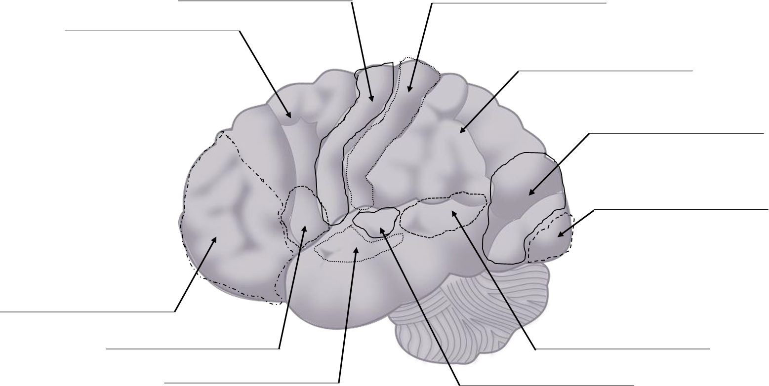
7. Match each structure with its description.
|
_____ Broca's area _____ dura mater _____ prefrontal cortex _____ thalamus _____ hippocampus _____ Wernicke's area _____ visual cortex _____ primary motor cortex _____ hypothalamus _____ cerebellum _____ pia mater _____ falx cerebri _____ arachnoid mater _____ pineal gland _____ amygdala _____ medulla oblongata |
A. Sends out signals causing contractions in voluntary skeletal muscles B. Middle meningeal layer C. Emotion center D. A fold in the dura mater positioned between the right and left hemispheres E. The speech center; where speech is formed F. The deepest meningeal layer, found lining the immediate surface of the brain G. Personality traits and social interaction center H. Center for automated muscular contractions such as sneezing, swallowing, and breathing I. Formation and storage of long-term memory J. Understanding writing or speech K. Collects visual information L. Outermost meningeal layer M. Governs transmission of sensory and motor information to and from the cerebral cortex N. Maintenance of a relatively constant internal body environment including blood pressure, heart rate, and body temperature O. Contributes to balance and coordination, smoothing out multiple motor signals from the primary motor cortex P. Produces melatonin to regulate sleep cycles |
Part 2: Review of Spinal Cord Anatomy
1. Label the following structures of the spinal cord and vertebra on the diagram below.
|
|
|
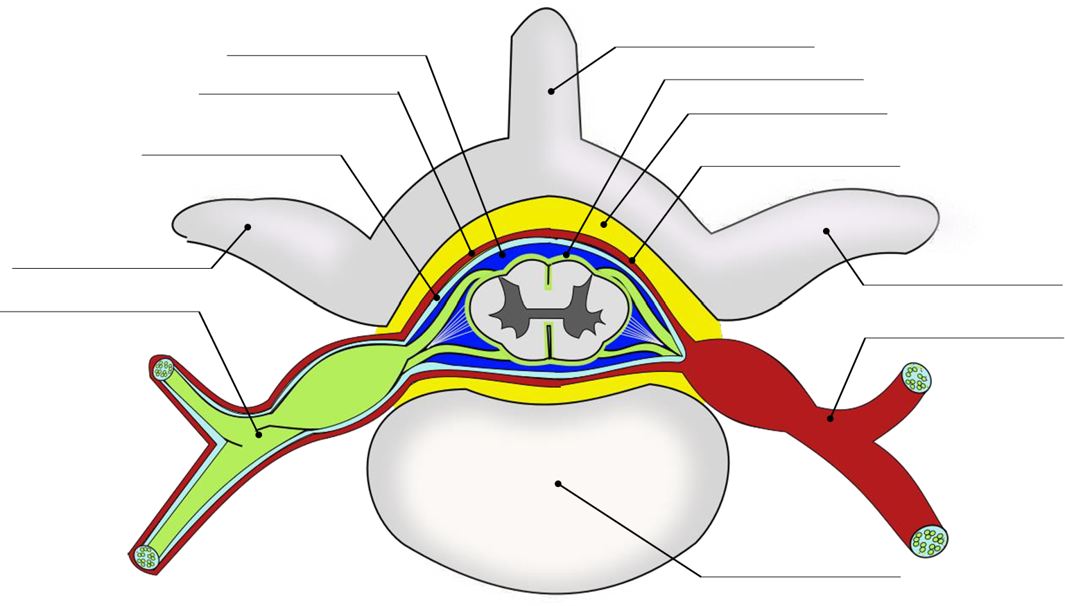
2. Label the following structures of the spinal cord and vertebra on the diagram below.
|
|
|
3. Match the functional regions of the regions of the gray horns.
|
_____ visceral sensory _____ visceral motor _____ somatic sensory _____ somatic motor |
A. Lateral gray horns B. Posterior aspect of posterior gray horn C. Anterior aspect of posterior gray horn D. Anterior gray horn |
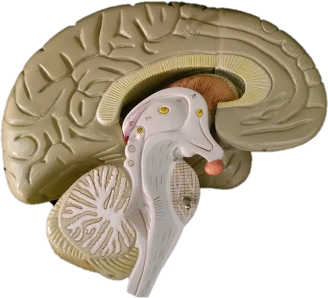
Part 3: Sheep Brain Dissection
Background Information
The sheep brain is remarkably similar to the human brain. One major difference, however, is in proportion. For example, the sheep brain has a proportionately smaller cerebrum. Another difference is in orientation of the spinal cord. The sheep spinal cord is orientated anterior to posterior, as in any four-legged animal. The human spinal cord is orientated superior to inferior. This orientation difference has a major affect on the location of the brain stem. The sheep brain stem is located more towards the rear (posteriorly). The sheep skull, in order to compensate for this, has the foramen magnum located more towards the rear of the skull. In humans, since we walk upright (bipedalism), the spinal cord is in a more vertical plane, thus the foramen magnum is located centrally on the bottom of the skull (inferiorly). By observing the movement of this major skull feature from the rear (as in very early human ancestors) to the current location (modern humans), scientists have been able to determine when the human species began to walk upright on two legs.
Dissection Instructions
- Collect a dissection tray, scalpel, scissors, probes, and gloves.
- Obtain a preserved sheep brain and place it on your dissection tray.
- Make a transverse section of the spinal cord (below the medulla oblongata) and set aside for Part 4: Sheep Spinal Cord Dissection.
- The sheep brain may be is enclosed in a tough outer covering called the dura mater. You can still see some structures on the brain before you remove the dura mater. Take special note of the pituitary gland and the optic chiasma. These two structures will likely be pulled off when you remove the dura mater.
- Make illustrations of the external sheep brain in the space below (alternatively, you may take photos and label the photos on the computer). Label the following structures on your illustrations or photos:
|
|
|
5. Using a scalpel and the longitudinal fissure as a guide, the brain is separated into the left and the right hemispheres. Sharp scalpels work best for this procedure. Always leave the specimen in the dissecting tray when cutting it, DO NOT hold it in your hand! If you are very careful, you will cleanly cut the brain into two halves and can see the internal structures, the most visible of them being the corpus callosum, which divides the left and right hemispheres. The cerebrum will still be visible as a wrinkled structurer, and you can even locate the "bumps" of the superior and inferior colliculi. These structures are located by pulling down the cerebellum. The cerebellum, when cut will have a very distinct tree-like white area within it. This is called the arbor vitae, or the tree of life. Once the brain is cut this way, the colliculi can also be seen from the inside and the pineal gland is revealed only if you made a very careful incision. Finally, a section of the brain is cut to examine the difference between white matter and gray matter.
6. Make illustrations of the dissected medial section of your sheep brain in the space below (alternatively, you may take photos and label the photos on the computer). Label the following structures on your illustrations or photos:
|
|
|
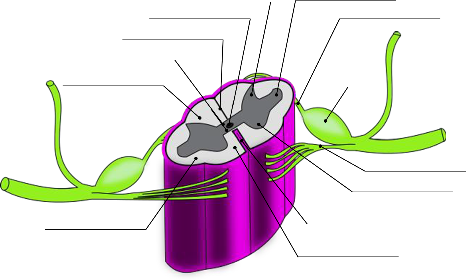
Part 4: Sheep Spinal Cord Dissection
Dissection Instructions
- Use transverse section of the spinal cord produced in step 3 of Part 3: Sheep Brain Dissection.
- Make an illustration of the superior view of the transverse section of your sheep spinal cord slice in the space below (alternatively, you may take a photo and label the photo on the computer). Label the following structures on your illustration or photo:
|
|
|
Attributions
Part 1: Review of Brain Anatomy
- "Anatomy and Physiology Lab Homework" by Laird C Sheldahl is licensed under CC BY-SA 4.0
Part 2: Review of Spinal Cord Anatomy
- "Anatomy and Physiology Lab Homework" by Laird C Sheldahl is licensed under CC BY-SA 4.0
Part 3: Sheep Brain Dissection
- "BIOL 250 Human Anatomy Lab Manual SU 19" by Yancy Aquino, Skyline College is licensed under CC BY-NC-SA 4.0