
14.1: Brain
- Page ID
- 53716

\( \newcommand{\vecs}[1]{\overset { \scriptstyle \rightharpoonup} {\mathbf{#1}} } \)
\( \newcommand{\vecd}[1]{\overset{-\!-\!\rightharpoonup}{\vphantom{a}\smash {#1}}} \)
\( \newcommand{\dsum}{\displaystyle\sum\limits} \)
\( \newcommand{\dint}{\displaystyle\int\limits} \)
\( \newcommand{\dlim}{\displaystyle\lim\limits} \)
\( \newcommand{\id}{\mathrm{id}}\) \( \newcommand{\Span}{\mathrm{span}}\)
( \newcommand{\kernel}{\mathrm{null}\,}\) \( \newcommand{\range}{\mathrm{range}\,}\)
\( \newcommand{\RealPart}{\mathrm{Re}}\) \( \newcommand{\ImaginaryPart}{\mathrm{Im}}\)
\( \newcommand{\Argument}{\mathrm{Arg}}\) \( \newcommand{\norm}[1]{\| #1 \|}\)
\( \newcommand{\inner}[2]{\langle #1, #2 \rangle}\)
\( \newcommand{\Span}{\mathrm{span}}\)
\( \newcommand{\id}{\mathrm{id}}\)
\( \newcommand{\Span}{\mathrm{span}}\)
\( \newcommand{\kernel}{\mathrm{null}\,}\)
\( \newcommand{\range}{\mathrm{range}\,}\)
\( \newcommand{\RealPart}{\mathrm{Re}}\)
\( \newcommand{\ImaginaryPart}{\mathrm{Im}}\)
\( \newcommand{\Argument}{\mathrm{Arg}}\)
\( \newcommand{\norm}[1]{\| #1 \|}\)
\( \newcommand{\inner}[2]{\langle #1, #2 \rangle}\)
\( \newcommand{\Span}{\mathrm{span}}\) \( \newcommand{\AA}{\unicode[.8,0]{x212B}}\)
\( \newcommand{\vectorA}[1]{\vec{#1}} % arrow\)
\( \newcommand{\vectorAt}[1]{\vec{\text{#1}}} % arrow\)
\( \newcommand{\vectorB}[1]{\overset { \scriptstyle \rightharpoonup} {\mathbf{#1}} } \)
\( \newcommand{\vectorC}[1]{\textbf{#1}} \)
\( \newcommand{\vectorD}[1]{\overrightarrow{#1}} \)
\( \newcommand{\vectorDt}[1]{\overrightarrow{\text{#1}}} \)
\( \newcommand{\vectE}[1]{\overset{-\!-\!\rightharpoonup}{\vphantom{a}\smash{\mathbf {#1}}}} \)
\( \newcommand{\vecs}[1]{\overset { \scriptstyle \rightharpoonup} {\mathbf{#1}} } \)
\(\newcommand{\longvect}{\overrightarrow}\)
\( \newcommand{\vecd}[1]{\overset{-\!-\!\rightharpoonup}{\vphantom{a}\smash {#1}}} \)
\(\newcommand{\avec}{\mathbf a}\) \(\newcommand{\bvec}{\mathbf b}\) \(\newcommand{\cvec}{\mathbf c}\) \(\newcommand{\dvec}{\mathbf d}\) \(\newcommand{\dtil}{\widetilde{\mathbf d}}\) \(\newcommand{\evec}{\mathbf e}\) \(\newcommand{\fvec}{\mathbf f}\) \(\newcommand{\nvec}{\mathbf n}\) \(\newcommand{\pvec}{\mathbf p}\) \(\newcommand{\qvec}{\mathbf q}\) \(\newcommand{\svec}{\mathbf s}\) \(\newcommand{\tvec}{\mathbf t}\) \(\newcommand{\uvec}{\mathbf u}\) \(\newcommand{\vvec}{\mathbf v}\) \(\newcommand{\wvec}{\mathbf w}\) \(\newcommand{\xvec}{\mathbf x}\) \(\newcommand{\yvec}{\mathbf y}\) \(\newcommand{\zvec}{\mathbf z}\) \(\newcommand{\rvec}{\mathbf r}\) \(\newcommand{\mvec}{\mathbf m}\) \(\newcommand{\zerovec}{\mathbf 0}\) \(\newcommand{\onevec}{\mathbf 1}\) \(\newcommand{\real}{\mathbb R}\) \(\newcommand{\twovec}[2]{\left[\begin{array}{r}#1 \\ #2 \end{array}\right]}\) \(\newcommand{\ctwovec}[2]{\left[\begin{array}{c}#1 \\ #2 \end{array}\right]}\) \(\newcommand{\threevec}[3]{\left[\begin{array}{r}#1 \\ #2 \\ #3 \end{array}\right]}\) \(\newcommand{\cthreevec}[3]{\left[\begin{array}{c}#1 \\ #2 \\ #3 \end{array}\right]}\) \(\newcommand{\fourvec}[4]{\left[\begin{array}{r}#1 \\ #2 \\ #3 \\ #4 \end{array}\right]}\) \(\newcommand{\cfourvec}[4]{\left[\begin{array}{c}#1 \\ #2 \\ #3 \\ #4 \end{array}\right]}\) \(\newcommand{\fivevec}[5]{\left[\begin{array}{r}#1 \\ #2 \\ #3 \\ #4 \\ #5 \\ \end{array}\right]}\) \(\newcommand{\cfivevec}[5]{\left[\begin{array}{c}#1 \\ #2 \\ #3 \\ #4 \\ #5 \\ \end{array}\right]}\) \(\newcommand{\mattwo}[4]{\left[\begin{array}{rr}#1 \amp #2 \\ #3 \amp #4 \\ \end{array}\right]}\) \(\newcommand{\laspan}[1]{\text{Span}\{#1\}}\) \(\newcommand{\bcal}{\cal B}\) \(\newcommand{\ccal}{\cal C}\) \(\newcommand{\scal}{\cal S}\) \(\newcommand{\wcal}{\cal W}\) \(\newcommand{\ecal}{\cal E}\) \(\newcommand{\coords}[2]{\left\{#1\right\}_{#2}}\) \(\newcommand{\gray}[1]{\color{gray}{#1}}\) \(\newcommand{\lgray}[1]{\color{lightgray}{#1}}\) \(\newcommand{\rank}{\operatorname{rank}}\) \(\newcommand{\row}{\text{Row}}\) \(\newcommand{\col}{\text{Col}}\) \(\renewcommand{\row}{\text{Row}}\) \(\newcommand{\nul}{\text{Nul}}\) \(\newcommand{\var}{\text{Var}}\) \(\newcommand{\corr}{\text{corr}}\) \(\newcommand{\len}[1]{\left|#1\right|}\) \(\newcommand{\bbar}{\overline{\bvec}}\) \(\newcommand{\bhat}{\widehat{\bvec}}\) \(\newcommand{\bperp}{\bvec^\perp}\) \(\newcommand{\xhat}{\widehat{\xvec}}\) \(\newcommand{\vhat}{\widehat{\vvec}}\) \(\newcommand{\uhat}{\widehat{\uvec}}\) \(\newcommand{\what}{\widehat{\wvec}}\) \(\newcommand{\Sighat}{\widehat{\Sigma}}\) \(\newcommand{\lt}{<}\) \(\newcommand{\gt}{>}\) \(\newcommand{\amp}{&}\) \(\definecolor{fillinmathshade}{gray}{0.9}\)Brain

Above: A human brain, lateral view of the left side.
The brain is a complex organ with integrated parts. It contains over 100 billion neurons that are specialized to form different regions of the brain. As the brain develops, its neurons develop in complexity, become specialized into specific brain areas and functions, create circuits or pathways based on repeated experiences with some pathways fading away when they are not used.
Anatomy of the Brain
The brain can be subdivided into four different regions (listed superior to inferior): the cerebrum, the diencephalon, the cerebellum, and the brainstem.

Above: Subdivisions of the brain (left) lateral view of the left side of the brain and (right) anterior view of the brain.
Structures of the brain, divided into each of the four brain subdivisions: cerebrum, diencephalon, cerebellum, and brain stem.
|
Brain structure |
Location / Associations |
Function |
|---|---|---|
|
cerebrum |
||
| amygdala | cerebrum, limbic system | emotions, memories, and decision-making |
|
caudate nucleus |
cerebrum, basal nuclei |
regulation of movements |
|
cerebral cortex (frontal, temporal, parietal, & occipital lobes) |
cerebrum |
receiving and processing sensory information, processing and issuing motor commands, personality, reasoning, decision-making, language interpretation, language formation, memory |
|
fornix |
cerebrum, limbic system |
involved in memory |
|
globus pallidus |
cerebrum, basal nuclei |
regulation of movements |
| hippocampus | cerebrum, limbic system | stores and retrieves long-term explicit memories |
|
insula |
cerebrum, cerebral cortex |
receiving visceral (organ) sensation and taste sensation |
|
putamen |
cerebrum, basal nuclei |
regulation of movements |
|
uncus |
cerebrum |
receiving olfactory (scent) sensory information, forming memories, and emotions |
|
diencephalon |
||
|
hypothalamus |
diencephalon, limbic system |
regulates homeostasis for body temperature, heart rate, thirst, blood pressure, influencing pituitary gland, sleep cycle, fluid balance; the hypothalamus is both nervous and endocrine |
|
pineal gland |
diencephalon |
controls sleep cycles, circadian rhythm, and seasonal cycles; produces melatonin; endocrine gland |
|
thalamus |
diencephalon, limbic system |
a "gate" between sensory and motor neurons and relaying these signals to the cerebral cortex, regulating alertness, sleep, and consciousness |
|
cerebellum |
||
|
cerebellum |
cerebellum |
outgoing motor commands are coordinated; maintaining balance, posture and coordination |
|
brainstem |
||
|
cerebral crus (cerebral peduncle) |
brainstem, midbrain |
location where motor neuron tracts pass from the cerebral cortex to pons and then ultimately to the spinal cord |
|
inferior colliculi |
brainstem, midbrain |
receiving auditory (sound) information |
|
medulla oblongata |
brainstem |
causing reflexes such as swallowing, coughing, sneezing, and vomiting, regulates breathing, regulates heart rate, regulates blood pressure |
|
midbrain / mesencephalon |
brainstem |
regulating alertness and sleep, involved in processing sensory perception (hearing, vision), and relaying motor controls |
|
pons |
brainstem |
contains tracts of neurons regulating motor reflexes such as swallowing, crying, and bladder control as well as breathing, sleep, posture, and facial expressions; tracts carry equilibrium sensation and auditory sensation signals |
|
pyramids of medulla oblongata |
brainstem, medulla oblongata |
contains tracts of motor neurons |
|
superior colliculi |
brainstem, midbrain |
producing behavioral responses to stimuli in the environment |
Note: The limbic system governs emotion, motivation, memory, and learning.
The cerebral cortex is a prominent part of the cerebrum that forms the outer layers of the brain located more superior and superficial to the rest of the brain. It is mainly composed of four lobes: the frontal lobe, parietal lobe, temporal lobe, and occipital lobe. The cerebral cortex has two hemispheres, the right hemisphere and the left hemisphere, divided by the longitudinal fissure . Each hemisphere receives sensory information and gives motor commands to the opposite side of the body, that is, the right hemisphere senses the left side of the body and issues motor commands to the left side of the body. This happens because most axons of sensory and motor neurons cross over from right to left or from left to right, often as they ascend up the spinal cord. The right and left hemispheres are connected by tracts of neural tissue located between them called the corpus callosum allowing communication between the two hemispheres.

Above: Superficial view of the brain showing the lobes of the cerebral cortex (frontal, parietal, temporal, and occipital). The frontal lobe is shown in yellow, parietal lobe in pink, temporal lobe in orange, occipital lobe in mint green, cerebellum in teal, pons in magenta, medulla oblongata in light red, and corpus callosum in blue. (Top) Lateral view of the left side of the brain, (bottom) lateral view of the right side of the brain, and (right) superior view of the brain.
|
Lobe of the Cerebral Cortex |
Location |
Function Regions in this Lobe |
|---|---|---|
|
frontal lobe |
medial, anterior cerebral cortex |
prefrontal cortex, premotor cortex, primary motor cortex (precentral gyrus), Broca's area (speech production) |
|
parietal lobe |
medial, intermediate cerebral cortex |
primary sensory cortex (postcentral gyrus), somatic sensory association area |
|
temporal lobe |
lateral cerebral cortex |
auditory association area, auditory cortex, Wernicke's area (understanding speech) |
|
occipital lobe |
medial, posterior cerebral cortex |
visual association area, visual cortex |
Each rounded ridge on the surface of the cerebral cortex is called a gyrus (pl. gyri) and are separated by indentations called sulci (sing. sulcus) if they are shallow indentations and called fissures if they are deep indentations. A slice through the cerebral cortex (see figure below) reveals that it is composed of both white matter and gray matter, neural tissues that have distinctly lighter and darker colorations, respectively. The gray matter of the cerebral cortex contains the cell bodies (part of the neuron with its nucleus and organelles) of neurons involved in the specific functions of that brain region and the white matter contains axons, surrounded by myelin sheaths, bringing signals to and from the cerebral cortex. There are additional islands of gray matter, positioned within the white matter of the cerebral cortex, that are called basal nuclei (not shown in the figure below).

Above: Frontal section of the cerebrum showing the gray and white matter of the cerebral cortex, the two hemispheres separated by the longitudinal fissure and the corpus callosum connecting them, and the distinctions between a gyrus, sulcus, and fissure.

Above: Superficial view of the brain showing the lobes of the cerebral cortex and the precentral gyri, postcentral gyri, and the central sulcus. (Top) Lateral view of the left side of the brain, (bottom) lateral view of the right side of the brain right, and (right) superior view.
Separating the frontal lobe and the parietal lobe is the central sulcus. The gyri on either side of the central sulcus are called the precentral gyrus anterior to the central sulcus and the postcentral gyrus posterior to the central sulcus. The precentral gyrus is also known as the primary motor cortex since it is responsible for movements of the skeletal muscle under conscious control of the individual. This means that raising your right arm will be controlled by the precentral gyrus in the left hemisphere and kicking your left leg will be controlled by the precentral gyrus in the right hemisphere. The postcentral gyrus is also known as the primary somatosensory cortex ("somato-" comes from the Greek "soma" referring to "the body") that receives sensory information (touch, temperature, pain, vibration, etc.) from the body including the skin and muscles.

Above: Functional regions of the cerebral cortex with a lateral view of the left side of the brain. Wernicke's area and Broca's area are found only in the dominant hemisphere of the cerebral cortex (usually the left hemisphere, but may be in the right hemisphere instead, especially if an individual is left-handed).
|
Functional Areas of the Cerebral Cortex |
Location in the Cerebral Cortex |
Functions |
|---|---|---|
|
auditory association area |
temporal lobe |
processing auditory (sound) information and auditory memory |
|
auditory cortex |
temporal lobe |
receiving auditory (sound) information from the ears |
|
Broca's area |
frontal lobe |
producing speech; located in the left temporal lobe only in most individuals; in some individuals located instead in the right temporal lobe |
|
prefrontal cortex |
frontal lobe |
executing executive and higher cognitive functions including decision-making, planning, memory, and social behaviors |
|
primary motor cortex |
frontal lobe, precentral gyrus |
causing voluntary movements of the skeletal muscles |
|
primary sensory cortex |
parietal lobe, postcentral gyrus |
receiving sensory information from the body |
|
somatic motor association area (premotor cortex) |
frontal lobe |
involved in timing and spatial relationships of movements |
|
somatic sensory association area |
parietal lobe |
combines multiple sensory signals |
|
visual association area |
occipital lobe |
processing visual information |
|
visual cortex |
occipital lobe |
receiving visual information |
|
Wernicke's area |
temporal lobe |
understanding language (both written and verbal); located in the left temporal lobe only in most individuals; in some individuals located instead in the right temporal lobe |
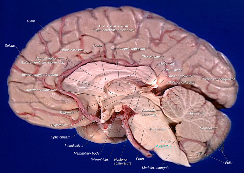
Above: Midsagittal view of a human brain.

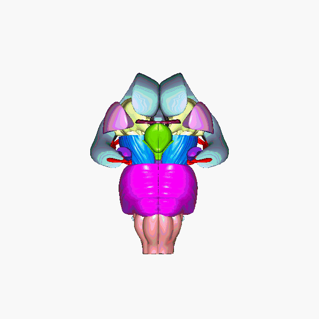
Above: Structures deeper in the brain with some of the left hemisphere of the cerebral cortex removed and a lateral view of the left side of the brain.

Above: The brain with the entire cerebral cortex removed (frontal lobe, parietal lobe, temporal lobe, and occipital lobe) to show deeper structures of the brain.

Above: The brain without the cerebral cortex, corpus callosum, cerebellum, amygdalae, caudate nuclei, and putamina.

Above: The brain without the cerebral cortex, corpus callosum, cerebellum, amygdalae, caudate nuclei, putamina, all of the ventricles (right and left lateral ventricles, third ventricle, fourth ventricle, and cerebral aqueduct), and globus pallidi.
Clinical Application: Memory
There are three main subdivisions of memory, each of which is processed in a different areas of the brain:
- Working memory happens in the front of the brain, in the prefrontal cortex. This type of memory coordinates long-term memories with sights, sounds, and feelings so you can respond to events as they happen.
- Short-term memory involves retaining information for short periods of time, such as a mental "to do" list. Short-term memory involves coordination between the prefrontal cortex and the parietal lobes.
- Long-term memory accesses an amazing range of information - from knowledge of yourself to your understanding of the world. Long-term memory for events and experiences are processed deep in the brain, in a spired area called the hippocampus. It is here that the brain transfers newly gained information into permanent memory. For example, when repeatedly saying a new anatomy term while you are studying, you are passing this information through your hippocampus over and over again which strengthens the associations in the brain with the anatomy term. After repeating it over and over, eventually the pathways in the brain become strong enough to form long-term memories.
Ventricles, Choroid Plexuses, and Cerebrospinal Fluid
The ventricles are a system of structures in the brain that produce and circulate cerebrospinal fluid (CSF). Each of the four ventricles (left lateral ventricle, right lateral ventricle, third ventricle, and fourth ventricle) contain regions called choroid plexuses with ependymal cells (glia cells) responsible for creating CSF. CSF is a clear fluid that cushions the delicate tissues of the central nervous system and is found circulating throughout the ventricles, central canal of the spinal cord, through the subarachnoid space surrounding the brain and spinal cord, and ultimately leaves the subarachnoid space and into the blood in the superior sagittal sinus and straight sinus (blood vessels). CSF also is extremely important in maintaining biochemical homeostasis throughout the central nervous system. It is 99% water but also contains ions, proteins, neurotransmitters, and glucose.

Above: Ventricles of the brain shown without other brain structures.

Above: The flow of CSF in and around the central nervous system. There are two step 5's since CSF can flow into the central canal, although most travels to the subarachnoid space.
Meninges
There are three layers of connective tissue of the central nervous system called meninges. These tissue layers from superficial to deep are: dura mater, arachnoid mater, and pia mater. The brain and spinal cord both are both covered with these same meningeal layers. That is, the dura mater of the brain is continuous with the dura mater of the spinal cord, the arachnoid mater of the brain is continuous with the arachnoid mater of the spinal cord, and the pia mater of the brain is continuous with the pia mater of the spinal cord. Inflammation of the meninges, in either the spinal cord, the brain, or both, is called meningitis.

Above: Frontal section of the skull, meninges, and superficial cerebral cortex. The three layers of meninges (dura mater, arachnoid mater, and pia mater) and their associated spaces (subdural cavity and subarachnoid cavity) are shown.
The dura mater in the brain forms a large fold, positioned within the longitudinal fissure of the cerebral cortex. This prominent fold largely separating the right and left hemispheres of the brain is called falx cerebri. Some of the large veins of the cranial cavity carrying blood back towards the heart, the superior sagittal sinus and straight sinus, are positioned within the falx cerebri.
Dura Mater
The dura mater is a thick, durable membrane, closest to the skull and vertebrae. The dura mater, the outermost part, is a loosely arranged, fibroelastic layer of cells, characterized by multiple interdigitating cell processes, no extracellular collagen, and significant extracellular spaces. The middle region is a mostly fibrous portion. It consists of two layers: the endosteal layer, which lies closest to the calvaria (skullcap), and the inner meningeal layer, which lies closer to the brain. It contains larger blood vessels that split into the capillaries in the pia mater. It is composed of dense fibrous tissue, and its inner surface is covered by flattened cells like those present on the surfaces of the pia mater and arachnoid mater. The dura mater is a sac that envelops the arachnoid mater and surrounds and supports the large dural sinuses carrying blood from the brain toward the heart.
Arachnoid Mater
The middle element of the meninges is the arachnoid mater, so named because of its spider web-like appearance. It cushions the central nervous system. This thin, transparent membrane is composed of fibrous tissue and, like the pia mater, is covered by flat cells also thought to be impermeable to fluid.
The shape of the arachnoid does not follow the convolutions of the surface of the brain and so looks like a loosely fitting sac. In particular, in the region of the brain a large number of fine filaments called arachnoid trabeculae pass from the arachnoid through the subarachnoid space to blend with the tissue of the pia mater. The arachnoid is composed of an outermost portion (arachnoid barrier cell layer) with tightly packed cells and no extracellular collagen; that is why it is considered to represent an effective morphological and physiological meningeal barrier between the cerebrospinal fluid and subarachnoid space and the blood circulation in the dura. The arachnoid barrier layer is characterized by a distinct continuous basal lamina on its inner surface toward the innermost collagenous portion of the arachnoid reticular layer

Above: Cadaver images of the dura mater, arachnoid mater, two of the meningeal layers, and falx cerebri with an anatomical illustration of the falx cerebri with the large venous sinuses superior sagittal sinus, straight sinus, and transverse sinus that carry blood from the brain towards the heart.
Pia Mater
The pia mater is a very delicate membrane. It is the meningeal envelope that firmly adheres to the surface of the brain and spinal cord, following all of the brain's contours (the gyri and sulci). It is a very thin membrane composed of fibrous tissue covered on its outer surface by a sheet of flat cells thought to be impermeable to fluid. The pia mater is pierced by blood vessels to the brain and spinal cord, and its capillaries nourish the brain.
Subarachnoid Space
The subarachnoid space is the space that normally exists between the arachnoid and the pia mater, which is filled with cerebrospinal fluid, and continues down the spinal cord. Spaces are formed from openings at different points along the subarachnoid space; these are the subarachnoid cisterns, which are filled with cerebrospinal fluid.
The dura mater is attached to the skull, whereas in the spinal cord, the dura mater is separated from the vertebrae by a space called the epidural space, which contains fat and blood vessels. The arachnoid is attached to the dura mater, while the pia mater is attached to the central nervous system tissue. When the dura mater and the arachnoid separate through injury or illness, the space between them is the subdural space. There is a subpial space underneath the pia mater that separates it from the glia limitans.
Attributions
- "Anatomy 204L: Laboratory Manual (Second Edition)" by Ethan Snow, University of North Dakota is licensed under CC BY-NC 4.0
- "Anatomy and Physiology I Lab" by Victoria Vidal is licensed under CC BY 4.0
- "Anatomy and Physiology Lab Reference" by Laird C Sheldahl, OpenOregonEducational Resources, Mt. Hood Community College is licensed under CC BY-SA 4.0
- "Blausen 0102 Brain Motor&Sensory.png" by BruceBlaus is licensed under CC BY 3.0
- "BodyParts3D/Anatomography" by The Database Center for Life Science is licensed under CC BY-SA 2.1
- "Brain dura-mater 1.jpg" by Anatomist90 is licensed under CC BY-SA 3.0
- "Cerebellum NIH.png" by NIH is in the Public Domain
- "Cerebellum small.gif" by Life Science Databases(LSDB) is licensed under CC BY-SA 2.1
- "Cerebrum animation small.gif" by Database Center for Life Science(DBCLS) is licensed under CC BY-SA 2.1
- "Diencephalon.png" by Life Science Databases(LSDB) is licensed under CC BY-SA 2.1
- "Gray's Anatomy plates" by Henry Vandyke Carte is in the Public Domain
- "Human brain arachnoid.JPG" by John A Beal, PhD is licensed under CC BY 2.5
- "Human brain dura mater (reflections).JPG" by John A Beal, PhD is licensed under CC BY 2.5
- "Memory" by Exploratorium is licensed under CC BY-NC-SA 4.0
- "Neurobiology of Trauma" by California Social Work Education Center (CalSWEC) is licensed under CC BY-NC-SA 3.0