
12.5: Muscles of the Upper Limbs
- Page ID
- 53688

\( \newcommand{\vecs}[1]{\overset { \scriptstyle \rightharpoonup} {\mathbf{#1}} } \)
\( \newcommand{\vecd}[1]{\overset{-\!-\!\rightharpoonup}{\vphantom{a}\smash {#1}}} \)
\( \newcommand{\dsum}{\displaystyle\sum\limits} \)
\( \newcommand{\dint}{\displaystyle\int\limits} \)
\( \newcommand{\dlim}{\displaystyle\lim\limits} \)
\( \newcommand{\id}{\mathrm{id}}\) \( \newcommand{\Span}{\mathrm{span}}\)
( \newcommand{\kernel}{\mathrm{null}\,}\) \( \newcommand{\range}{\mathrm{range}\,}\)
\( \newcommand{\RealPart}{\mathrm{Re}}\) \( \newcommand{\ImaginaryPart}{\mathrm{Im}}\)
\( \newcommand{\Argument}{\mathrm{Arg}}\) \( \newcommand{\norm}[1]{\| #1 \|}\)
\( \newcommand{\inner}[2]{\langle #1, #2 \rangle}\)
\( \newcommand{\Span}{\mathrm{span}}\)
\( \newcommand{\id}{\mathrm{id}}\)
\( \newcommand{\Span}{\mathrm{span}}\)
\( \newcommand{\kernel}{\mathrm{null}\,}\)
\( \newcommand{\range}{\mathrm{range}\,}\)
\( \newcommand{\RealPart}{\mathrm{Re}}\)
\( \newcommand{\ImaginaryPart}{\mathrm{Im}}\)
\( \newcommand{\Argument}{\mathrm{Arg}}\)
\( \newcommand{\norm}[1]{\| #1 \|}\)
\( \newcommand{\inner}[2]{\langle #1, #2 \rangle}\)
\( \newcommand{\Span}{\mathrm{span}}\) \( \newcommand{\AA}{\unicode[.8,0]{x212B}}\)
\( \newcommand{\vectorA}[1]{\vec{#1}} % arrow\)
\( \newcommand{\vectorAt}[1]{\vec{\text{#1}}} % arrow\)
\( \newcommand{\vectorB}[1]{\overset { \scriptstyle \rightharpoonup} {\mathbf{#1}} } \)
\( \newcommand{\vectorC}[1]{\textbf{#1}} \)
\( \newcommand{\vectorD}[1]{\overrightarrow{#1}} \)
\( \newcommand{\vectorDt}[1]{\overrightarrow{\text{#1}}} \)
\( \newcommand{\vectE}[1]{\overset{-\!-\!\rightharpoonup}{\vphantom{a}\smash{\mathbf {#1}}}} \)
\( \newcommand{\vecs}[1]{\overset { \scriptstyle \rightharpoonup} {\mathbf{#1}} } \)
\(\newcommand{\longvect}{\overrightarrow}\)
\( \newcommand{\vecd}[1]{\overset{-\!-\!\rightharpoonup}{\vphantom{a}\smash {#1}}} \)
\(\newcommand{\avec}{\mathbf a}\) \(\newcommand{\bvec}{\mathbf b}\) \(\newcommand{\cvec}{\mathbf c}\) \(\newcommand{\dvec}{\mathbf d}\) \(\newcommand{\dtil}{\widetilde{\mathbf d}}\) \(\newcommand{\evec}{\mathbf e}\) \(\newcommand{\fvec}{\mathbf f}\) \(\newcommand{\nvec}{\mathbf n}\) \(\newcommand{\pvec}{\mathbf p}\) \(\newcommand{\qvec}{\mathbf q}\) \(\newcommand{\svec}{\mathbf s}\) \(\newcommand{\tvec}{\mathbf t}\) \(\newcommand{\uvec}{\mathbf u}\) \(\newcommand{\vvec}{\mathbf v}\) \(\newcommand{\wvec}{\mathbf w}\) \(\newcommand{\xvec}{\mathbf x}\) \(\newcommand{\yvec}{\mathbf y}\) \(\newcommand{\zvec}{\mathbf z}\) \(\newcommand{\rvec}{\mathbf r}\) \(\newcommand{\mvec}{\mathbf m}\) \(\newcommand{\zerovec}{\mathbf 0}\) \(\newcommand{\onevec}{\mathbf 1}\) \(\newcommand{\real}{\mathbb R}\) \(\newcommand{\twovec}[2]{\left[\begin{array}{r}#1 \\ #2 \end{array}\right]}\) \(\newcommand{\ctwovec}[2]{\left[\begin{array}{c}#1 \\ #2 \end{array}\right]}\) \(\newcommand{\threevec}[3]{\left[\begin{array}{r}#1 \\ #2 \\ #3 \end{array}\right]}\) \(\newcommand{\cthreevec}[3]{\left[\begin{array}{c}#1 \\ #2 \\ #3 \end{array}\right]}\) \(\newcommand{\fourvec}[4]{\left[\begin{array}{r}#1 \\ #2 \\ #3 \\ #4 \end{array}\right]}\) \(\newcommand{\cfourvec}[4]{\left[\begin{array}{c}#1 \\ #2 \\ #3 \\ #4 \end{array}\right]}\) \(\newcommand{\fivevec}[5]{\left[\begin{array}{r}#1 \\ #2 \\ #3 \\ #4 \\ #5 \\ \end{array}\right]}\) \(\newcommand{\cfivevec}[5]{\left[\begin{array}{c}#1 \\ #2 \\ #3 \\ #4 \\ #5 \\ \end{array}\right]}\) \(\newcommand{\mattwo}[4]{\left[\begin{array}{rr}#1 \amp #2 \\ #3 \amp #4 \\ \end{array}\right]}\) \(\newcommand{\laspan}[1]{\text{Span}\{#1\}}\) \(\newcommand{\bcal}{\cal B}\) \(\newcommand{\ccal}{\cal C}\) \(\newcommand{\scal}{\cal S}\) \(\newcommand{\wcal}{\cal W}\) \(\newcommand{\ecal}{\cal E}\) \(\newcommand{\coords}[2]{\left\{#1\right\}_{#2}}\) \(\newcommand{\gray}[1]{\color{gray}{#1}}\) \(\newcommand{\lgray}[1]{\color{lightgray}{#1}}\) \(\newcommand{\rank}{\operatorname{rank}}\) \(\newcommand{\row}{\text{Row}}\) \(\newcommand{\col}{\text{Col}}\) \(\renewcommand{\row}{\text{Row}}\) \(\newcommand{\nul}{\text{Nul}}\) \(\newcommand{\var}{\text{Var}}\) \(\newcommand{\corr}{\text{corr}}\) \(\newcommand{\len}[1]{\left|#1\right|}\) \(\newcommand{\bbar}{\overline{\bvec}}\) \(\newcommand{\bhat}{\widehat{\bvec}}\) \(\newcommand{\bperp}{\bvec^\perp}\) \(\newcommand{\xhat}{\widehat{\xvec}}\) \(\newcommand{\vhat}{\widehat{\vvec}}\) \(\newcommand{\uhat}{\widehat{\uvec}}\) \(\newcommand{\what}{\widehat{\wvec}}\) \(\newcommand{\Sighat}{\widehat{\Sigma}}\) \(\newcommand{\lt}{<}\) \(\newcommand{\gt}{>}\) \(\newcommand{\amp}{&}\) \(\definecolor{fillinmathshade}{gray}{0.9}\)Muscles of the Upper Limbs
Arm (Shoulder to Elbow)
Anatomists refer to the upper arm as just the arm or the brachium. (The lower arm is the forearm or antebrachium.) There are three muscles on the upper arm that are parallel to the long axis of the humerus, the biceps brachii, the brachialis, and the triceps brachii.
The biceps brachii is on the anterior side of the humerus and is the prime mover (agonist) responsible for flexing the forearm. It has two orgins (hence the “biceps” part of its name), both of which attach to the scapula bone. Biceps brachii has two heads, called the short head of biceps brachii on the medial aspect and the long head of biceps brachii on the lateral aspect. It inserts on the radius bone. The biceps brachii has two synergist muscles that assist it in flexing the forearm. Both are found on the anterior side of the arm and forearm. One of these is the brachioradialis muscle which is largely on the forearm (see the next section) and the other is the brachialis, which is largely on the upper arm. The brachialis muscle is deep to the biceps brachii and both its origin and its insertion are more distal to the shoulder than its equivalents on the biceps brachii. Like the biceps brachii the origin of the brachialis is on the humerus. Parts on the brachialis can be seen peeking out from under the biceps brachii, especially lower on the arm. The locations of these three muscles are shown in.
On the posterior side of the arm is the triceps brachii muscle. It the antagonist to the biceps brachii. When the triceps brachii contracts it extends the forearm, undoing any flexing brought about by contractions of the biceps brachii. As a result, when the triceps brachii is contracted, the biceps brachii and its synergists must be relaxed, and vice versa. The triceps brachii has three origins, called the long head (medial aspect), the lateral head (lateral aspect), and the medial head (deep to the long and lateral heads). It is easiest to view the triceps brachii from the posterior, but the medial head and its origin are deep to the lateral head and the long head, and so is the medial head of the triceps brachii is partially obscured from the posterior.

Above: Superficial muscles of the arm (shoulder to elbow) with a (left) lateral view, (middle) anterior view, and (right) anterolateral view.

Above: Anterior upper limb with some muscles removed and deep muscles of the arm labeled (left) without biceps brachii and (right) without biceps brachii and brachialis.

Above: Posterior upper limb with muscles of the arm labeled. (Left) The superficial muscles of the posterior arm and (right) muscles of the posterior arm without the lateral head and long head of triceps brachii.

Above: Cadaver images of the muscles of the arm.
Forearm
Anatomists refer to the lower arm as the forearm or antebrachium. The musculature of the forearm is complicated in order to provide the dexterity possible with the forearm and some of the movements of the hand. Many of the muscles of the anterior forearm are "flexor" muscles since they cause flexion. Likewise, many of the muscles of the posterior forearm are "extensor" muscles since they cause extension. The names of the forearm muscles reflect attachments, locations, and actions they cause including "digitorum" (mover of the digits aka fingers), "carpi" (relating to the carpals/wrist bones), "radialis" (relating to the radius), "ulnaris" (relating to the ulna), "pronator" (causes pronation), "supinator" (causes supination), "pollicis" (relating to the thumb/pollex) and "abductor" (causing abduction).

Above: Superficial muscles of the forearm (elbow to wrist) with a (left) lateral view, (middle) anterior view, and (right) anterolateral view.
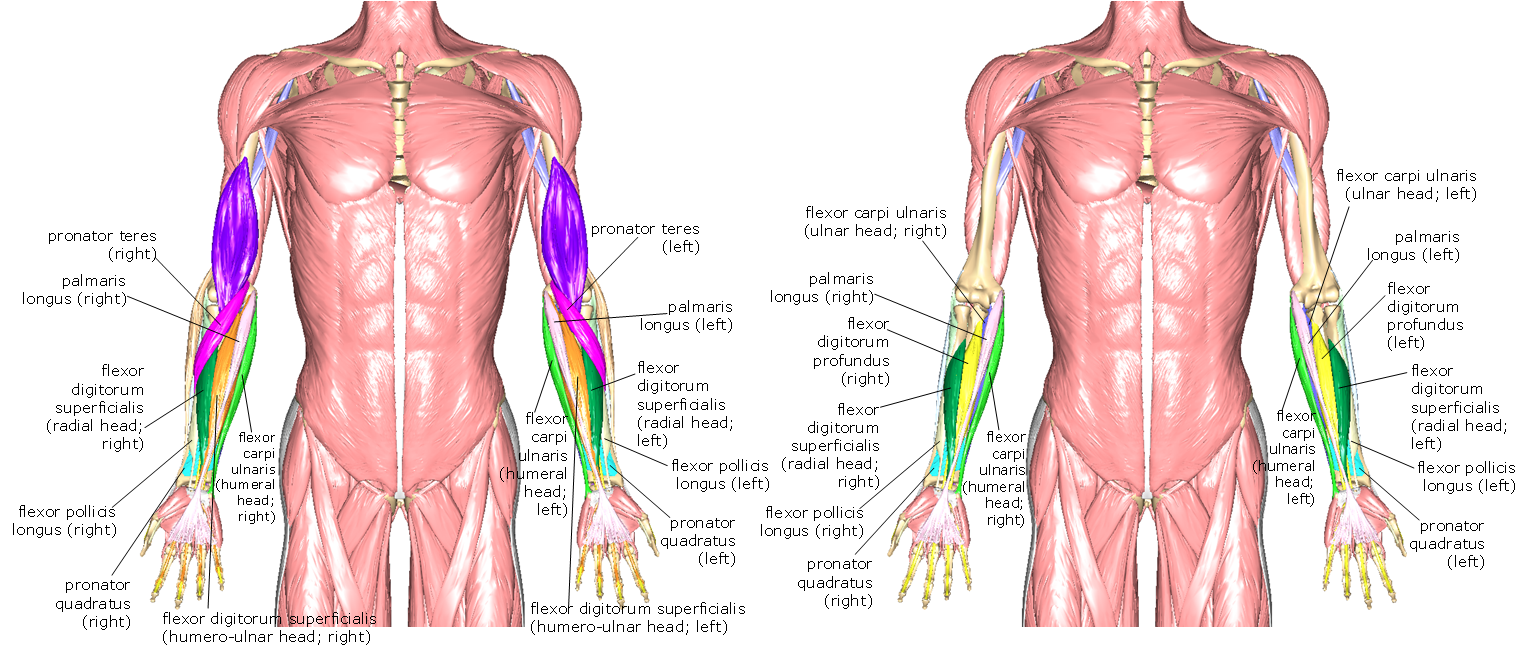
Above: Anterior upper limb with some muscles removed with deep muscles of the forearm labeled. (Left) without brachioradialis and flexor carpi radialis and (right) without muscles in the left figure and also without pronator teres, humero-ulnar head of flexor digitorum, and extensor carpi radialis longus.
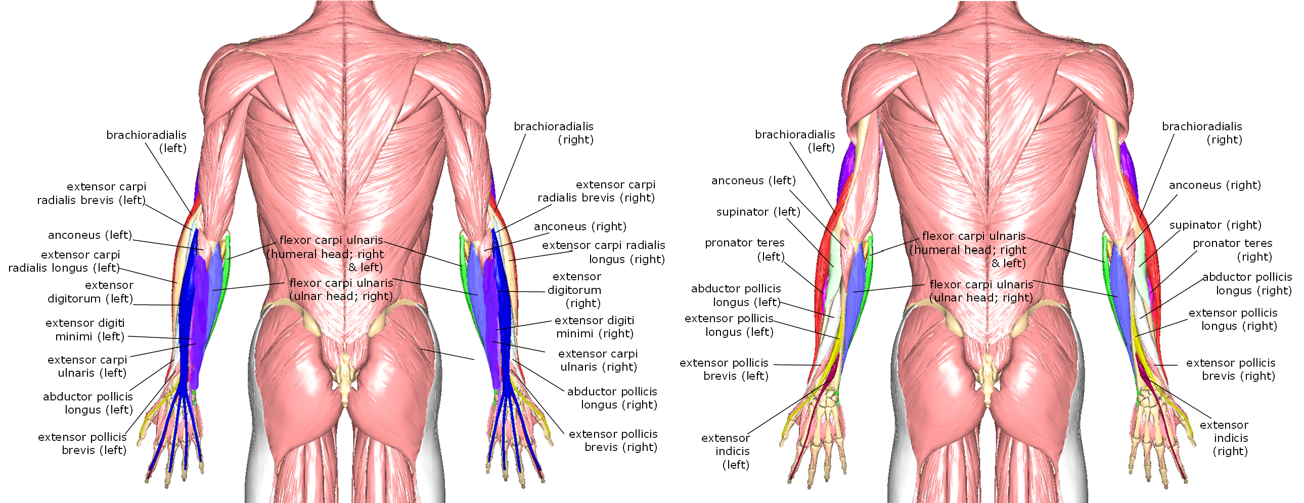
Above: Posterior upper limb with muscles of the forearm labeled. (Right) The posterior forearm and (left) without extensor digitorum, extensor carpi radialis longus, extensor carpi radialis brevis, extensor carpi ulnaris, and extensor digiti minimi.

Above: Cadaver images of the muscles of the forearm.
Hand
Humans are capable of very fine movements as well as powerful movements with their hands in a variety of directions. These abilities require a host of muscles to create the high amount of dexterity possible with the hands. A number of muscles of the forearm attach to the hands including palmaris longus, flexor digitorum (humero-ulnar head), and flexor pollicis longus. These attachments may be seen in the figure below.
Lateral muscles of the palm include abductor pollicis brevis, opponens pollicis, flexor pollicis brevis, and adductor pollicis. The medial muscles of the palm include palmaris brevis, flexor digiti minimi brevis, abductor digiti minimi, and opponens digiti minimi.
There are multiple lumbricals and interossei are muscles that are intermediate in the hand with palmar interossei located on the palmar or anterior aspect of the hand and dorsal interossei located on the dorsal aspect of the hand. The lumbricals are named first lumbrical (lateral edge of the second phalanx), second lumbrical (lateral edge of the third phalanx), third lumbrical (lateral edge of the fourth phalanx), and fourth lumbrical (lateral edge of the fifth phalanx)

Above: Palm of the hand (anterior aspect) (top) with all the muscles and (bottom) without palmaris longus.

Above: Deeper muscles of the palm of the hand (anterior aspect) (top) without palmaris longus, flexor digitorum (humero-ulnar head), abductor digiti minimi, adductor pollicis, and abductor pollicis brevis. (Bottom) The palm without the muscles in the top diagram and also without flexor digitorum profundus, lumbricals, flexor retinaculum of the wrist, abductor pollicis brevis, and opponens pollicis.

Above: Muscles of the posterior hand. (Top) All of the muscles of the posterior hand and (bottom) without extensor digitorum.
Attributions
- "Anatomy 204L: Laboratory Manual (Second Edition)" by Ethan Snow, University of North Dakota is licensed under CC BY-NC 4.0
- "Anatomy and Physiology" by J. Gordon Betts et al., OpenStax is licensed under CC BY 4.0
- "Anatomy of the Human Body" by Henry Gray is in the Public Domain
- "BIOL 250 Human Anatomy Lab Manual SU 19" by Yancy Aquino, Skyline College is licensed under CC BY-NC-SA 4.0
- "BodyParts3D/Anatomography" by The Database Center for Life Science is licensed under CC BY-SA 2.1
- "Gray's Anatomy plates" by Henry Vandyke Carte is in the Public Domain