
11.2: Microscopic Anatomy of Skeletal Muscles
- Page ID
- 53678

\( \newcommand{\vecs}[1]{\overset { \scriptstyle \rightharpoonup} {\mathbf{#1}} } \)
\( \newcommand{\vecd}[1]{\overset{-\!-\!\rightharpoonup}{\vphantom{a}\smash {#1}}} \)
\( \newcommand{\id}{\mathrm{id}}\) \( \newcommand{\Span}{\mathrm{span}}\)
( \newcommand{\kernel}{\mathrm{null}\,}\) \( \newcommand{\range}{\mathrm{range}\,}\)
\( \newcommand{\RealPart}{\mathrm{Re}}\) \( \newcommand{\ImaginaryPart}{\mathrm{Im}}\)
\( \newcommand{\Argument}{\mathrm{Arg}}\) \( \newcommand{\norm}[1]{\| #1 \|}\)
\( \newcommand{\inner}[2]{\langle #1, #2 \rangle}\)
\( \newcommand{\Span}{\mathrm{span}}\)
\( \newcommand{\id}{\mathrm{id}}\)
\( \newcommand{\Span}{\mathrm{span}}\)
\( \newcommand{\kernel}{\mathrm{null}\,}\)
\( \newcommand{\range}{\mathrm{range}\,}\)
\( \newcommand{\RealPart}{\mathrm{Re}}\)
\( \newcommand{\ImaginaryPart}{\mathrm{Im}}\)
\( \newcommand{\Argument}{\mathrm{Arg}}\)
\( \newcommand{\norm}[1]{\| #1 \|}\)
\( \newcommand{\inner}[2]{\langle #1, #2 \rangle}\)
\( \newcommand{\Span}{\mathrm{span}}\) \( \newcommand{\AA}{\unicode[.8,0]{x212B}}\)
\( \newcommand{\vectorA}[1]{\vec{#1}} % arrow\)
\( \newcommand{\vectorAt}[1]{\vec{\text{#1}}} % arrow\)
\( \newcommand{\vectorB}[1]{\overset { \scriptstyle \rightharpoonup} {\mathbf{#1}} } \)
\( \newcommand{\vectorC}[1]{\textbf{#1}} \)
\( \newcommand{\vectorD}[1]{\overrightarrow{#1}} \)
\( \newcommand{\vectorDt}[1]{\overrightarrow{\text{#1}}} \)
\( \newcommand{\vectE}[1]{\overset{-\!-\!\rightharpoonup}{\vphantom{a}\smash{\mathbf {#1}}}} \)
\( \newcommand{\vecs}[1]{\overset { \scriptstyle \rightharpoonup} {\mathbf{#1}} } \)
\( \newcommand{\vecd}[1]{\overset{-\!-\!\rightharpoonup}{\vphantom{a}\smash {#1}}} \)
\(\newcommand{\avec}{\mathbf a}\) \(\newcommand{\bvec}{\mathbf b}\) \(\newcommand{\cvec}{\mathbf c}\) \(\newcommand{\dvec}{\mathbf d}\) \(\newcommand{\dtil}{\widetilde{\mathbf d}}\) \(\newcommand{\evec}{\mathbf e}\) \(\newcommand{\fvec}{\mathbf f}\) \(\newcommand{\nvec}{\mathbf n}\) \(\newcommand{\pvec}{\mathbf p}\) \(\newcommand{\qvec}{\mathbf q}\) \(\newcommand{\svec}{\mathbf s}\) \(\newcommand{\tvec}{\mathbf t}\) \(\newcommand{\uvec}{\mathbf u}\) \(\newcommand{\vvec}{\mathbf v}\) \(\newcommand{\wvec}{\mathbf w}\) \(\newcommand{\xvec}{\mathbf x}\) \(\newcommand{\yvec}{\mathbf y}\) \(\newcommand{\zvec}{\mathbf z}\) \(\newcommand{\rvec}{\mathbf r}\) \(\newcommand{\mvec}{\mathbf m}\) \(\newcommand{\zerovec}{\mathbf 0}\) \(\newcommand{\onevec}{\mathbf 1}\) \(\newcommand{\real}{\mathbb R}\) \(\newcommand{\twovec}[2]{\left[\begin{array}{r}#1 \\ #2 \end{array}\right]}\) \(\newcommand{\ctwovec}[2]{\left[\begin{array}{c}#1 \\ #2 \end{array}\right]}\) \(\newcommand{\threevec}[3]{\left[\begin{array}{r}#1 \\ #2 \\ #3 \end{array}\right]}\) \(\newcommand{\cthreevec}[3]{\left[\begin{array}{c}#1 \\ #2 \\ #3 \end{array}\right]}\) \(\newcommand{\fourvec}[4]{\left[\begin{array}{r}#1 \\ #2 \\ #3 \\ #4 \end{array}\right]}\) \(\newcommand{\cfourvec}[4]{\left[\begin{array}{c}#1 \\ #2 \\ #3 \\ #4 \end{array}\right]}\) \(\newcommand{\fivevec}[5]{\left[\begin{array}{r}#1 \\ #2 \\ #3 \\ #4 \\ #5 \\ \end{array}\right]}\) \(\newcommand{\cfivevec}[5]{\left[\begin{array}{c}#1 \\ #2 \\ #3 \\ #4 \\ #5 \\ \end{array}\right]}\) \(\newcommand{\mattwo}[4]{\left[\begin{array}{rr}#1 \amp #2 \\ #3 \amp #4 \\ \end{array}\right]}\) \(\newcommand{\laspan}[1]{\text{Span}\{#1\}}\) \(\newcommand{\bcal}{\cal B}\) \(\newcommand{\ccal}{\cal C}\) \(\newcommand{\scal}{\cal S}\) \(\newcommand{\wcal}{\cal W}\) \(\newcommand{\ecal}{\cal E}\) \(\newcommand{\coords}[2]{\left\{#1\right\}_{#2}}\) \(\newcommand{\gray}[1]{\color{gray}{#1}}\) \(\newcommand{\lgray}[1]{\color{lightgray}{#1}}\) \(\newcommand{\rank}{\operatorname{rank}}\) \(\newcommand{\row}{\text{Row}}\) \(\newcommand{\col}{\text{Col}}\) \(\renewcommand{\row}{\text{Row}}\) \(\newcommand{\nul}{\text{Nul}}\) \(\newcommand{\var}{\text{Var}}\) \(\newcommand{\corr}{\text{corr}}\) \(\newcommand{\len}[1]{\left|#1\right|}\) \(\newcommand{\bbar}{\overline{\bvec}}\) \(\newcommand{\bhat}{\widehat{\bvec}}\) \(\newcommand{\bperp}{\bvec^\perp}\) \(\newcommand{\xhat}{\widehat{\xvec}}\) \(\newcommand{\vhat}{\widehat{\vvec}}\) \(\newcommand{\uhat}{\widehat{\uvec}}\) \(\newcommand{\what}{\widehat{\wvec}}\) \(\newcommand{\Sighat}{\widehat{\Sigma}}\) \(\newcommand{\lt}{<}\) \(\newcommand{\gt}{>}\) \(\newcommand{\amp}{&}\) \(\definecolor{fillinmathshade}{gray}{0.9}\)Microscopic Anatomy of Skeletal Muscles
Skeletal muscle is found attached to bones. It consists of long multinucleate fibers. The fibers run the entire length of the muscle they come from and so are usually too long to have their ends visible when viewed under the microscope. The fibers are relatively wide and very long, but unbranched. Fibers are formed from the fusion of thousands of precursor cells. This is why they are so long and why individual fibers are multinucleate (a single fiber has many nuclei). The nuclei are usually up against the edge of the fiber. There are striations in skeletal muscle. These are alternating dark and light bands perpendicular to the edge of the fiber that are present all along the fiber.

Above: Skeletal muscle fibers, teased, longitudinal view. Tissue magnified by 200x.
Skeletal muscles are bundles of fascicles (bundles of muscle fibers) surrounded by epimysium (fibrous elastic tissue). Fascicles are bundles of muscle fibers surrounded by perimysium (connective tissue). A single muscle fiber is surrounded by a layer of endomysium (areolar connective tissue). The plasma membrane of a muscle fiber is called the sarcolemma and these cells contain specialized endoplasmic reticulum called sarcoplasmic reticulum. Each muscle fiber is a multinucleated structure composed myofibrils, long cylinder-shaped cell structures composed of actin and myosin filaments, protein fibers capable of contraction. Myofibrils have repeating sarcomeres causing the striations (banding pattern) observed with a microscope. Sarcomeres are the functional units of skeletal muscles enabling contraction to occur when actin and myosin filaments cross-link and the myosin heads bend in a power stroke causing an increased overlap in the actin and myosin filaments resulting in shortening of the sarcomere (contraction).
Muscle contraction occurs as a result of many sarcomeres within many myofibrils cross-linking and pulling on each other to shorten the sarcomere. The dark region of a striation (made of a sarcomere) is composed of the thicker myosin filaments with some actin overlap (the A band). The lighter region of a striation is where only actin filaments, the thinner filaments, are present (the I band). Myosin filaments connect with each other at the M line and actin filaments connect with each other at the Z disc. A single sarcomere unit begins at one Z disc and ends at another Z disc with the M line in the center of the sarcomere. The H zone in the center of the A band is a region where only myosin filaments, and no actin filaments, are present. For this reason, the H zone appears as a light band in the center of each dark band in the striations of skeletal muscle (see microscopic image of skeletal muscle below).

Above: The structure of a skeletal muscle and its muscle fibers.

Above: Muscle fibers are cells composed of multiple myofibrils, bundles of actin and myosin filaments that cross-link for contraction. Each myofibril is composed of units called sarcomeres, repeating structures of actin and myosin responsible for muscular contraction. In this diagram, myosin is shown in purple and actin is shown in green.
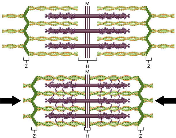
Above: A single sarcomere (top) relaxed and (bottom) contracted. The purple filaments are myosin and the green are actin. When the myosin heads cross-link with actin and undergo the power stroke (myosin heads move), the overlap between actin and myosin increases and the entire sarcomere structure becomes smaller (contraction).

Above: (Bottom) Diagram shows the structure of a myofibril with repeating sarcomeres and how this corresponds with the striations of skeletal muscle (top and middle; middle is top image highly zoomed into striations).
A series of events resulting in muscular contraction begins with an action potential from a motor neuron at a neuromuscular junction. When the action potential reaches the synaptic terminal at the neuromuscular junction, acetylcholine, a neurotransmitter, is released from vesicles from the neuron triggering a chain reaction that orchestrates sodium channels in the sarcolemma (plasma membrane of a muscle fiber) to trigger release of calcium from sarcoplasmic reticulum enabling myosin heads to crosslink with actin to initiate contraction. This causes the sarcomeres in nearby myofibrils to crosslink and initiate the power stroke causing the myofibrils to become shorter, thereby pulling the entire muscle and causing it to become shorter.

Above: Sequence of events beginning with an action potential from a neuron resulting in muscular contraction.

Above: Microscopic image of skeletal muscle fibers with multiple axons and neuromuscular junctions.

Above: (Middle and bottom) Microscopic images of muscle cross sections at 100x (middle) and at 400x (bottom). The structures in these microscopic images correspond with the illustration showing structures of skeletal muscle (top).
Attributions
- "Anatomy and Physiology" by J. Gordon Betts et al., OpenStax is licensed under CC BY 4.0
- "Anatomy and Physiology Lab Homework" by Laird C Sheldahl is licensed under CC BY-SA 4.0
- "Animal Tissues and Organs" by Berkshire Community College Bioscience Image Library is in the Public Domain, CC0
- "BIOL 250 Human Anatomy Lab Manual SU 19" by Yancy Aquino, Skyline College is licensed under CC BY-NC-SA 4.0
- "Digital Histology" by Department of Anatomy and Neurobiology and the Office of Faculty Affairs, Virginia Commonwealth University School of Medicine and the ALT Lab at Virginia Commonwealth University is licensed under CC BY 4.0
- "Skeletal muscle and fiber.jpg" by Abdulrahman112 is in the Public Domain, CC0 / A derivative from the original work