
8.5.4: Thoracic Vertebrae
- Page ID
- 53927

Thoracic Vertebrae
The bodies of the thoracic vertebrae are larger than those of cervical vertebrae. The characteristic feature for a typical thoracic vertebra is the spinous process, which is long and has a pronounced downward angle that causes it to overlap the next inferior vertebra. The superior articular processes of thoracic vertebrae face anteriorly and the inferior processes face posteriorly. These orientations are important determinants for the type and range of movements available to the thoracic region of the vertebral column.

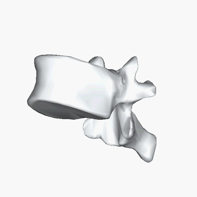
Above: A thoracic vertebra with (A) an anterior view, (B) an anterolateral view, (C) a lateral view of the left side of the vertebra, and (D) a superior view with anterior being at the bottom of the image. Click here to see rotating gif of a thoracic vertebra.
Thoracic vertebrae have several additional articulation sites (facets), where a rib is attached. Most thoracic vertebrae have two facets located on the lateral sides of the body, each of which is called a costal facet (costal = “rib”). These are for articulation with the head (end) of a rib. An additional facet is located on the transverse process for articulation with the tubercle of a rib.

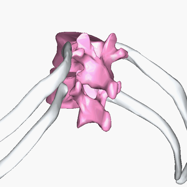
Above: T11 and T12 (pink) articulating with right and left ribs 11 and ribs 12 (white), respectively. Ribs articulate with superior, inferior, and transverse costal facets located on the lateral aspects of each vertebra.
Attributions (All Vertebral Column Sections)
- "Anatomy 204L: Laboratory Manual (Second Edition)" by Ethan Snow, University of North Dakota is licensed under CC BY-NC 4.0
- "Anatomy and Physiology" by J. Gordon Betts et al., OpenStax is licensed under CC BY 4.0
- "BIOL 250 Human Anatomy Lab Manual SU 19" by Yancy Aquino, Skyline College is licensed under CC BY-NC-SA 4.0
- "BodyParts3D/Anatomography" by The Database Center for Life Science is licensed under CC BY-SA 2.1
- "Cervical vertebra english.png" by user:debivort is licensed under CC BY-SA 3.0
- "Sacrum - animation02.gif" by BodyParts3D is made by DBCLS is licensed under CC BY-SA 2.1