
3.2: Subcellular Structures
- Page ID
- 53551

\( \newcommand{\vecs}[1]{\overset { \scriptstyle \rightharpoonup} {\mathbf{#1}} } \)
\( \newcommand{\vecd}[1]{\overset{-\!-\!\rightharpoonup}{\vphantom{a}\smash {#1}}} \)
\( \newcommand{\id}{\mathrm{id}}\) \( \newcommand{\Span}{\mathrm{span}}\)
( \newcommand{\kernel}{\mathrm{null}\,}\) \( \newcommand{\range}{\mathrm{range}\,}\)
\( \newcommand{\RealPart}{\mathrm{Re}}\) \( \newcommand{\ImaginaryPart}{\mathrm{Im}}\)
\( \newcommand{\Argument}{\mathrm{Arg}}\) \( \newcommand{\norm}[1]{\| #1 \|}\)
\( \newcommand{\inner}[2]{\langle #1, #2 \rangle}\)
\( \newcommand{\Span}{\mathrm{span}}\)
\( \newcommand{\id}{\mathrm{id}}\)
\( \newcommand{\Span}{\mathrm{span}}\)
\( \newcommand{\kernel}{\mathrm{null}\,}\)
\( \newcommand{\range}{\mathrm{range}\,}\)
\( \newcommand{\RealPart}{\mathrm{Re}}\)
\( \newcommand{\ImaginaryPart}{\mathrm{Im}}\)
\( \newcommand{\Argument}{\mathrm{Arg}}\)
\( \newcommand{\norm}[1]{\| #1 \|}\)
\( \newcommand{\inner}[2]{\langle #1, #2 \rangle}\)
\( \newcommand{\Span}{\mathrm{span}}\) \( \newcommand{\AA}{\unicode[.8,0]{x212B}}\)
\( \newcommand{\vectorA}[1]{\vec{#1}} % arrow\)
\( \newcommand{\vectorAt}[1]{\vec{\text{#1}}} % arrow\)
\( \newcommand{\vectorB}[1]{\overset { \scriptstyle \rightharpoonup} {\mathbf{#1}} } \)
\( \newcommand{\vectorC}[1]{\textbf{#1}} \)
\( \newcommand{\vectorD}[1]{\overrightarrow{#1}} \)
\( \newcommand{\vectorDt}[1]{\overrightarrow{\text{#1}}} \)
\( \newcommand{\vectE}[1]{\overset{-\!-\!\rightharpoonup}{\vphantom{a}\smash{\mathbf {#1}}}} \)
\( \newcommand{\vecs}[1]{\overset { \scriptstyle \rightharpoonup} {\mathbf{#1}} } \)
\( \newcommand{\vecd}[1]{\overset{-\!-\!\rightharpoonup}{\vphantom{a}\smash {#1}}} \)
\(\newcommand{\avec}{\mathbf a}\) \(\newcommand{\bvec}{\mathbf b}\) \(\newcommand{\cvec}{\mathbf c}\) \(\newcommand{\dvec}{\mathbf d}\) \(\newcommand{\dtil}{\widetilde{\mathbf d}}\) \(\newcommand{\evec}{\mathbf e}\) \(\newcommand{\fvec}{\mathbf f}\) \(\newcommand{\nvec}{\mathbf n}\) \(\newcommand{\pvec}{\mathbf p}\) \(\newcommand{\qvec}{\mathbf q}\) \(\newcommand{\svec}{\mathbf s}\) \(\newcommand{\tvec}{\mathbf t}\) \(\newcommand{\uvec}{\mathbf u}\) \(\newcommand{\vvec}{\mathbf v}\) \(\newcommand{\wvec}{\mathbf w}\) \(\newcommand{\xvec}{\mathbf x}\) \(\newcommand{\yvec}{\mathbf y}\) \(\newcommand{\zvec}{\mathbf z}\) \(\newcommand{\rvec}{\mathbf r}\) \(\newcommand{\mvec}{\mathbf m}\) \(\newcommand{\zerovec}{\mathbf 0}\) \(\newcommand{\onevec}{\mathbf 1}\) \(\newcommand{\real}{\mathbb R}\) \(\newcommand{\twovec}[2]{\left[\begin{array}{r}#1 \\ #2 \end{array}\right]}\) \(\newcommand{\ctwovec}[2]{\left[\begin{array}{c}#1 \\ #2 \end{array}\right]}\) \(\newcommand{\threevec}[3]{\left[\begin{array}{r}#1 \\ #2 \\ #3 \end{array}\right]}\) \(\newcommand{\cthreevec}[3]{\left[\begin{array}{c}#1 \\ #2 \\ #3 \end{array}\right]}\) \(\newcommand{\fourvec}[4]{\left[\begin{array}{r}#1 \\ #2 \\ #3 \\ #4 \end{array}\right]}\) \(\newcommand{\cfourvec}[4]{\left[\begin{array}{c}#1 \\ #2 \\ #3 \\ #4 \end{array}\right]}\) \(\newcommand{\fivevec}[5]{\left[\begin{array}{r}#1 \\ #2 \\ #3 \\ #4 \\ #5 \\ \end{array}\right]}\) \(\newcommand{\cfivevec}[5]{\left[\begin{array}{c}#1 \\ #2 \\ #3 \\ #4 \\ #5 \\ \end{array}\right]}\) \(\newcommand{\mattwo}[4]{\left[\begin{array}{rr}#1 \amp #2 \\ #3 \amp #4 \\ \end{array}\right]}\) \(\newcommand{\laspan}[1]{\text{Span}\{#1\}}\) \(\newcommand{\bcal}{\cal B}\) \(\newcommand{\ccal}{\cal C}\) \(\newcommand{\scal}{\cal S}\) \(\newcommand{\wcal}{\cal W}\) \(\newcommand{\ecal}{\cal E}\) \(\newcommand{\coords}[2]{\left\{#1\right\}_{#2}}\) \(\newcommand{\gray}[1]{\color{gray}{#1}}\) \(\newcommand{\lgray}[1]{\color{lightgray}{#1}}\) \(\newcommand{\rank}{\operatorname{rank}}\) \(\newcommand{\row}{\text{Row}}\) \(\newcommand{\col}{\text{Col}}\) \(\renewcommand{\row}{\text{Row}}\) \(\newcommand{\nul}{\text{Nul}}\) \(\newcommand{\var}{\text{Var}}\) \(\newcommand{\corr}{\text{corr}}\) \(\newcommand{\len}[1]{\left|#1\right|}\) \(\newcommand{\bbar}{\overline{\bvec}}\) \(\newcommand{\bhat}{\widehat{\bvec}}\) \(\newcommand{\bperp}{\bvec^\perp}\) \(\newcommand{\xhat}{\widehat{\xvec}}\) \(\newcommand{\vhat}{\widehat{\vvec}}\) \(\newcommand{\uhat}{\widehat{\uvec}}\) \(\newcommand{\what}{\widehat{\wvec}}\) \(\newcommand{\Sighat}{\widehat{\Sigma}}\) \(\newcommand{\lt}{<}\) \(\newcommand{\gt}{>}\) \(\newcommand{\amp}{&}\) \(\definecolor{fillinmathshade}{gray}{0.9}\)Subcellular Structures
A generalized diagram of an animal cell is shown below. Closely examine the shapes and locations of the organelles that may be found within animal cells and their functions:
|
Subcellular Structure |
Function of the Structure |
Form of the Structure |
|
centrosome
|
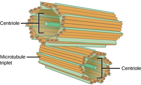
The centrosome is a microtubule-organizing center found near the nuclei of animal cells. Centrosomes play a major role in cell division by organizing the microtubules that move and separate chromosomes into separate cells. |
Two small barrel-like structures composed of straight fibers. These two barrels (each called a centriole) cross each other. |
|
chromatin
|
Chromatin describes the material that makes up the chromosomes both when condensed (packed into tight structures for cell division) and decondensed (loose; chromatin is loose most of the time). |
Thin winding fibers located within the nucleus. Think of it as spaghetti within a bowl (the bowl is the nucleus). |
|
cilium (pl. cilia)
|
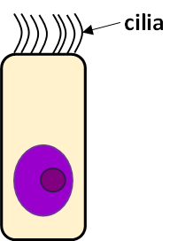
Cilia are protein fibers anchored inside of a cell with the majority of these hair-like fibers on the exterior of a cell. In humans, cilia move substances along the cell's outer surface. For example, the cilia lining the Fallopian tubes move the ovum (egg) toward the uterus. Another example is the cilia on the cells lining the respiratory tract that moves mucus, and debris that gets trapped in the mucus, away from the lungs. |
Small hairs lining the outside of the cell. Cilia differ from microvilli because they are hair-like fibers outside of the cell whereas microvilli describe a way the plasma membrane is wrapped. |
|
cytoplasm |
The cytoplasm is the cell's entire region between the plasma membrane and the nuclear envelope. It is comprised of organelles suspended in the gel-like cytosol, the cytoskeleton, and various chemicals. Even though the cytoplasm consists of 70 to 80 percent water, it has a semi- solid consistency, which comes from the proteins within it. Glucose and other simple sugars, polysaccharides, amino acids, nucleic acids, fatty acids, and derivatives of glycerol are also found in the cytoplasm. Many metabolic reactions, including protein synthesis, take place in the cytoplasm. |
All of the semi-fluid surrounding all of the other organelles as well as the organelles except for the nucleus. If a cell is a swimming pool with the organelles being rafts in the pool, the cytoplasm is the water and the rafts and the cytosol is the water. |
|
cytoskeleton
|
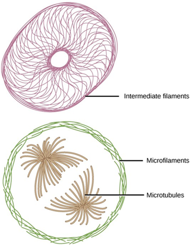
A network of protein fibers creating support, structure, shape, and movement for cells. The cytoskeleton can be thought of as the "skeleton" of the cell. There are three types of fibers within the cytoskeleton: microfilaments, intermediate filaments, and microtubules.
|
Long thin fibers scattered throughout the cytoplasm that attach to the plasma membrane and the organelles. |
|
flagellum (pl. flagella)
|
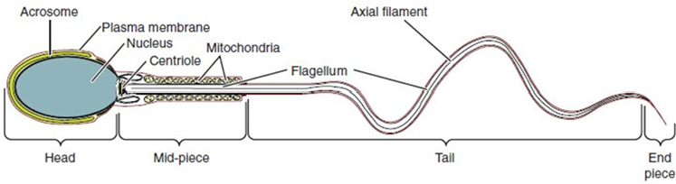
Flagella are long, hair-like structures that extend from the plasma membrane and enable an entire cell to move. The only human cell type that has a flagellum is a sperm cell. |
A long thin whip-like tail with one end embedded inside of the cell. The majority of a flagellum located outside of the cell. |
|
Golgi apparatus / Golgi body / Golgi complex
|
Sorting, tagging, packaging, and distributing lipids and proteins takes place in the Golgi apparatus. The Golgi can be thought of as the "post office" of a cell. |
A series of roughly dumbbell-shaped membranes stacked next to each other. |
|
lysosome
|
Lysosomes are the cell’s “garbage disposal.” Enzymes within the lysosomes aid in breaking down proteins, polysaccharides, lipids, nucleic acids, and even worn-out organelles. These enzymes are active at a much lower pH than the cytoplasm's. Therefore, the pH within lysosomes is more acidic than the cytoplasm's pH. Many reactions that take place in the cytoplasm could not occur at a low pH. |
A spherical structure smaller than Golgi and ER membranes. |
|
microvillus (pl. microvilli)
|
The plasma membranes of cells that specialize in absorption fold into fingerlike projections that we call microvilli. Such cells typically line the small intestine, the organ that absorbs nutrients from digested food. This is an excellent example of form following function. People with celiac disease have an immune response to gluten, which is a protein in what, barley, and rye. The immune response damages microvilli, and thus, afflicted individuals cannot absorb nutrients. This leads to malnutrition, cramping, and diarrhea. |
The plasma membrane forms finger-like structures on one of the cell's surfaces. Microvilli resemble Bart Simpson's (the cartoon character's) hair. |
|
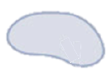
mitochondrion (pl. mitochondria)
|
Mitochondria are often called the “powerhouses” or “energy factories” of a cell because they are responsible for making adenosine triphosphate (ATP), the cell’s main energy-carrying molecule. ATP represents the short-term stored energy of the cell. Cellular respiration is the process of making ATP using the chemical energy found in glucose and other nutrients. In mitochondria, this process uses oxygen and produces carbon dioxide as a waste product. |
Often kidney-bean shaped structure with membranes pointing into its interior. |
|
nucleus (pl. nuclei)
|
Houses the cell’s DNA and directs the synthesis of proteins (gene expression) and the synthesis of ribosomes. Composed of the chromatin, nucleolus or nucleoli, a nuclear envelope, and nuclear pores. Nucleoplasm is essentially the cytoplasm of the nucleus. |
A large and often prominent structure that may be roughly spherical, but not always. |
|
nucleolus (pl. nucleoli)
|
A darkly staining area within the nucleus called the nucleolus aggregates the ribosomal RNA with associated proteins to assemble the ribosomal subunits that are then transported out through the pores in the nuclear envelope to the cytoplasm. |
A dark and dense region within the nucleus. There may be more than one nucleolus or none. |
|
peroxisome
|
Peroxisomes are small, round organelles enclosed by single membranes. They carry out oxidation reactions that break down fatty acids and amino acids. They also detoxify many poisons that may enter the body. (Many of these oxidation reactions release hydrogen peroxide, H2O2, which would be damaging to cells; however, when these reactions are confined to peroxisomes, enzymes safely break down the H2O2 into oxygen and water.) For example, alcohol is detoxified by peroxisomes in liver cells. |
A spherical structure smaller than Golgi and ER membranes. |
|
plasma membrane (cell membrane)
|
The plasma membrane is a phospholipid bilayer with embedded proteins that separates the internal contents of the cell from its surrounding environment. The plasma membrane controls the passage of organic molecules, ions, water, and oxygen into and out of the cell. Wastes (such as carbon dioxide and ammonia) also leave the cell by passing through the plasma membrane.
|
A sphere that surrounds the entire cell. It is shown as a line that separates the cell from its environment. |
|
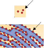
ribosome
|
Ribosomes are the cellular structures responsible for protein synthesis. When viewed through an electron microscope, ribosomes appear either as clusters (polyribosomes) or single, tiny dots that float freely in the cytoplasm. They may also be attached to the cytoplasmic side of the plasma membrane, the exterior of the rough endoplasmic reticulum and the outer membrane of the nuclear envelope. |
Very small dot-like structures that are smaller than lysosomes, peroxisomes, and vesicles. Ribosomes may be found in the cytoplasm or attached to the RER. |
|
rough endoplasmic reticulum (RER)
|
Ribosomes attached to the RER transfer their newly synthesized proteins into the RER's interior where they undergo structural modifications, such as folding or acquiring side chains. These modified proteins incorporate into cellular membranes. The proteins synthesized with the RER can also be secreted from the cell (such as protein hormones and enzymes). The RER also makes phospholipids for cellular membranes. |
Thin long ovals and circles of membrane that often stack irregularly on top of each other. RER has ribosomes attached to the outside giving it a "rough" appearance. |
|
smooth endoplasmic reticulum (SER)
|
SER functions include synthesis of carbohydrates, lipids, and steroid hormones; detoxification of medications and poisons; and storing calcium ions. In muscle cells, a specialized SER, called the sarcoplasmic reticulum, is responsible for storing calcium ions that are needed to trigger the muscle cells' coordinated contractions. |
Thin long ovals and circles of membrane that often stack irregularly on top of each other. SER does not have ribosomes attached giving it a "smooth" appearance. |
|
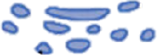
vacuole / vesicle / storage vesicles
|
Vesicles and vacuoles are membrane-bound sacs that function in storage and transport. Other than the fact that vacuoles are somewhat larger than vesicles, there is a very subtle distinction between them: The membranes of vesicles can fuse with either the plasma membrane or other membrane systems within the cell and vacuoles cannot. Storage vesicles can store materials, such as lipid droplets in adipose cells (fat cells). |
A spherical or irregularly shaped structure. Sizes vary widely. |


Attributions
- "Anatomy and Physiology" by J. Gordon Betts et al., OpenStax is licensed under CC BY 4.0
- "Biology 2e" by Mary Ann Clark, Matthew Douglas, Jung Choi, OpenStax is licensed under CC BY-NC 4.0