4.5: MR-VP Tests
- Page ID
- 149780
\( \newcommand{\vecs}[1]{\overset { \scriptstyle \rightharpoonup} {\mathbf{#1}} } \)
\( \newcommand{\vecd}[1]{\overset{-\!-\!\rightharpoonup}{\vphantom{a}\smash {#1}}} \)
\( \newcommand{\dsum}{\displaystyle\sum\limits} \)
\( \newcommand{\dint}{\displaystyle\int\limits} \)
\( \newcommand{\dlim}{\displaystyle\lim\limits} \)
\( \newcommand{\id}{\mathrm{id}}\) \( \newcommand{\Span}{\mathrm{span}}\)
( \newcommand{\kernel}{\mathrm{null}\,}\) \( \newcommand{\range}{\mathrm{range}\,}\)
\( \newcommand{\RealPart}{\mathrm{Re}}\) \( \newcommand{\ImaginaryPart}{\mathrm{Im}}\)
\( \newcommand{\Argument}{\mathrm{Arg}}\) \( \newcommand{\norm}[1]{\| #1 \|}\)
\( \newcommand{\inner}[2]{\langle #1, #2 \rangle}\)
\( \newcommand{\Span}{\mathrm{span}}\)
\( \newcommand{\id}{\mathrm{id}}\)
\( \newcommand{\Span}{\mathrm{span}}\)
\( \newcommand{\kernel}{\mathrm{null}\,}\)
\( \newcommand{\range}{\mathrm{range}\,}\)
\( \newcommand{\RealPart}{\mathrm{Re}}\)
\( \newcommand{\ImaginaryPart}{\mathrm{Im}}\)
\( \newcommand{\Argument}{\mathrm{Arg}}\)
\( \newcommand{\norm}[1]{\| #1 \|}\)
\( \newcommand{\inner}[2]{\langle #1, #2 \rangle}\)
\( \newcommand{\Span}{\mathrm{span}}\) \( \newcommand{\AA}{\unicode[.8,0]{x212B}}\)
\( \newcommand{\vectorA}[1]{\vec{#1}} % arrow\)
\( \newcommand{\vectorAt}[1]{\vec{\text{#1}}} % arrow\)
\( \newcommand{\vectorB}[1]{\overset { \scriptstyle \rightharpoonup} {\mathbf{#1}} } \)
\( \newcommand{\vectorC}[1]{\textbf{#1}} \)
\( \newcommand{\vectorD}[1]{\overrightarrow{#1}} \)
\( \newcommand{\vectorDt}[1]{\overrightarrow{\text{#1}}} \)
\( \newcommand{\vectE}[1]{\overset{-\!-\!\rightharpoonup}{\vphantom{a}\smash{\mathbf {#1}}}} \)
\( \newcommand{\vecs}[1]{\overset { \scriptstyle \rightharpoonup} {\mathbf{#1}} } \)
\(\newcommand{\longvect}{\overrightarrow}\)
\( \newcommand{\vecd}[1]{\overset{-\!-\!\rightharpoonup}{\vphantom{a}\smash {#1}}} \)
\(\newcommand{\avec}{\mathbf a}\) \(\newcommand{\bvec}{\mathbf b}\) \(\newcommand{\cvec}{\mathbf c}\) \(\newcommand{\dvec}{\mathbf d}\) \(\newcommand{\dtil}{\widetilde{\mathbf d}}\) \(\newcommand{\evec}{\mathbf e}\) \(\newcommand{\fvec}{\mathbf f}\) \(\newcommand{\nvec}{\mathbf n}\) \(\newcommand{\pvec}{\mathbf p}\) \(\newcommand{\qvec}{\mathbf q}\) \(\newcommand{\svec}{\mathbf s}\) \(\newcommand{\tvec}{\mathbf t}\) \(\newcommand{\uvec}{\mathbf u}\) \(\newcommand{\vvec}{\mathbf v}\) \(\newcommand{\wvec}{\mathbf w}\) \(\newcommand{\xvec}{\mathbf x}\) \(\newcommand{\yvec}{\mathbf y}\) \(\newcommand{\zvec}{\mathbf z}\) \(\newcommand{\rvec}{\mathbf r}\) \(\newcommand{\mvec}{\mathbf m}\) \(\newcommand{\zerovec}{\mathbf 0}\) \(\newcommand{\onevec}{\mathbf 1}\) \(\newcommand{\real}{\mathbb R}\) \(\newcommand{\twovec}[2]{\left[\begin{array}{r}#1 \\ #2 \end{array}\right]}\) \(\newcommand{\ctwovec}[2]{\left[\begin{array}{c}#1 \\ #2 \end{array}\right]}\) \(\newcommand{\threevec}[3]{\left[\begin{array}{r}#1 \\ #2 \\ #3 \end{array}\right]}\) \(\newcommand{\cthreevec}[3]{\left[\begin{array}{c}#1 \\ #2 \\ #3 \end{array}\right]}\) \(\newcommand{\fourvec}[4]{\left[\begin{array}{r}#1 \\ #2 \\ #3 \\ #4 \end{array}\right]}\) \(\newcommand{\cfourvec}[4]{\left[\begin{array}{c}#1 \\ #2 \\ #3 \\ #4 \end{array}\right]}\) \(\newcommand{\fivevec}[5]{\left[\begin{array}{r}#1 \\ #2 \\ #3 \\ #4 \\ #5 \\ \end{array}\right]}\) \(\newcommand{\cfivevec}[5]{\left[\begin{array}{c}#1 \\ #2 \\ #3 \\ #4 \\ #5 \\ \end{array}\right]}\) \(\newcommand{\mattwo}[4]{\left[\begin{array}{rr}#1 \amp #2 \\ #3 \amp #4 \\ \end{array}\right]}\) \(\newcommand{\laspan}[1]{\text{Span}\{#1\}}\) \(\newcommand{\bcal}{\cal B}\) \(\newcommand{\ccal}{\cal C}\) \(\newcommand{\scal}{\cal S}\) \(\newcommand{\wcal}{\cal W}\) \(\newcommand{\ecal}{\cal E}\) \(\newcommand{\coords}[2]{\left\{#1\right\}_{#2}}\) \(\newcommand{\gray}[1]{\color{gray}{#1}}\) \(\newcommand{\lgray}[1]{\color{lightgray}{#1}}\) \(\newcommand{\rank}{\operatorname{rank}}\) \(\newcommand{\row}{\text{Row}}\) \(\newcommand{\col}{\text{Col}}\) \(\renewcommand{\row}{\text{Row}}\) \(\newcommand{\nul}{\text{Nul}}\) \(\newcommand{\var}{\text{Var}}\) \(\newcommand{\corr}{\text{corr}}\) \(\newcommand{\len}[1]{\left|#1\right|}\) \(\newcommand{\bbar}{\overline{\bvec}}\) \(\newcommand{\bhat}{\widehat{\bvec}}\) \(\newcommand{\bperp}{\bvec^\perp}\) \(\newcommand{\xhat}{\widehat{\xvec}}\) \(\newcommand{\vhat}{\widehat{\vvec}}\) \(\newcommand{\uhat}{\widehat{\uvec}}\) \(\newcommand{\what}{\widehat{\wvec}}\) \(\newcommand{\Sighat}{\widehat{\Sigma}}\) \(\newcommand{\lt}{<}\) \(\newcommand{\gt}{>}\) \(\newcommand{\amp}{&}\) \(\definecolor{fillinmathshade}{gray}{0.9}\)
- Describe the purpose and usefulness of the MR-VP test.
- Compare and contrast the MR test and the VP test.
- Tell how the MR test and VP test are conducted and what the results mean.
- Define fermentation and describe its importance.
- Name the metabolic pathway that occurs in MR positive bacterial species.
- Name the metabolic pathway that occurs in VP positive bacterial species.
- Successfully conduct and interpret the MR-VP test.
Methyl Red–Voges Proskauer (MR-VP) Test
The MR-VP test is actually a set of two separate tests that uses one broth medium containing glucose to determine the possible types of glucose fermentation being conducted by a bacterial species. Some bacterial species may ferment glucose a using mixed acid fermentation pathway (detected in the MR test or methyl red test) while some other bacterial species may ferment glucose via the butanediol fermentation pathway (detected in the VP test or Voges-Proskauer test).
Since the type of fermentation conducted differs by bacterial species (or strain), testing the type of fermentation pathway is useful for characterizing bacteria and identifying them. Metabolic reactions are dependent on the enzymes that species (or strain) has, which is dependent on the genes that species (or strain) carries in its DNA. Therefore, testing the types of metabolisms species conduct provides insights into differences in their genetics (their DNA).
Fermentation
Fermentation is a type of metabolic pathway some bacterial species conduct to produce ATP (ATP is used as an energy source to keep the cell alive). Fermentation is an anaerobic process (no O2 is used) and species that conduct fermentation will produce a small amount of ATP that has been derived from the energy in a glucose molecule.
Fermentation begins with glycolysis, the metabolic pathway that breaks down glucose into two pyruvate (or pyruvic acid) molecules and will produce a net gain (overall gain) of 2 ATP. Then, in a fermentation pathway (the pathways differ depending on the species), pyruvate is converted into other molecules to regenerate NAD+ so the glycolysis pathway can continue (NAD+ is a cofactor in a glycolysis reaction that must be available for glycolysis to occur). Additionally, fermentation pathways utilize organic molecules, derived from pyruvate, as final electron acceptors.

Methyl Red (MR) Test Detects Mixed Acid Fermentation
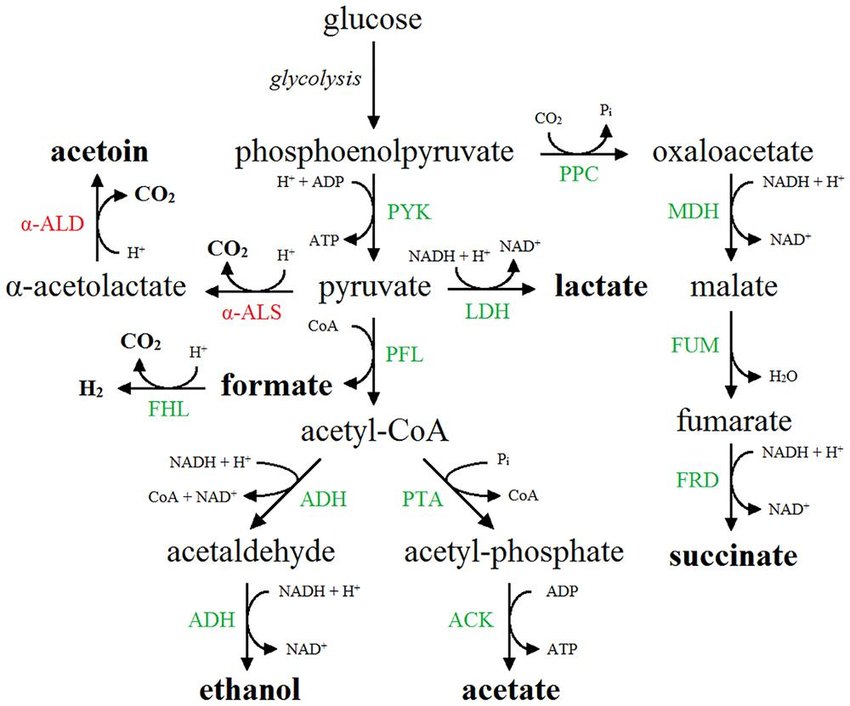
Mixed-acid fermentation pathways are fermentation pathways that some bacterial species conduct to produce a mixture of fermentation products. There is not just one way bacteria conduct mixed acid fermentation, but a number of them. The end-products produced through mixed-acid fermentation is species-dependent and sometimes strain-dependent (depends on the genes that the species has; genes dictate the enzymes produced and therefore the chemical reactions occurring in the species' or strains' metabolism). Below shows one example of glycolysis with a mixed acid fermentation pathway that produces acetoin, formate, lactate, succinate, acetate, and ethanol as byproducts of the process.

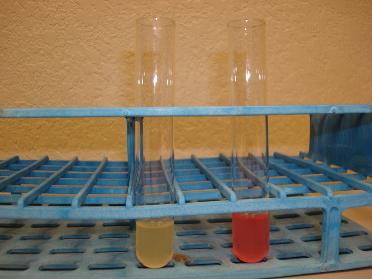
The methyl red test (MR) utilizes a liquid medium containing glucose. After this medium is inoculated and incubated to enable bacteria to grow and conduct fermentation, the reagent methyl red is added to the medium. Methyl red is a pH indicator that will detect mixed acid production by changing the medium to a red color (methyl red positive). In the absence of mixed acid fermentation, methyl red will not produce a red color (methyl red negative).
MR-VP Broth (glucose) –> pyruvate –> mixed acid fermentation pathway (red color with methyl red)

Voges-Proskauer (VP) Test Detects Butanediol Fermentation
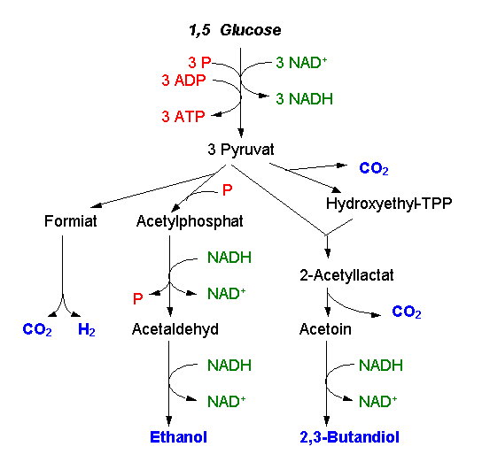
The Voges-Proskauer test determines if 2,3-butanediol is a product of glucose fermentation by a bacterial species. This fermentation byproduct typically is produced by species of Klebsiella and Enterobacter.

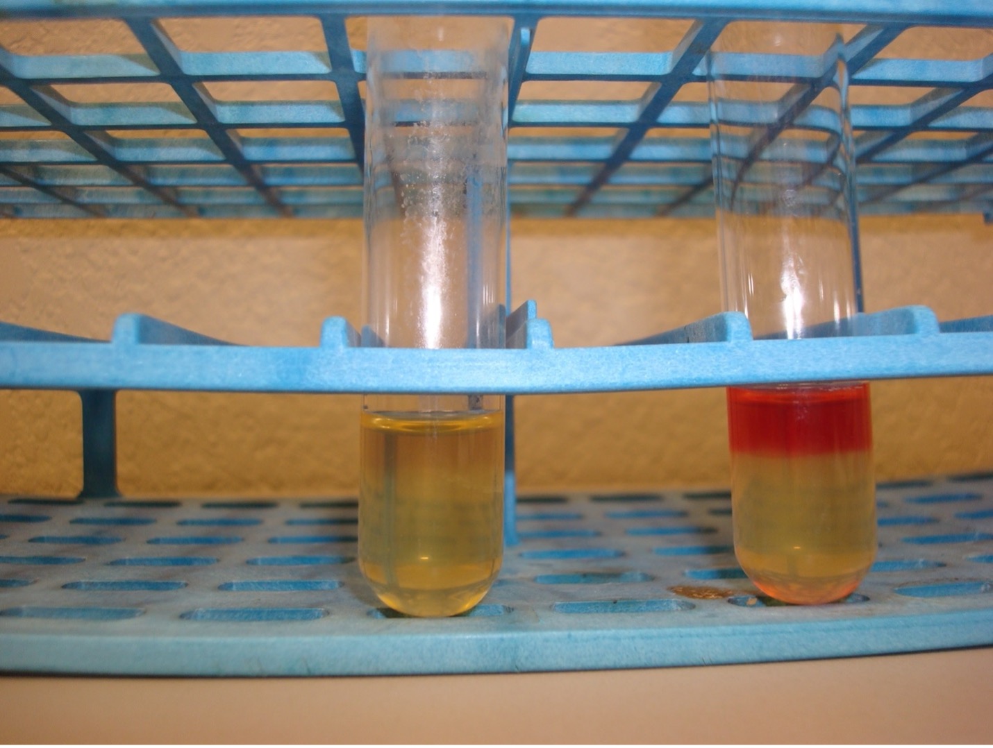
The Voges-Proskauer test removes some of the MR-VP medium into a separate test tube prior to conducting the methyl red test and adds Barritt’s A reagent followed by Barritt's B reagent to the medium. After mixing and a rest period, a liquid layer forms at the top of the test tube indicating the test results. If the top liquid layer is red or red-brown, the bacteria is Voges-Proskauer positive indicating it produces 2,3-butanediol as a byproduct of fermentation. If the top liquid layer is yellow, the bacteria is Voges-Proskauer negative indicating it does not produce 2,3-butanediol as a byproduct of fermentation.
MR-VP Broth (glucose) –> pyruvate –>butanediol fermentation pathway (red color in top layers of medium after adding reagents, mixing, and resting)

Laboratory Instructions
- Obtain a MR-VP broth tube. Use tape to label the tube with your name, your assigned organism(s) and the name of the test media.
- Aseptically transfer bacteria with a loop to inoculate the MR-VP broth.
- Incubate the inoculated tube in the class test tube rack until next lab session.
- After incubation, using a transfer pipette, transfer half of the inoculated MR-VP broth to a clean test tube. Label this tube “VP.” Leave the other half of the inoculated broth in the original MR-VP broth test tube. Label this tube “MR.”
- Methyl red test:
- Add 10 drops of methyl red reagent to “MR” tube.
- Examine the color of the medium.
- Record the results.
- Voges-Proskauer test:
- First add 15 drops of Barritt’s A reagent (alpha-napththol) to the "VP" tube.
- Add 5 drops of Barritt’s B reagent (40% KOH) to the “VP” tube. NOTE: A reversal in the order of the reagents may result in a weak-positive or false-negative reaction.
- Hold the test tube from the glass and mix the tube well by flicking it with your fingers.
- Let the test tube sit undisturbed in a test tube rack for 20 minutes.
- Examine the color of the top layer of liquid and record the results.
Results & Questions
| bacterial species tested | color of medium for MR test | results from methyl red test (+/-) | species conducts mixed-acid fermentation (+/-) | color of top liquid layer for VP test | results from Voges-Proskauer test (+/-) | species conducts butanediol fermentation (+/-) |
|---|---|---|---|---|---|---|
| Escherichia coli | ||||||
| Enterobacter aerogenes |
- Complete the table above to summarize results and interpretations of those results.
- What is fermentation and why is it important for bacterial species that conduct fermentation?
- What are the interrelationships between glycolysis and fermentation (there are two main ones)?
- What is mixed-acid fermentation?
- What is butanediol fermentation?
- Explain why the MR-VP test is useful for characterizing and identifying bacterial species.
Attributions
- 2,3-Butandiol-Gaerung Schema.png by Brudersohn is licensed under CC BY-SA 3.0
- Formate hydrogen lyase mediates stationary-phase deacidification and increases survival during sugar fermentation in acetoin-producing enterobacteria by Bram Vivijs et al. is licensed under CC BY 4.0
- Red Mountain Microbiology by Jill Raymond Ph.D.; Graham Boorse, Ph.D.; and Anne Mason M.S. is licensed under CC BY-NC 4.0


