
3.2: Comparison of Sizes and Shapes of Microorganisms
- Page ID
- 52236
\( \newcommand{\vecs}[1]{\overset { \scriptstyle \rightharpoonup} {\mathbf{#1}} } \)
\( \newcommand{\vecd}[1]{\overset{-\!-\!\rightharpoonup}{\vphantom{a}\smash {#1}}} \)
\( \newcommand{\dsum}{\displaystyle\sum\limits} \)
\( \newcommand{\dint}{\displaystyle\int\limits} \)
\( \newcommand{\dlim}{\displaystyle\lim\limits} \)
\( \newcommand{\id}{\mathrm{id}}\) \( \newcommand{\Span}{\mathrm{span}}\)
( \newcommand{\kernel}{\mathrm{null}\,}\) \( \newcommand{\range}{\mathrm{range}\,}\)
\( \newcommand{\RealPart}{\mathrm{Re}}\) \( \newcommand{\ImaginaryPart}{\mathrm{Im}}\)
\( \newcommand{\Argument}{\mathrm{Arg}}\) \( \newcommand{\norm}[1]{\| #1 \|}\)
\( \newcommand{\inner}[2]{\langle #1, #2 \rangle}\)
\( \newcommand{\Span}{\mathrm{span}}\)
\( \newcommand{\id}{\mathrm{id}}\)
\( \newcommand{\Span}{\mathrm{span}}\)
\( \newcommand{\kernel}{\mathrm{null}\,}\)
\( \newcommand{\range}{\mathrm{range}\,}\)
\( \newcommand{\RealPart}{\mathrm{Re}}\)
\( \newcommand{\ImaginaryPart}{\mathrm{Im}}\)
\( \newcommand{\Argument}{\mathrm{Arg}}\)
\( \newcommand{\norm}[1]{\| #1 \|}\)
\( \newcommand{\inner}[2]{\langle #1, #2 \rangle}\)
\( \newcommand{\Span}{\mathrm{span}}\) \( \newcommand{\AA}{\unicode[.8,0]{x212B}}\)
\( \newcommand{\vectorA}[1]{\vec{#1}} % arrow\)
\( \newcommand{\vectorAt}[1]{\vec{\text{#1}}} % arrow\)
\( \newcommand{\vectorB}[1]{\overset { \scriptstyle \rightharpoonup} {\mathbf{#1}} } \)
\( \newcommand{\vectorC}[1]{\textbf{#1}} \)
\( \newcommand{\vectorD}[1]{\overrightarrow{#1}} \)
\( \newcommand{\vectorDt}[1]{\overrightarrow{\text{#1}}} \)
\( \newcommand{\vectE}[1]{\overset{-\!-\!\rightharpoonup}{\vphantom{a}\smash{\mathbf {#1}}}} \)
\( \newcommand{\vecs}[1]{\overset { \scriptstyle \rightharpoonup} {\mathbf{#1}} } \)
\(\newcommand{\longvect}{\overrightarrow}\)
\( \newcommand{\vecd}[1]{\overset{-\!-\!\rightharpoonup}{\vphantom{a}\smash {#1}}} \)
\(\newcommand{\avec}{\mathbf a}\) \(\newcommand{\bvec}{\mathbf b}\) \(\newcommand{\cvec}{\mathbf c}\) \(\newcommand{\dvec}{\mathbf d}\) \(\newcommand{\dtil}{\widetilde{\mathbf d}}\) \(\newcommand{\evec}{\mathbf e}\) \(\newcommand{\fvec}{\mathbf f}\) \(\newcommand{\nvec}{\mathbf n}\) \(\newcommand{\pvec}{\mathbf p}\) \(\newcommand{\qvec}{\mathbf q}\) \(\newcommand{\svec}{\mathbf s}\) \(\newcommand{\tvec}{\mathbf t}\) \(\newcommand{\uvec}{\mathbf u}\) \(\newcommand{\vvec}{\mathbf v}\) \(\newcommand{\wvec}{\mathbf w}\) \(\newcommand{\xvec}{\mathbf x}\) \(\newcommand{\yvec}{\mathbf y}\) \(\newcommand{\zvec}{\mathbf z}\) \(\newcommand{\rvec}{\mathbf r}\) \(\newcommand{\mvec}{\mathbf m}\) \(\newcommand{\zerovec}{\mathbf 0}\) \(\newcommand{\onevec}{\mathbf 1}\) \(\newcommand{\real}{\mathbb R}\) \(\newcommand{\twovec}[2]{\left[\begin{array}{r}#1 \\ #2 \end{array}\right]}\) \(\newcommand{\ctwovec}[2]{\left[\begin{array}{c}#1 \\ #2 \end{array}\right]}\) \(\newcommand{\threevec}[3]{\left[\begin{array}{r}#1 \\ #2 \\ #3 \end{array}\right]}\) \(\newcommand{\cthreevec}[3]{\left[\begin{array}{c}#1 \\ #2 \\ #3 \end{array}\right]}\) \(\newcommand{\fourvec}[4]{\left[\begin{array}{r}#1 \\ #2 \\ #3 \\ #4 \end{array}\right]}\) \(\newcommand{\cfourvec}[4]{\left[\begin{array}{c}#1 \\ #2 \\ #3 \\ #4 \end{array}\right]}\) \(\newcommand{\fivevec}[5]{\left[\begin{array}{r}#1 \\ #2 \\ #3 \\ #4 \\ #5 \\ \end{array}\right]}\) \(\newcommand{\cfivevec}[5]{\left[\begin{array}{c}#1 \\ #2 \\ #3 \\ #4 \\ #5 \\ \end{array}\right]}\) \(\newcommand{\mattwo}[4]{\left[\begin{array}{rr}#1 \amp #2 \\ #3 \amp #4 \\ \end{array}\right]}\) \(\newcommand{\laspan}[1]{\text{Span}\{#1\}}\) \(\newcommand{\bcal}{\cal B}\) \(\newcommand{\ccal}{\cal C}\) \(\newcommand{\scal}{\cal S}\) \(\newcommand{\wcal}{\cal W}\) \(\newcommand{\ecal}{\cal E}\) \(\newcommand{\coords}[2]{\left\{#1\right\}_{#2}}\) \(\newcommand{\gray}[1]{\color{gray}{#1}}\) \(\newcommand{\lgray}[1]{\color{lightgray}{#1}}\) \(\newcommand{\rank}{\operatorname{rank}}\) \(\newcommand{\row}{\text{Row}}\) \(\newcommand{\col}{\text{Col}}\) \(\renewcommand{\row}{\text{Row}}\) \(\newcommand{\nul}{\text{Nul}}\) \(\newcommand{\var}{\text{Var}}\) \(\newcommand{\corr}{\text{corr}}\) \(\newcommand{\len}[1]{\left|#1\right|}\) \(\newcommand{\bbar}{\overline{\bvec}}\) \(\newcommand{\bhat}{\widehat{\bvec}}\) \(\newcommand{\bperp}{\bvec^\perp}\) \(\newcommand{\xhat}{\widehat{\xvec}}\) \(\newcommand{\vhat}{\widehat{\vvec}}\) \(\newcommand{\uhat}{\widehat{\uvec}}\) \(\newcommand{\what}{\widehat{\wvec}}\) \(\newcommand{\Sighat}{\widehat{\Sigma}}\) \(\newcommand{\lt}{<}\) \(\newcommand{\gt}{>}\) \(\newcommand{\amp}{&}\) \(\definecolor{fillinmathshade}{gray}{0.9}\)Learning Outcomes
- Recognize the shapes and arrangements of some common types of bacteria.
A. BACTERIAL SHAPES, ARRANGEMENTS, AND FORMS
Bacteria are unicellular prokaryotic microorganisms that divide by binary fission, a process by which one bacterium splits into two.
There are three common shapes of bacteria:
- coccus (singular) or cocci (plural)
- bacillus or rod (singular) or bacilli, rods (plural)
- spiral (example of a spirochete, a type of spiral is shown in image 1)
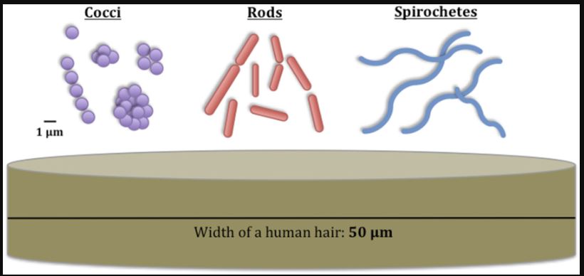
Image 1: These are the different shapes of bacteria and their sizes compared with the width of a human hair.
The unit “μm” is a measurement of length, the “micrometer,” or commonly known as the micron. It equals 1×10⁻⁶ meters that is, one millionth of a meter. Image courtesy of Kestin Schulz, Mariya W. Smit, Lydie Herfort and Holly M. Simon URL: https://commons.wikimedia.org/wiki/F...omparisons.jpg
The cocci come in 5 different arrangements; the bacilli in 3 different arrangements; and the spirals in 3 different forms.
1. Coccus
A coccus-shaped bacterium is usually spherical, although some appear oval, elongated, or flattened on one side. Most cocci are approximately 0.5 - 1.0 micrometer (µm) in diameter and may be seen, based on their planes of division and tendency to remain attached after replication, in one of the following arrangements (see Fig. 1A):
a. Division in one plane produces either a diplococcus (see Fig. 1A and Fig. 1B) or streptococcus (see Fig. 1A and Fig. 1C) arrangement.
1. diplococcus: a pair of cocci
- Photomicrograph of a diplococcus
- Scanning electron micrograph of a Streptococcus pneumoniae, a diplococcus
- Scanning electron micrograph of a Neisseria, a diplococcus; courtesy of Dennis Kunkel's Microscopy
2. streptococcus: a chain of cocci
- Photomicrograph of a streptococcus
- Scanning electron micrograph of Streptococcus pyogenes; courtesy of Dennis Kunkel's Microscopy
- Transmission electron micrograph of Streptococcus
-Scanning Electron Micrograph of Enterococcus
b. Division in two planes produces a tetrad arrangement (see Fig. 1A and Fig. 1D).
- tetrad: a square of 4 cocci
- Photomicrograph of a tetrad
- Scanning electron micrograph of Micrococcus luteus
c. Division in three planes produces a sarcina arrangement (see Fig. 1A).
- sarcina: a cube of 8 cocci
- Photomicrograph of a sarcina
It is difficult with a conventional light microscope to tell a tetrad arrangement (square of four cocci) from a sarcina arrangement (cube of eight) so in our lab, anytime you see ba square of four cocci, say it is either a tetrad or a sarcina arrangement.
d. Division in random planes produces a staphylococcus arrangement (see Fig. 1A and Fig. 1E).
- Photomicrograph of a staphylococcus, negative image
- Scanning electron micrograph of Staphylococcus aureus; courtesy of Dennis Kunkel's Microscopy
- Scanning electron micrograph of methicillin-resistant Staphylococcus aureus (MRSA); courtesy of CDC
As you observe these different cocci, keep in mind that the procedures used in slide preparation may cause some arrangements to break apart or clump together (see Figs. 1D and 1E). The correct form, however, should predominate. Also remember that each coccus in an arrangement represents a complete, individual, one-celled organism.
2. Bacillus (rod)
A bacillus or rod is a hotdog-shaped bacterium having one of the following arrangements (see Fig 2A):
a. bacillus: a single bacillus (see Fig 2A and Fig 2B)
- Photomicrograph of a bacillus
- Scanning electron micrograph of a bacillus
- Scanning electron micrograph of Escherichia coli O157H7, a bacillus; courtesy of CDC
b. streptobacillus: bacilli in chains (see Fig 2A and Fig 2C)
- Photomicrograph of a streptobacillus
c. coccobacillus: oval and similar to a coccus (see Fig 2A, 2D, and 2E)
A single bacillus is typically 0.5-1.0 µm wide and from 1- 4 µm long. Small bacilli or bacilli that are dividing or have just divided by binary fission may at first glance be confused for diplococci or cocci (see Fig. 2A) so they must be observed carefully. You will, however, be able to see bacilli that have not divided and are definitely rod-shaped as well as bacilli in the process of dividing.
3. Spiral
Spiral-shaped bacteria occur in one of three forms (see Fig. 3A):
a. vibrio: an incomplete spiral or comma-shaped (see Fig. 3A and Fig. 3B)
- Photomicrograph of a vibrio
- Scanning electron micrograph of Vibrio cholerae; courtesy of Dennis Kunkel's Microscopy
b. spirillum: a thick, rigid spiral (see Fig. 3A and Fig. 3C)
- Photomicrograph of a spirillum
c. spirochete: a thin, flexible spiral (see Fig. 3A and Fig. 3D)
- Photomicrograph of a spirochete
- Scanning electron micrograph of the spirochete Leptospira; courtesy of CDC
- scanning electron micrograph of the spirochete Treponema pallidum; courtesy of CDC
The spirals you will observe range from 5-40 µm long but some are over 100 µm in length. The spirochetes are the thinnest of the bacteria, often having a width of only 0.25-0.5 µm.
To view a nice interactive illustration comparing size of cells and microbes, see the Cell Size and Scale Resource at the University of Utah.
B. YEASTS
Yeasts, such as the common baker's yeast Saccharomyces cerevisiae (see Fig. 4), are unicellular fungi. They usually appear spherical and have a diameter of 3 - 5 µm. Yeasts commonly reproduce asexually by a process called budding. Unlike bacteria, which are prokaryotic, yeasts are eukaryotic.
- Scanning electron micrograph of Saccharomyces; courtesy of Dennis Kunkel's Microscopy
To view a nice interactive illustration comparing size of cells and microbes, see the Cell Size and Scale Resource at the University of Utah.
C. MEASUREMENT OF MICROORGANISMS
The approximate size of a microorganism can be determined using an ocular micrometer (see Fig. 5) , an eyepiece that contains a scale that will appear superimposed upon the focused specimen.
To view a nice interactive illustration comparing size of cells and microbes, see the Cell size & scale resource at the University of Utah's Learn.genetics site, URL: https://learn.genetics.utah.edu/content/cells/scale/