Biology LibreTexts

15.1: Embryogenesis

  • Page ID
    123973
  • \( \newcommand{\vecs}[1]{\overset { \scriptstyle \rightharpoonup} {\mathbf{#1}} } \)

    \( \newcommand{\vecd}[1]{\overset{-\!-\!\rightharpoonup}{\vphantom{a}\smash {#1}}} \)

    \( \newcommand{\id}{\mathrm{id}}\) \( \newcommand{\Span}{\mathrm{span}}\)

    ( \newcommand{\kernel}{\mathrm{null}\,}\) \( \newcommand{\range}{\mathrm{range}\,}\)

    \( \newcommand{\RealPart}{\mathrm{Re}}\) \( \newcommand{\ImaginaryPart}{\mathrm{Im}}\)

    \( \newcommand{\Argument}{\mathrm{Arg}}\) \( \newcommand{\norm}[1]{\| #1 \|}\)

    \( \newcommand{\inner}[2]{\langle #1, #2 \rangle}\)

    \( \newcommand{\Span}{\mathrm{span}}\)

    \( \newcommand{\id}{\mathrm{id}}\)

    \( \newcommand{\Span}{\mathrm{span}}\)

    \( \newcommand{\kernel}{\mathrm{null}\,}\)

    \( \newcommand{\range}{\mathrm{range}\,}\)

    \( \newcommand{\RealPart}{\mathrm{Re}}\)

    \( \newcommand{\ImaginaryPart}{\mathrm{Im}}\)

    \( \newcommand{\Argument}{\mathrm{Arg}}\)

    \( \newcommand{\norm}[1]{\| #1 \|}\)

    \( \newcommand{\inner}[2]{\langle #1, #2 \rangle}\)

    \( \newcommand{\Span}{\mathrm{span}}\) \( \newcommand{\AA}{\unicode[.8,0]{x212B}}\)

    \( \newcommand{\vectorA}[1]{\vec{#1}}      % arrow\)

    \( \newcommand{\vectorAt}[1]{\vec{\text{#1}}}      % arrow\)

    \( \newcommand{\vectorB}[1]{\overset { \scriptstyle \rightharpoonup} {\mathbf{#1}} } \)

    \( \newcommand{\vectorC}[1]{\textbf{#1}} \)

    \( \newcommand{\vectorD}[1]{\overrightarrow{#1}} \)

    \( \newcommand{\vectorDt}[1]{\overrightarrow{\text{#1}}} \)

    \( \newcommand{\vectE}[1]{\overset{-\!-\!\rightharpoonup}{\vphantom{a}\smash{\mathbf {#1}}}} \)

    \( \newcommand{\vecs}[1]{\overset { \scriptstyle \rightharpoonup} {\mathbf{#1}} } \)

    \( \newcommand{\vecd}[1]{\overset{-\!-\!\rightharpoonup}{\vphantom{a}\smash {#1}}} \)

    \(\newcommand{\avec}{\mathbf a}\) \(\newcommand{\bvec}{\mathbf b}\) \(\newcommand{\cvec}{\mathbf c}\) \(\newcommand{\dvec}{\mathbf d}\) \(\newcommand{\dtil}{\widetilde{\mathbf d}}\) \(\newcommand{\evec}{\mathbf e}\) \(\newcommand{\fvec}{\mathbf f}\) \(\newcommand{\nvec}{\mathbf n}\) \(\newcommand{\pvec}{\mathbf p}\) \(\newcommand{\qvec}{\mathbf q}\) \(\newcommand{\svec}{\mathbf s}\) \(\newcommand{\tvec}{\mathbf t}\) \(\newcommand{\uvec}{\mathbf u}\) \(\newcommand{\vvec}{\mathbf v}\) \(\newcommand{\wvec}{\mathbf w}\) \(\newcommand{\xvec}{\mathbf x}\) \(\newcommand{\yvec}{\mathbf y}\) \(\newcommand{\zvec}{\mathbf z}\) \(\newcommand{\rvec}{\mathbf r}\) \(\newcommand{\mvec}{\mathbf m}\) \(\newcommand{\zerovec}{\mathbf 0}\) \(\newcommand{\onevec}{\mathbf 1}\) \(\newcommand{\real}{\mathbb R}\) \(\newcommand{\twovec}[2]{\left[\begin{array}{r}#1 \\ #2 \end{array}\right]}\) \(\newcommand{\ctwovec}[2]{\left[\begin{array}{c}#1 \\ #2 \end{array}\right]}\) \(\newcommand{\threevec}[3]{\left[\begin{array}{r}#1 \\ #2 \\ #3 \end{array}\right]}\) \(\newcommand{\cthreevec}[3]{\left[\begin{array}{c}#1 \\ #2 \\ #3 \end{array}\right]}\) \(\newcommand{\fourvec}[4]{\left[\begin{array}{r}#1 \\ #2 \\ #3 \\ #4 \end{array}\right]}\) \(\newcommand{\cfourvec}[4]{\left[\begin{array}{c}#1 \\ #2 \\ #3 \\ #4 \end{array}\right]}\) \(\newcommand{\fivevec}[5]{\left[\begin{array}{r}#1 \\ #2 \\ #3 \\ #4 \\ #5 \\ \end{array}\right]}\) \(\newcommand{\cfivevec}[5]{\left[\begin{array}{c}#1 \\ #2 \\ #3 \\ #4 \\ #5 \\ \end{array}\right]}\) \(\newcommand{\mattwo}[4]{\left[\begin{array}{rr}#1 \amp #2 \\ #3 \amp #4 \\ \end{array}\right]}\) \(\newcommand{\laspan}[1]{\text{Span}\{#1\}}\) \(\newcommand{\bcal}{\cal B}\) \(\newcommand{\ccal}{\cal C}\) \(\newcommand{\scal}{\cal S}\) \(\newcommand{\wcal}{\cal W}\) \(\newcommand{\ecal}{\cal E}\) \(\newcommand{\coords}[2]{\left\{#1\right\}_{#2}}\) \(\newcommand{\gray}[1]{\color{gray}{#1}}\) \(\newcommand{\lgray}[1]{\color{lightgray}{#1}}\) \(\newcommand{\rank}{\operatorname{rank}}\) \(\newcommand{\row}{\text{Row}}\) \(\newcommand{\col}{\text{Col}}\) \(\renewcommand{\row}{\text{Row}}\) \(\newcommand{\nul}{\text{Nul}}\) \(\newcommand{\var}{\text{Var}}\) \(\newcommand{\corr}{\text{corr}}\) \(\newcommand{\len}[1]{\left|#1\right|}\) \(\newcommand{\bbar}{\overline{\bvec}}\) \(\newcommand{\bhat}{\widehat{\bvec}}\) \(\newcommand{\bperp}{\bvec^\perp}\) \(\newcommand{\xhat}{\widehat{\xvec}}\) \(\newcommand{\vhat}{\widehat{\vvec}}\) \(\newcommand{\uhat}{\widehat{\uvec}}\) \(\newcommand{\what}{\widehat{\wvec}}\) \(\newcommand{\Sighat}{\widehat{\Sigma}}\) \(\newcommand{\lt}{<}\) \(\newcommand{\gt}{>}\) \(\newcommand{\amp}{&}\) \(\definecolor{fillinmathshade}{gray}{0.9}\)
    Learning Objectives
    • Explain the steps of embryogensis in eudicots.
    • Compare embryogenesis in eudicots versus monocots.

    The Seed Plants chapter discussed the fertilization of the egg within the ovule. The zygote ultimately divides to produce the mature embryo, the ovule develops into a seed, and the ovary that contained one or more ovules develops into a fruit. A typical seed contains a seed coat, cotyledons, endosperm, and a single embryo. The development of the embryo occurs through a process called embryogenesis.

    Embryogenesis in Eudicots

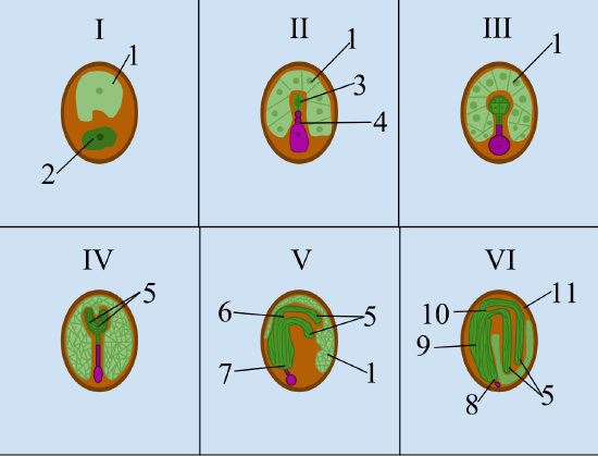
    After fertilization in eudicots, the zygote (Figure \(\PageIndex{1-I}\)) divides to form two cells: the upper apical cell and the lower basal cell . The division of the basal cell gives rise to the suspensor, which attaches the embryo to the micropyle (the pore through which the pollen tube original entered). The suspensor provides a route for nutrition to be transported from the mother plant to the growing embryo. The apical cell also divides, initially producing a proembryo (figure \(\PageIndex{1-II}\)).

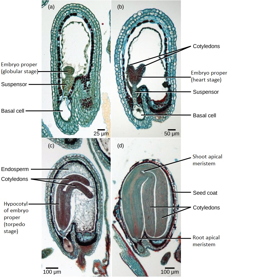
    As the proembryo continues to divide, it takes a spherical form, called the globular stage (Figures \(\PageIndex{1-III}\) and \(\PageIndex{2a}\)). The globular is the first stage that is considered the embryo proper. Next, cotyledons arise from the embryo proper, forming the heart stage (Figures \(\PageIndex{1-IV}\) and \(\PageIndex{2-4}\)). Cotyledons are embryonic leaf-life structures that function in food storage, food absorption and/or photosynthesis. As the cotyledons elongate, and the base of the embryo thickens, it results a torpedo. (This stage is called the torpedo stage; Figures \(\PageIndex{1-V}\) and \(\PageIndex{2c}\)). Cell division is concentrated at the shoot apical meristem, located at the shoot tip in between the cotyledons, and the root apical meristem at the most basal (bottom) part of the embryo. Most of the suspensor deteriorates during the torpedo stage.

    The final stage of embryogenesis results in the mature embryo (Figures \(\PageIndex{1-VI}\) and \(\PageIndex{2d}\)). The mature embryo includes an embryonic root called the radicle. The embryo becomes dormant at this point, halting metabolic activity and cell division. At this point, the seed is ready for dispersal. Growth resumes when the seed germinates and the embryo develops into a seedling.

    In some eudicots, the endosperm (triploid tissue that was formed when a sperm fertilized the two polar nuclei) cells divide, and endosperm fills a substantial portion of the mature seed. The endosperm stores nutrients. In other (non-endospermic) eudicots, such as Capsella bursa-pastoris, the endosperm develops initially, but is then digested, and the nutrients are moved into the two cotyledons (Figure \(\PageIndex{2}\)). After germination, the developing seedling relies on these food reserves stored in the endosperm or cotyledons until the first set of leaves begin photosynthesis.

    The six stages of embryogenesis within a developing eudicot seed
    Figure \(\PageIndex{1}\): The stages of embryogenesis in eudicots. I. The developing seed contains the triploid endosperm (1) and the diploid zygote (2). II. The zygote divides, forming the proembryo (3), which is anchored to the micropyle by suspensor (4), which has a large basal cell. The endosperm has also divided. III. The embryo proper is in the globular stage, now a spherical mass of cells. IV. The embryo proper is heart stage. The heart shape results from the two cotyledons (5). V. The embryo proper is in the torpedo stage, which is cylindrical with a rounded base. The shoot apical meristem (6) is between the cotyledons, and the root apical meristem (7) is on the opposite end, anchored to the suspensor. VI. The mature embryo includes the radicle (8), the hypocotyl (9) is the stem-like axis below the cotyledons. The epicotyl (10) is the stem-like axis above the cotyledons. The integument of the ovule has developed into the seed coat (11), which surrounds the seed. Image by TheLAW14 (CC-BY-SA).
    Four of the stages of embryogensis in the eudicot Capsella bursa-pastoris
    Figure \(\PageIndex{2}\): Selected stages of embryogenesis in the ovule of shepherd’s purse (Capsella bursa-pastoris). After fertilization, the zygote divides to form an apical cell and basal cell. The apical cell divides to first produce a proembryo, and then form the embryo proper. The basal cell also divides, giving rise to the suspensor. The embryo proper is initially in the globular stage (a), which is a spherical mass of cells seen growing inside the oval-shaped ovule. As the cotyledons emerge the embryo proper enters the heart stage (b), which resembles a heart due to the two cotyledons. The base of the embryo widens to form a cylindrical hypocotyl, and the cotyledons start to bend in the torpedo stage (c). Cells inside the embryo grow in vertical columns. Finally, the mature embryo fills the seed and becomes dormant (d). The seed coat now surrounds the ovule. (credit: modification of work by Robert R. Wise; scale-bar data from Matt Russell; labels modified by Melissa Ha)

    Embryogenesis in Monocots

    The process of embryogenesis in monocots is similar to that of eudicots, but as there is only a single cotyledon, no heart stage occurs. Instead, the embryo proper of the monocot becomes cylindrical at this point in development. The shoot apical meristem, while still present at the shoot tip, is not in between cotyledons in the monocot (because there is only a single cotyledon).

    Attribution

    Curated and authored by Melissa Ha using 32.2 Pollination and Fertilization from Biology 2e by OpenStax (licensed CC-BY). Access for free at openstax.org.


    This page titled 15.1: Embryogenesis is shared under a CC BY-SA 4.0 license and was authored, remixed, and/or curated by Teresa Friedrich Finnern.

    • Was this article helpful?