

3.2: Plant Cells
- Page ID
- 229231

\( \newcommand{\vecs}[1]{\overset { \scriptstyle \rightharpoonup} {\mathbf{#1}} } \)
\( \newcommand{\vecd}[1]{\overset{-\!-\!\rightharpoonup}{\vphantom{a}\smash {#1}}} \)
\( \newcommand{\dsum}{\displaystyle\sum\limits} \)
\( \newcommand{\dint}{\displaystyle\int\limits} \)
\( \newcommand{\dlim}{\displaystyle\lim\limits} \)
\( \newcommand{\id}{\mathrm{id}}\) \( \newcommand{\Span}{\mathrm{span}}\)
( \newcommand{\kernel}{\mathrm{null}\,}\) \( \newcommand{\range}{\mathrm{range}\,}\)
\( \newcommand{\RealPart}{\mathrm{Re}}\) \( \newcommand{\ImaginaryPart}{\mathrm{Im}}\)
\( \newcommand{\Argument}{\mathrm{Arg}}\) \( \newcommand{\norm}[1]{\| #1 \|}\)
\( \newcommand{\inner}[2]{\langle #1, #2 \rangle}\)
\( \newcommand{\Span}{\mathrm{span}}\)
\( \newcommand{\id}{\mathrm{id}}\)
\( \newcommand{\Span}{\mathrm{span}}\)
\( \newcommand{\kernel}{\mathrm{null}\,}\)
\( \newcommand{\range}{\mathrm{range}\,}\)
\( \newcommand{\RealPart}{\mathrm{Re}}\)
\( \newcommand{\ImaginaryPart}{\mathrm{Im}}\)
\( \newcommand{\Argument}{\mathrm{Arg}}\)
\( \newcommand{\norm}[1]{\| #1 \|}\)
\( \newcommand{\inner}[2]{\langle #1, #2 \rangle}\)
\( \newcommand{\Span}{\mathrm{span}}\) \( \newcommand{\AA}{\unicode[.8,0]{x212B}}\)
\( \newcommand{\vectorA}[1]{\vec{#1}} % arrow\)
\( \newcommand{\vectorAt}[1]{\vec{\text{#1}}} % arrow\)
\( \newcommand{\vectorB}[1]{\overset { \scriptstyle \rightharpoonup} {\mathbf{#1}} } \)
\( \newcommand{\vectorC}[1]{\textbf{#1}} \)
\( \newcommand{\vectorD}[1]{\overrightarrow{#1}} \)
\( \newcommand{\vectorDt}[1]{\overrightarrow{\text{#1}}} \)
\( \newcommand{\vectE}[1]{\overset{-\!-\!\rightharpoonup}{\vphantom{a}\smash{\mathbf {#1}}}} \)
\( \newcommand{\vecs}[1]{\overset { \scriptstyle \rightharpoonup} {\mathbf{#1}} } \)
\(\newcommand{\longvect}{\overrightarrow}\)
\( \newcommand{\vecd}[1]{\overset{-\!-\!\rightharpoonup}{\vphantom{a}\smash {#1}}} \)
\(\newcommand{\ket}[1]{\left| #1 \right>}\)
\(\newcommand{\bra}[1]{\left< #1 \right|}\)
\(\newcommand{\braket}[2]{\left< #1 \vphantom{#2} \right| \left. #2 \vphantom{#1} \right>}\)
\(\newcommand{\braopket}[3]{\left< #1 \vphantom{#2}\vphantom{#3} \right| #2 \vphantom{#1}\vphantom{#3} \left| #3 \vphantom{#1}\vphantom{#2} \right>}\)
\(\newcommand{\qmvec}[1]{\mathbf{\vec{#1}}}\)
\(\newcommand{\op}[1]{\hat{\mathbf{#1}}}\)
\(\newcommand{\expect}[1]{\langle #1 \rangle}\)
\(\newcommand{\dfn}[1]{\emph{\textbf{#1}}}\)
A Generic Plant Cell
A plant cell is a eukaryotic cell (a cell with a nucleus) that is the basic structural and functional unit of a plant. Plants share many characteristics with other eukaryotic cells such as the cells of animals and fungi. There are differences as well. The simplified drawing of a plant cell (Figure 3-2) will guide us as we explore the fundamental organelles of a plant cell.
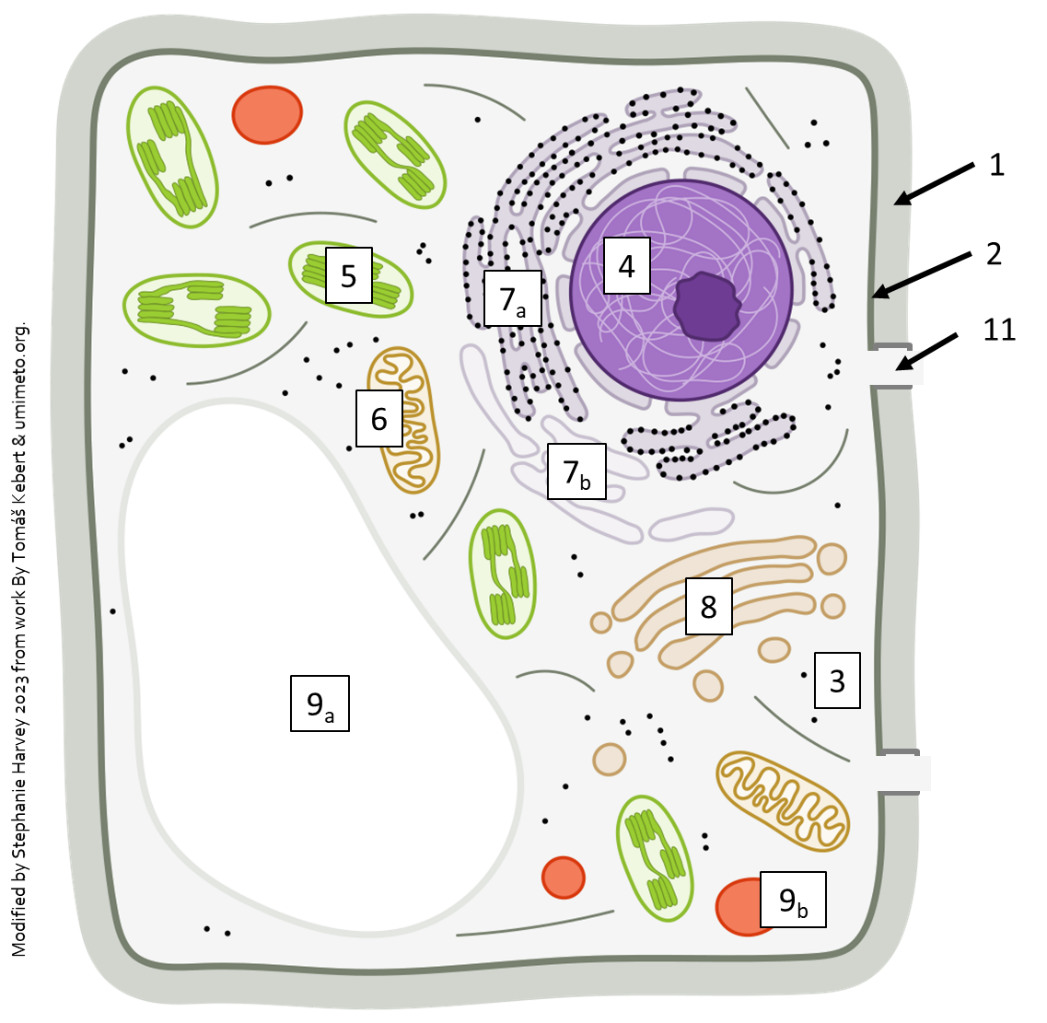
1. Cell Wall: Plant cells have a rigid cell wall surrounding the cell membrane. Fungi also have cell walls but while the fungi walls are primarily composed of chitin, the plant cell wall is composed mainly of cellulose. In both cases the walls provide strength, support, and protection for the cells.
2. Cell Membrane: All cells have a cell membrane, also known as the plasma membrane. It is a thin, flexible layer (a phospholipid bilayer) that encloses the contents of the cell. It controls the movement of substances into and out of the cell and plays a crucial role in maintaining cell homeostasis.
3. Cytoplasm: The cytoplasm is the gel-like fluid that fills the cell. It contains various organelles, including the nucleus, as well as cytosol, which consists of water, enzymes, and other molecules necessary for cell metabolism. All cells, both prokaryotic (without a nuclei) and eukaryotic cells have this.
4. Nucleus: The nucleus, found in all eukaryotic cells, is a membrane-bound organelle that houses the cell's genetic material, DNA. It acts as the control center of the cell, regulating gene expression and coordinating cellular activities. The nucleus is typically located near the center of the cell and is surrounded by a nuclear envelope, which separates it from the cytoplasm. The nucleolus is often visible with the aid of a light microscope. It is a dense structure within the nucleus that is responsible for ribosomal RNA synthesis.
5. Chloroplasts: Some plant cells contain chloroplasts, which are organelles responsible for photosynthesis. Chloroplasts contain chlorophyll, a pigment that captures sunlight and converts it into chemical energy. They have a double membrane and contain a 3rd membrane called the thylakoid membrane. The thylakoid is where chlorophylls are found and is where the light-dependent reactions of photosynthesis occur. Structures similar to chloroplast (called plastids) are found in non-photosynthetic plant cells.
6. Mitochondria: Mitochondria are organelles responsible for cellular respiration, where energy is generated in the form of ATP. Plant cells, like animal cells, have mitochondria to meet their energy needs. Mitochondria have a double membrane and contain their own DNA. Even plant cells with chloroplast have mitochondria.
7. Endoplasmic Reticulum (ER): The endoplasmic reticulum is a network of membranous tubules and sacs involved in protein synthesis, lipid metabolism, and the transport of molecules within the cell. The rough endoplasmic reticulum (RER) (7a) is studded with ribosomes and involved in protein synthesis, while the smooth endoplasmic reticulum (SER) (7b) lacks ribosomes and plays a role in lipid metabolism.
8. Golgi Apparatus: The Golgi apparatus is responsible for modifying, sorting, and packaging proteins and lipids for transport to their final destinations within or outside the cell. It consists of a stack of flattened, membrane-bound sacs called cisternae. While animal cells typically have one, plants often have many and they are sometimes referred to as dictosomes.
9. Vacuole: Plant cells typically have a large central vacuole (9a) that occupies a significant portion of the cell. The vacuole stores water, nutrients, ions, and waste products. It also helps maintain turgor pressure, which contributes to cell rigidity and support. Smaller vacuoles (vesicles; 9b) also have a single membrane and are important for moving material throughout the cell.
10. Plasmodesmata: Plasmodesmata are small channels that connect adjacent plant cells. The plasma membrane continues through the plasmodesmata and into the next cell, allowing for the exchange of materials, such as nutrients and signaling molecules. They enable communication and transport between cells within a plant tissue. There is some regulation as to what moves between cells – it is not just an open hallway.
Not labeled in the image are the ribosome, seen here as dots in the cytoplasm and on the RER. These structures made of protein and ribosomal RNA are part of the “factory” that assembles new proteins.
Of these structures, the chloroplast, central vacuole and plasmodesmata are unique to plants. Both plants and fungi have cell walls. The remaining structures are common to all eukaryotic cells.
These are some of the key structures found in a typical plant cell. While there may be variations in the structure and composition of plant cells depending on the specific cell type and function, these structures provide the foundation for plant growth, metabolism, and reproduction. Prokaryotic cells, simpler in structure have plasma membranes, cell walls (different type from that of eukaroytic cells), cytoplasms, DNA (not in a nucleus) and ribosomes. They completely lack internal membrane-structures.
Types of Plant Cells
All cells start as unspecialized units. They can specialize (differentiate) as they mature to perform specific tasks for the organism. This is often referred to as division of labor. There are three primary types of cells found in plants: parenchyma, collenchyma, and sclerenchyma.
All cells start their lives as parenchyma (Figure 3-3). They have thin, primary cell walls and are living at maturity. The primary cell wall is the flexible layer formed while the cell is growing, and many cells never have more than a primary wall. Due to its thinness, materials can more easily move in and out of parenchyma cells. This is needed in metabolically active cells.

Most of the metabolic functions of a plant are carried out by parenchyma cells. These cells can specialize to provide specific functions: water storage, carbohydrate storage, photosynthesis, or chemical production. New, undifferentiated cells would also be considered parenchyma. The first photomicrograph (Figure 3-3) is from potato tuber – note the evenly thin walls.
Collenchyma cells live at maturity and only have a primary cell wall (Figure 3-4). But, unlike parenchyma cells, these have thick walls, often unevenly in deposition. The primary function for the plant is to provide support, particularly in growing regions of the plant. Collections of these cells make up the resilient strands in the stalks of celery. In Figure 3-4 the white (unstained) areas are the inside of the cell, and the darker material is cell wall. Note that the thickening primarily happens where three or more cells touch.

As Sclerenchyma cells develop from undifferentiated cells, a thick, secondary wall is produced (Figure 3-5). Additionally, a chemical called lignin is added to the cell wall components. This produces an extraordinarily strong cell wall that can be used for structural support.

Unlike collenchyma, mature sclerenchyma cells are dead at maturity, and the thick secondary wall can make up to 90% of the cell volume. There are two types of sclerenchyma cells commonly found in plants. Fibers are long, slender cells that are typically found in bundles where they add strength. Sclereids (Figure 3-6) in contrast are more cuboidal and participate in protection and often form hard durable layers such as the woody “stones” around the seeds of peaches and cherries.

Some specialized sclerenchyma cells play a vital role in water transport in vascular plants. The transport cells of the xylem are sclerenchyma cells. The photomicrograph Figure 3-5 shows the transport xylem cells. The cell walls show up red due to a stain that interacts with the lignin. The thickened secondary walls allow the structural support needed to prevent collapse and the lack of cytoplasm (since the cells are dead) allow for bulk flow of water.