
5.5: Cytoplasm and Cytoskeleton
- Page ID
- 16743

\( \newcommand{\vecs}[1]{\overset { \scriptstyle \rightharpoonup} {\mathbf{#1}} } \)
\( \newcommand{\vecd}[1]{\overset{-\!-\!\rightharpoonup}{\vphantom{a}\smash {#1}}} \)
\( \newcommand{\dsum}{\displaystyle\sum\limits} \)
\( \newcommand{\dint}{\displaystyle\int\limits} \)
\( \newcommand{\dlim}{\displaystyle\lim\limits} \)
\( \newcommand{\id}{\mathrm{id}}\) \( \newcommand{\Span}{\mathrm{span}}\)
( \newcommand{\kernel}{\mathrm{null}\,}\) \( \newcommand{\range}{\mathrm{range}\,}\)
\( \newcommand{\RealPart}{\mathrm{Re}}\) \( \newcommand{\ImaginaryPart}{\mathrm{Im}}\)
\( \newcommand{\Argument}{\mathrm{Arg}}\) \( \newcommand{\norm}[1]{\| #1 \|}\)
\( \newcommand{\inner}[2]{\langle #1, #2 \rangle}\)
\( \newcommand{\Span}{\mathrm{span}}\)
\( \newcommand{\id}{\mathrm{id}}\)
\( \newcommand{\Span}{\mathrm{span}}\)
\( \newcommand{\kernel}{\mathrm{null}\,}\)
\( \newcommand{\range}{\mathrm{range}\,}\)
\( \newcommand{\RealPart}{\mathrm{Re}}\)
\( \newcommand{\ImaginaryPart}{\mathrm{Im}}\)
\( \newcommand{\Argument}{\mathrm{Arg}}\)
\( \newcommand{\norm}[1]{\| #1 \|}\)
\( \newcommand{\inner}[2]{\langle #1, #2 \rangle}\)
\( \newcommand{\Span}{\mathrm{span}}\) \( \newcommand{\AA}{\unicode[.8,0]{x212B}}\)
\( \newcommand{\vectorA}[1]{\vec{#1}} % arrow\)
\( \newcommand{\vectorAt}[1]{\vec{\text{#1}}} % arrow\)
\( \newcommand{\vectorB}[1]{\overset { \scriptstyle \rightharpoonup} {\mathbf{#1}} } \)
\( \newcommand{\vectorC}[1]{\textbf{#1}} \)
\( \newcommand{\vectorD}[1]{\overrightarrow{#1}} \)
\( \newcommand{\vectorDt}[1]{\overrightarrow{\text{#1}}} \)
\( \newcommand{\vectE}[1]{\overset{-\!-\!\rightharpoonup}{\vphantom{a}\smash{\mathbf {#1}}}} \)
\( \newcommand{\vecs}[1]{\overset { \scriptstyle \rightharpoonup} {\mathbf{#1}} } \)
\(\newcommand{\longvect}{\overrightarrow}\)
\( \newcommand{\vecd}[1]{\overset{-\!-\!\rightharpoonup}{\vphantom{a}\smash {#1}}} \)
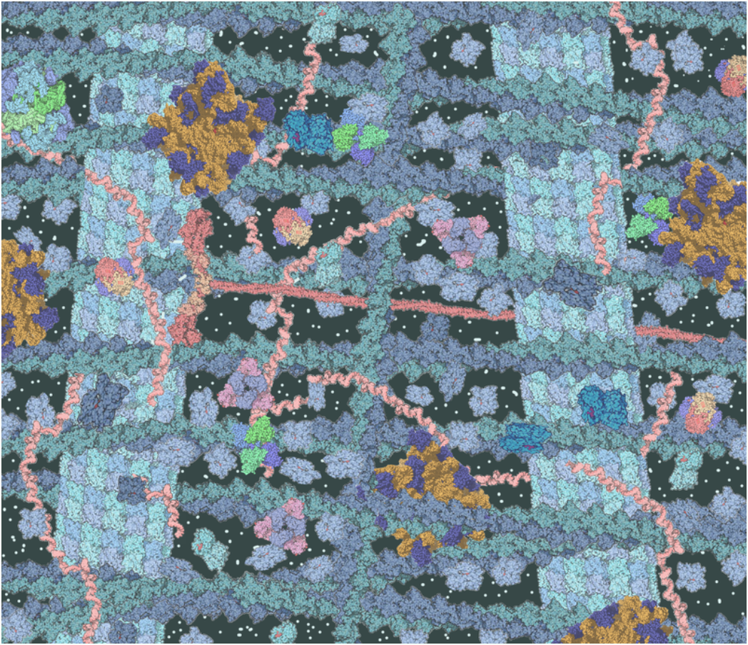
\(\newcommand{\avec}{\mathbf a}\) \(\newcommand{\bvec}{\mathbf b}\) \(\newcommand{\cvec}{\mathbf c}\) \(\newcommand{\dvec}{\mathbf d}\) \(\newcommand{\dtil}{\widetilde{\mathbf d}}\) \(\newcommand{\evec}{\mathbf e}\) \(\newcommand{\fvec}{\mathbf f}\) \(\newcommand{\nvec}{\mathbf n}\) \(\newcommand{\pvec}{\mathbf p}\) \(\newcommand{\qvec}{\mathbf q}\) \(\newcommand{\svec}{\mathbf s}\) \(\newcommand{\tvec}{\mathbf t}\) \(\newcommand{\uvec}{\mathbf u}\) \(\newcommand{\vvec}{\mathbf v}\) \(\newcommand{\wvec}{\mathbf w}\) \(\newcommand{\xvec}{\mathbf x}\) \(\newcommand{\yvec}{\mathbf y}\) \(\newcommand{\zvec}{\mathbf z}\) \(\newcommand{\rvec}{\mathbf r}\) \(\newcommand{\mvec}{\mathbf m}\) \(\newcommand{\zerovec}{\mathbf 0}\) \(\newcommand{\onevec}{\mathbf 1}\) \(\newcommand{\real}{\mathbb R}\) \(\newcommand{\twovec}[2]{\left[\begin{array}{r}#1 \\ #2 \end{array}\right]}\) \(\newcommand{\ctwovec}[2]{\left[\begin{array}{c}#1 \\ #2 \end{array}\right]}\) \(\newcommand{\threevec}[3]{\left[\begin{array}{r}#1 \\ #2 \\ #3 \end{array}\right]}\) \(\newcommand{\cthreevec}[3]{\left[\begin{array}{c}#1 \\ #2 \\ #3 \end{array}\right]}\) \(\newcommand{\fourvec}[4]{\left[\begin{array}{r}#1 \\ #2 \\ #3 \\ #4 \end{array}\right]}\) \(\newcommand{\cfourvec}[4]{\left[\begin{array}{c}#1 \\ #2 \\ #3 \\ #4 \end{array}\right]}\) \(\newcommand{\fivevec}[5]{\left[\begin{array}{r}#1 \\ #2 \\ #3 \\ #4 \\ #5 \\ \end{array}\right]}\) \(\newcommand{\cfivevec}[5]{\left[\begin{array}{c}#1 \\ #2 \\ #3 \\ #4 \\ #5 \\ \end{array}\right]}\) \(\newcommand{\mattwo}[4]{\left[\begin{array}{rr}#1 \amp #2 \\ #3 \amp #4 \\ \end{array}\right]}\) \(\newcommand{\laspan}[1]{\text{Span}\{#1\}}\) \(\newcommand{\bcal}{\cal B}\) \(\newcommand{\ccal}{\cal C}\) \(\newcommand{\scal}{\cal S}\) \(\newcommand{\wcal}{\cal W}\) \(\newcommand{\ecal}{\cal E}\) \(\newcommand{\coords}[2]{\left\{#1\right\}_{#2}}\) \(\newcommand{\gray}[1]{\color{gray}{#1}}\) \(\newcommand{\lgray}[1]{\color{lightgray}{#1}}\) \(\newcommand{\rank}{\operatorname{rank}}\) \(\newcommand{\row}{\text{Row}}\) \(\newcommand{\col}{\text{Col}}\) \(\renewcommand{\row}{\text{Row}}\) \(\newcommand{\nul}{\text{Nul}}\) \(\newcommand{\var}{\text{Var}}\) \(\newcommand{\corr}{\text{corr}}\) \(\newcommand{\len}[1]{\left|#1\right|}\) \(\newcommand{\bbar}{\overline{\bvec}}\) \(\newcommand{\bhat}{\widehat{\bvec}}\) \(\newcommand{\bperp}{\bvec^\perp}\) \(\newcommand{\xhat}{\widehat{\xvec}}\) \(\newcommand{\vhat}{\widehat{\vvec}}\) \(\newcommand{\uhat}{\widehat{\uvec}}\) \(\newcommand{\what}{\widehat{\wvec}}\) \(\newcommand{\Sighat}{\widehat{\Sigma}}\) \(\newcommand{\lt}{<}\) \(\newcommand{\gt}{>}\) \(\newcommand{\amp}{&}\) \(\definecolor{fillinmathshade}{gray}{0.9}\)Figure \(\PageIndex{1}\) may look like a colorful work of abstract art or maybe an ultra-modern carpet design, but it's neither. It is actually a model of the interior of a cell. It's an artist's representation of what you might see if you could take a peek inside one of these basic building blocks of living things. A cell's interior is obviously a crowded and busy space. It contains cytoplasm, dissolved substances, and many structures; and it's a hive of countless biochemical activities all going on at once.
Cytoplasm
The cytoplasm is a thick, usually colorless solution that fills each cell and is enclosed by the cell membrane. Cytoplasm presses against the cell membrane, filling out the cell and giving it its shape. Sometimes cytoplasm acts like a watery solution and sometimes it takes on a more gel-like consistency. In eukaryotic cells, the cytoplasm includes all of the material inside the cell but outside the nucleus, which contains its own watery substance called nucleoplasm. All of the organelles in eukaryotic cells, such as the endoplasmic reticulum and mitochondria, are located in the cytoplasm. The cytoplasm helps to keep them in place. It is also the site of most metabolic activities in the cell, and it allows materials to pass easily throughout the cell.
The portion of the cytoplasm surrounding organelles is called cytosol, which is the liquid part of the cytoplasm. It is composed of about 80 percent water and also contains dissolved salts, fatty acids, sugars, amino acids, and proteins such as enzymes. These dissolved substances are needed to keep the cell alive and carry out metabolic processes. For example, enzymes dissolved in cytosol break down larger molecules into smaller products that can then be used by organelles of the cell. Waste products are also dissolved in the cytosol before they are taken in by vacuoles or expelled from the cell.
Though prokaryotic cells do not have organelles (they do have ribosomes), they still have cytoplasm. It is within the cytoplasm that most cellular activities occur, including the many metabolic pathways that occur within organelles, such as photosynthesis and aerobic respiration.
Cytoskeleton
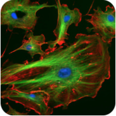
Although cytoplasm may appear to have no form or structure, it is actually highly organized. A framework of protein scaffolds called the cytoskeleton provides the cytoplasm and the cell with structure. The cytoskeleton consists of thread-like filaments and tubules that criss-cross the cytoplasm. You can see these filaments and tubules in the cells in Figure \(\PageIndex{2}\). As its name suggests, the cytoskeleton is like a cellular “skeleton.” It helps the cell maintain its shape and also helps to hold cell structures such as organelles in place within the cytoplasm.
The eukaryotic cytoskeleton is made up of a network of long, thin protein fibers. These threadlike proteins continually rebuild to adapt to the cell's constantly changing needs. Three main kinds of cytoskeleton fibers are microtubules, intermediate filaments, and microfilaments (Table \(\PageIndex{1}\)).
- Microtubules are the thickest of the cytoskeleton structures. They are most commonly made of filaments which are polymers of alpha and beta-tubulin and radiate outwards from an area near the nucleus called the centrosome. Two forms of tubulin form dimers (pairs) which come together to form the hollow cylinders. The cylinders are twisted around each other to form the microtubules. Microtubules help the cell keep its shape. They hold organelles in place and allow them to move around the cell, and they form the mitotic spindle during cell division. Microtubules also make up parts of cilia and flagella, the organelles that help a cell move.
- Microfilaments are made of two thin actin chains that are twisted around one another. Microfilaments are mostly concentrated just beneath the cell membrane, where they support the cell and help the cell keep its shape. Microfilaments form cytoplasmatic extensions, such as microvilli and pseudopodia, which allow certain cells to move. The actin and myosin protein interact to cause a contraction in muscle cells. Microfilaments are found in almost every cell and are numerous in muscle cells and in cells that move by changing shape, such as phagocytes (white blood cells that search the body for bacteria and other invaders).
- Intermediate filaments (IF) differ in make-up from one cell type to another. The IF may be composed of vimentin, keratin, desmin, or lamin. Each cell type can have a unique combination of IFs. For example, intermediate filaments made of keratin are found in skin, hair, and nail cells. IFs organize the inside structure of the cell by holding organelles and providing strength. They are also structural components of the nuclear envelope. Intermediate filaments made of the protein keratin are found in skin, hair, and nail cells.
| Characteristic | Microtubules | Intermediate Filaments | Microfilaments |
|---|---|---|---|
| Fiber Diameter | About 25 nm | 8 to 11 nm | Around 7 nm |
| Protein Composition | Tubulin with two subunits, alpha, and beta-tubulin | One of the different types of proteins such as lamin, vimentin, desmin, and keratin | Actin |
| Shape | Hollow cylinders made of two protein chains twisted around each other | Protein fiber coils twisted into each other | Two actin chains twisted around one another |
| Main Functions | Organelle and vesicle movement; form mitotic spindles during cell reproduction; cell motility (in cilia and flagella) | Organize cell shape; positions organelles in cytoplasm structural support of the nuclear envelope and sarcomeres; involved in cell-to-cell and cell-to-matrix junctions | Keep cellular shape; allows movement of certain cells by forming cytoplasmatic extensions or contraction of actin fibers; involved in some cell-to-cell or cell-to-matrix junctions |
News about an important study of the cytoplasm of eukaryotic cells appeared early in 2016. Researchers in Dresden, Germany discovered that when cells are deprived of adequate nutrients, they may essentially shut down and become dormant. Specifically, when cells do not get enough nutrients, they shut down their metabolism, their energy level drops, and the pH of their cytoplasm decreases. Their normally liquid cytoplasm also assumes a solid state. The cells appear dead and as though a kind of rigor mortis has set in. The researchers think that these changes protect the sensitive structures inside the cells and allow the cells to survive difficult conditions. If nutrients are returned to the cells, they can emerge from their dormant state unharmed. They will continue to grow and multiply when conditions improve.
This important basic science research was carried out on a nonhuman organism: one-celled fungi called yeasts. Nonetheless, it may have important implications for humans because yeasts have eukaryotic cells with many of the same structures as human cells. Yeast cells appear to be able to "trick" death by shutting down all life processes in a controlled way. Researchers hope to learn with the continued research on whether human cells can be taught this "trick" as well.
Review
- Describe the composition of cytoplasm.
- What are some of the functions of cytoplasm?
- Outline the structure and functions of the cytoskeleton.
- Is the cytoplasm made of cells? Why or why not?
- Name two types of cytoskeletal structures.
- True or False. The cytoplasm is usually green.
- True or False. The nucleus of a cell is filled with cytoplasm.
- In Figure \(\PageIndex{2}\) of the different cytoskeletal structures above (shown in red and green), what do you notice about these different structures?
- Describe one example of a metabolic process that occurs in the cytosol.
- In eukaryotic cells, all of the material inside of the cell but outside of the nucleus is called the ___________.
- What is the liquid part of the cytoplasm called?
- What chemical substance makes up most of the cytosol?
- When yeast cells deprived of nutrients go dormant, their cytoplasm assumes a solid-state. What effect do you think a solid cytoplasm would have on normal cellular processes? Explain your answer.
- What is the difference between cytoplasm and cytosol?
- Name the three main parts of the cytoskeleton.
- List two functions of the eukaryotic cytoskeleton
Explore More
Watch the video below to learn about motor proteins, which transport cellular material by the cytoskeleton.
Attributions
- Crowded cytosol by TimVickers, released into the public domain via Wikimedia Commons
- Fluorescent cells by NIH, released into the public domain via Wikimedia Commons
- Text adapted from Human Biology by CK-12 licensed CC BY-NC 3.0