2.2: Observing Mitosis
- Page ID
- 59375
\( \newcommand{\vecs}[1]{\overset { \scriptstyle \rightharpoonup} {\mathbf{#1}} } \)
\( \newcommand{\vecd}[1]{\overset{-\!-\!\rightharpoonup}{\vphantom{a}\smash {#1}}} \)
\( \newcommand{\dsum}{\displaystyle\sum\limits} \)
\( \newcommand{\dint}{\displaystyle\int\limits} \)
\( \newcommand{\dlim}{\displaystyle\lim\limits} \)
\( \newcommand{\id}{\mathrm{id}}\) \( \newcommand{\Span}{\mathrm{span}}\)
( \newcommand{\kernel}{\mathrm{null}\,}\) \( \newcommand{\range}{\mathrm{range}\,}\)
\( \newcommand{\RealPart}{\mathrm{Re}}\) \( \newcommand{\ImaginaryPart}{\mathrm{Im}}\)
\( \newcommand{\Argument}{\mathrm{Arg}}\) \( \newcommand{\norm}[1]{\| #1 \|}\)
\( \newcommand{\inner}[2]{\langle #1, #2 \rangle}\)
\( \newcommand{\Span}{\mathrm{span}}\)
\( \newcommand{\id}{\mathrm{id}}\)
\( \newcommand{\Span}{\mathrm{span}}\)
\( \newcommand{\kernel}{\mathrm{null}\,}\)
\( \newcommand{\range}{\mathrm{range}\,}\)
\( \newcommand{\RealPart}{\mathrm{Re}}\)
\( \newcommand{\ImaginaryPart}{\mathrm{Im}}\)
\( \newcommand{\Argument}{\mathrm{Arg}}\)
\( \newcommand{\norm}[1]{\| #1 \|}\)
\( \newcommand{\inner}[2]{\langle #1, #2 \rangle}\)
\( \newcommand{\Span}{\mathrm{span}}\) \( \newcommand{\AA}{\unicode[.8,0]{x212B}}\)
\( \newcommand{\vectorA}[1]{\vec{#1}} % arrow\)
\( \newcommand{\vectorAt}[1]{\vec{\text{#1}}} % arrow\)
\( \newcommand{\vectorB}[1]{\overset { \scriptstyle \rightharpoonup} {\mathbf{#1}} } \)
\( \newcommand{\vectorC}[1]{\textbf{#1}} \)
\( \newcommand{\vectorD}[1]{\overrightarrow{#1}} \)
\( \newcommand{\vectorDt}[1]{\overrightarrow{\text{#1}}} \)
\( \newcommand{\vectE}[1]{\overset{-\!-\!\rightharpoonup}{\vphantom{a}\smash{\mathbf {#1}}}} \)
\( \newcommand{\vecs}[1]{\overset { \scriptstyle \rightharpoonup} {\mathbf{#1}} } \)
\(\newcommand{\longvect}{\overrightarrow}\)
\( \newcommand{\vecd}[1]{\overset{-\!-\!\rightharpoonup}{\vphantom{a}\smash {#1}}} \)
\(\newcommand{\avec}{\mathbf a}\) \(\newcommand{\bvec}{\mathbf b}\) \(\newcommand{\cvec}{\mathbf c}\) \(\newcommand{\dvec}{\mathbf d}\) \(\newcommand{\dtil}{\widetilde{\mathbf d}}\) \(\newcommand{\evec}{\mathbf e}\) \(\newcommand{\fvec}{\mathbf f}\) \(\newcommand{\nvec}{\mathbf n}\) \(\newcommand{\pvec}{\mathbf p}\) \(\newcommand{\qvec}{\mathbf q}\) \(\newcommand{\svec}{\mathbf s}\) \(\newcommand{\tvec}{\mathbf t}\) \(\newcommand{\uvec}{\mathbf u}\) \(\newcommand{\vvec}{\mathbf v}\) \(\newcommand{\wvec}{\mathbf w}\) \(\newcommand{\xvec}{\mathbf x}\) \(\newcommand{\yvec}{\mathbf y}\) \(\newcommand{\zvec}{\mathbf z}\) \(\newcommand{\rvec}{\mathbf r}\) \(\newcommand{\mvec}{\mathbf m}\) \(\newcommand{\zerovec}{\mathbf 0}\) \(\newcommand{\onevec}{\mathbf 1}\) \(\newcommand{\real}{\mathbb R}\) \(\newcommand{\twovec}[2]{\left[\begin{array}{r}#1 \\ #2 \end{array}\right]}\) \(\newcommand{\ctwovec}[2]{\left[\begin{array}{c}#1 \\ #2 \end{array}\right]}\) \(\newcommand{\threevec}[3]{\left[\begin{array}{r}#1 \\ #2 \\ #3 \end{array}\right]}\) \(\newcommand{\cthreevec}[3]{\left[\begin{array}{c}#1 \\ #2 \\ #3 \end{array}\right]}\) \(\newcommand{\fourvec}[4]{\left[\begin{array}{r}#1 \\ #2 \\ #3 \\ #4 \end{array}\right]}\) \(\newcommand{\cfourvec}[4]{\left[\begin{array}{c}#1 \\ #2 \\ #3 \\ #4 \end{array}\right]}\) \(\newcommand{\fivevec}[5]{\left[\begin{array}{r}#1 \\ #2 \\ #3 \\ #4 \\ #5 \\ \end{array}\right]}\) \(\newcommand{\cfivevec}[5]{\left[\begin{array}{c}#1 \\ #2 \\ #3 \\ #4 \\ #5 \\ \end{array}\right]}\) \(\newcommand{\mattwo}[4]{\left[\begin{array}{rr}#1 \amp #2 \\ #3 \amp #4 \\ \end{array}\right]}\) \(\newcommand{\laspan}[1]{\text{Span}\{#1\}}\) \(\newcommand{\bcal}{\cal B}\) \(\newcommand{\ccal}{\cal C}\) \(\newcommand{\scal}{\cal S}\) \(\newcommand{\wcal}{\cal W}\) \(\newcommand{\ecal}{\cal E}\) \(\newcommand{\coords}[2]{\left\{#1\right\}_{#2}}\) \(\newcommand{\gray}[1]{\color{gray}{#1}}\) \(\newcommand{\lgray}[1]{\color{lightgray}{#1}}\) \(\newcommand{\rank}{\operatorname{rank}}\) \(\newcommand{\row}{\text{Row}}\) \(\newcommand{\col}{\text{Col}}\) \(\renewcommand{\row}{\text{Row}}\) \(\newcommand{\nul}{\text{Nul}}\) \(\newcommand{\var}{\text{Var}}\) \(\newcommand{\corr}{\text{corr}}\) \(\newcommand{\len}[1]{\left|#1\right|}\) \(\newcommand{\bbar}{\overline{\bvec}}\) \(\newcommand{\bhat}{\widehat{\bvec}}\) \(\newcommand{\bperp}{\bvec^\perp}\) \(\newcommand{\xhat}{\widehat{\xvec}}\) \(\newcommand{\vhat}{\widehat{\vvec}}\) \(\newcommand{\uhat}{\widehat{\uvec}}\) \(\newcommand{\what}{\widehat{\wvec}}\) \(\newcommand{\Sighat}{\widehat{\Sigma}}\) \(\newcommand{\lt}{<}\) \(\newcommand{\gt}{>}\) \(\newcommand{\amp}{&}\) \(\definecolor{fillinmathshade}{gray}{0.9}\)Mitosis In A Whitefish Embryo
The Cell Cycle and Mitosis:
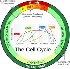
The cell cycle refers to a series of events that describe the metabolic processes of growth and replication of cells. The bulk of the cell cycle is spent in the “living phase”, known as interphase. Interphase is further broken down in to 3 distinct phases: G1 (Gap 1), S (Synthesis) and G2 (Gap 2). G1 is the phase of growth when the cell is accumulating resources to live and grow. After attaining a certain size and having amassed enough raw materials, a checkpoint is reached where the cell uses biochemical markers to decide if the next phase should be entered. If the cell is in an environment with enough nutrients in the environment, enough space and having reached the appropriate size, the cell will enter the S phase. S phase is when metabolism is shifted towards the replication (or synthesis) of the genetic material. During S phase, the amount of DNA in the nucleus is doubled and copied exactly in preparation to divide. The chromosomes at the end of G1 consist of a single chromatid. At the end of S phase, each chromosome consists of two identical sister chromatids joined at the centromere.
When the DNA synthesis is complete, the cell continues on to the second growth phase called G2. Another checkpoint takes place at the end of G2 to ensure the fidelity of the replicated DNA and to re- establish the success of the cell’s capacity to divide in the environment. If conditions are favorable, the cell continues on to mitosis.
.gif?revision=1)
Follow this link above to view a mitosis video.
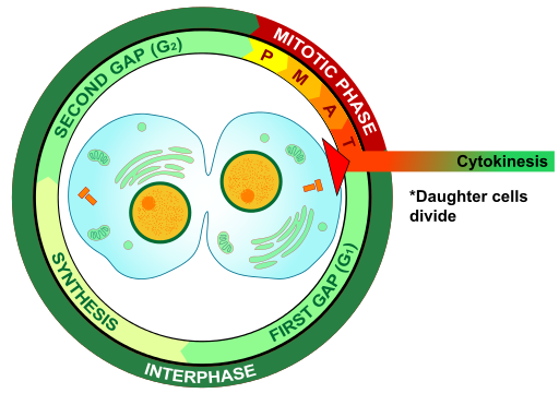
Mitosis is the process of nuclear division used in conjunction with cytokinesis to produce 2 identical daughter cells. Cytokinesis is the actual separation of these two cells enclosed in their own cellular membranes. Unicellular organisms utilize this process of division in order to reproduce asexually.
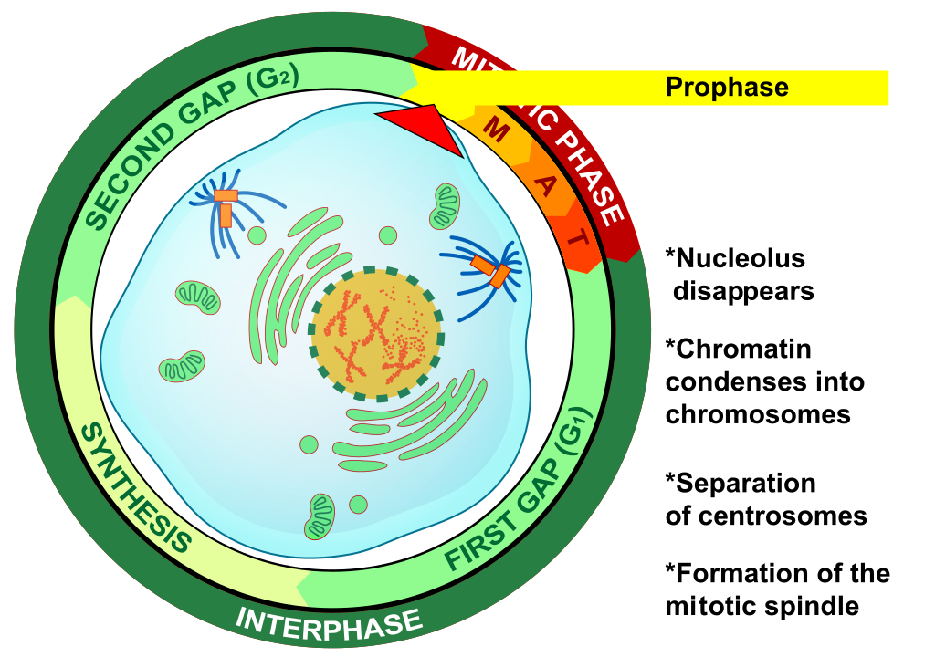
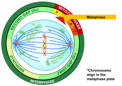
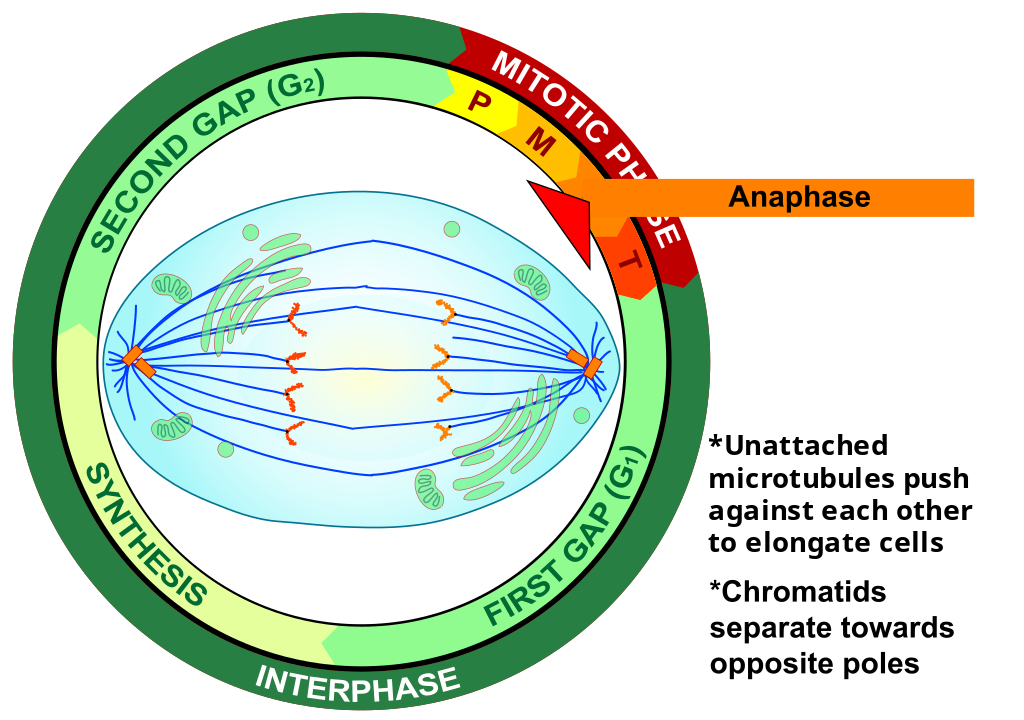
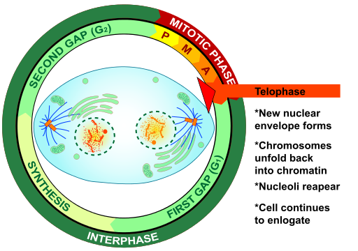
Prokaryotic organisms lack a nucleus, therefore they undergo a different process called binary fission. Multicellular eukaryotes undergo mitosis for repairing tissue and for growth. The process of mitosis is only a short period of the lifespan of cells. Mitosis is traditionally divided into four stages: prophase, metaphase, anaphase and telophase.
The actual events of mitosis are not discreet but occur in a continuous sequence—separation of mitosis into four stages is merely convenient for our discussion and organization. During these stages important cellular structures are synthesized and perform the mechanics of mitosis. For example, in animal cells two microtubule organizing centers called centrioles replicate. The pairs of centrioles move apart and form an axis of proteinaceous microtubules between them called spindle fibers.
These spindle fibers act as motors that pull at the centromeres of chromosomes and separate the sister chromatids into newly recognized chromosomes. The spindles also push against each other to stretch the cell in preparation of forming two new nuclei and separate cells. In animal cells, a contractile ring of actin fibers cinch together around the midline of the cell to coordinate cytokinesis. This cinching of the cell membrane creates a structure called the cleavage furrow.
Eventually, the cinching of the membrane completely separates into two daughter cells. Plant cells require the production of new cell wall material between daughter cells. Instead of a cleavage furrow, the two cells are separated by a series of vesicles derived from the Golgi. These vesicles fuse together along the midline and simultaneously secrete cellulose into the space between the two cells. This series of vesicles is called the cell plate.
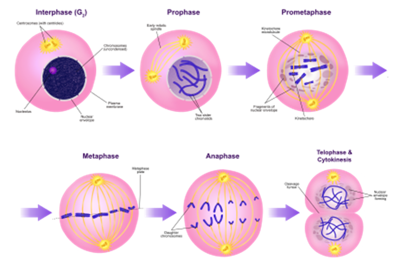
Figure \(\PageIndex{2}\): Mitosis stages (CC-BY-SA, Ali Zifan, Wikimedia)
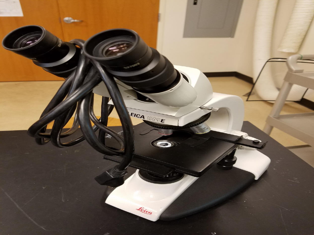
Review Setting up Your Microscope
![]() 1. Plug in the microscope & turn on light source.
1. Plug in the microscope & turn on light source.
![]() 2. Pick up microscope by carrying arm, position it so it is accessible to your seat, with open side of the stage facing you
2. Pick up microscope by carrying arm, position it so it is accessible to your seat, with open side of the stage facing you
![]() 3. Rotate the objectives so that the lowest power objective (smallest in size) clicks into place.
3. Rotate the objectives so that the lowest power objective (smallest in size) clicks into place.
![]() 4. Look at the slide with your naked eye and find the location of the specimen.
4. Look at the slide with your naked eye and find the location of the specimen.
![]() 5. Clip the slide into place with the stage clips. The cover slip on the slide must face up. Find the stage controls and make sure that, when they are turned, the slide moves smoothly left and right or up and down, depending on the knob.
5. Clip the slide into place with the stage clips. The cover slip on the slide must face up. Find the stage controls and make sure that, when they are turned, the slide moves smoothly left and right or up and down, depending on the knob.
![]() 6. Use the stage controls to move the slide so that the light source is shining directly on to the specimen to be magnified.
6. Use the stage controls to move the slide so that the light source is shining directly on to the specimen to be magnified.
![]() 7. Find the coarse and fine focus knobs. Watching the stage and objective, use the coarse focus knob to bring the low power objective as close to the slide as it will go.
7. Find the coarse and fine focus knobs. Watching the stage and objective, use the coarse focus knob to bring the low power objective as close to the slide as it will go.
![]() 8. Put your eye to the eyepiece (or eyepieces, if the microscope is binocular) and rotate the coarse focus knob in the lowering direction until some aspect of the specimen comes into focus.
8. Put your eye to the eyepiece (or eyepieces, if the microscope is binocular) and rotate the coarse focus knob in the lowering direction until some aspect of the specimen comes into focus.
![]() 9. Move your hand to the fine focus knob and get the specimen into perfect focus for your eyes. Do NOT touch the coarse focus knob again.
9. Move your hand to the fine focus knob and get the specimen into perfect focus for your eyes. Do NOT touch the coarse focus knob again.
![]() 10. Use the stage control knobs to move you specimen to close to the exact center of your field of view
10. Use the stage control knobs to move you specimen to close to the exact center of your field of view
![]() 11. Move to the next highest power objective (do not skip the individual objectives) and use only the fine focus to get your image into perfect focus for your eyes.
11. Move to the next highest power objective (do not skip the individual objectives) and use only the fine focus to get your image into perfect focus for your eyes.
![]() 12. If you need further magnification, move to the next highest power objective and use only the fine focus to get your image into perfect focus for your eyes.
12. If you need further magnification, move to the next highest power objective and use only the fine focus to get your image into perfect focus for your eyes.
![]() 13. Do not use the 100x objective (if you have one) in this course. It must be used with immersion oil and we won’t have students doing that.
13. Do not use the 100x objective (if you have one) in this course. It must be used with immersion oil and we won’t have students doing that.
Lab 2 Exercise \(\PageIndex{1}\)
- Obtain a slide of a whitefish embryo (blastula) from the slide box at your table.
- Follow the checklist above to set up your slide for viewing.
- View the slide on the objective which provides the best view. Find the representative object.
- In the circles below the name, draw a representative sample of the stage of mitosis, taking care to correctly and clearly draw the true shape in the slide. Draw your structures proportionately to their size in your microscope’s field of view.
- Fill in the blanks next to your drawing.
- Repeat this for each of the mitosis phases seen below.
A) Prophase
 |
Total Magnification: _________________
Type of eptihelium: _________________
Source of tissue: ____________________
Function of this phase: _______________
__________________________________ |
Function of this phase:
B) Metaphase
|
Total Magnification: _________________
Type of eptihelium: _________________
Source of tissue: ____________________
Function of this phase: _______________
__________________________________ |
C) Anaphase
|
Total Magnification: _________________
Type of eptihelium: _________________
Source of tissue: ____________________
Function of this phase: _______________
__________________________________ |
D) Telophase/cytokinesis
|
Total Magnification: _________________
Type of eptihelium: _________________
Source of tissue: ____________________
Function of this phase: _______________
__________________________________ |
Function of this phase:
Storing the microscope
When storing a microscope you should always follow this list:
- Remove any slide found on the stage and return it to the slide box.
- Rotate the smallest lens or no lens into place above the stage. Lower the stage a few turns.
- Loosely coil the cord in your hand starting near the microscope and working toward the plug.
- Hang the coiled cord over one ocular lens.
- Look at the number on the back of the microscope, return that scope to its numbered box.
- If there’s already a microscope in that numbered box, check its number and move it. If it is not numbered simply push it to the back of the box and place yours closer to the front. We have a few extra microscopes which we store in this fashion.