22.5: Gnetophytes and Conifers
- Page ID
- 33914
\( \newcommand{\vecs}[1]{\overset { \scriptstyle \rightharpoonup} {\mathbf{#1}} } \)
\( \newcommand{\vecd}[1]{\overset{-\!-\!\rightharpoonup}{\vphantom{a}\smash {#1}}} \)
\( \newcommand{\dsum}{\displaystyle\sum\limits} \)
\( \newcommand{\dint}{\displaystyle\int\limits} \)
\( \newcommand{\dlim}{\displaystyle\lim\limits} \)
\( \newcommand{\id}{\mathrm{id}}\) \( \newcommand{\Span}{\mathrm{span}}\)
( \newcommand{\kernel}{\mathrm{null}\,}\) \( \newcommand{\range}{\mathrm{range}\,}\)
\( \newcommand{\RealPart}{\mathrm{Re}}\) \( \newcommand{\ImaginaryPart}{\mathrm{Im}}\)
\( \newcommand{\Argument}{\mathrm{Arg}}\) \( \newcommand{\norm}[1]{\| #1 \|}\)
\( \newcommand{\inner}[2]{\langle #1, #2 \rangle}\)
\( \newcommand{\Span}{\mathrm{span}}\)
\( \newcommand{\id}{\mathrm{id}}\)
\( \newcommand{\Span}{\mathrm{span}}\)
\( \newcommand{\kernel}{\mathrm{null}\,}\)
\( \newcommand{\range}{\mathrm{range}\,}\)
\( \newcommand{\RealPart}{\mathrm{Re}}\)
\( \newcommand{\ImaginaryPart}{\mathrm{Im}}\)
\( \newcommand{\Argument}{\mathrm{Arg}}\)
\( \newcommand{\norm}[1]{\| #1 \|}\)
\( \newcommand{\inner}[2]{\langle #1, #2 \rangle}\)
\( \newcommand{\Span}{\mathrm{span}}\) \( \newcommand{\AA}{\unicode[.8,0]{x212B}}\)
\( \newcommand{\vectorA}[1]{\vec{#1}} % arrow\)
\( \newcommand{\vectorAt}[1]{\vec{\text{#1}}} % arrow\)
\( \newcommand{\vectorB}[1]{\overset { \scriptstyle \rightharpoonup} {\mathbf{#1}} } \)
\( \newcommand{\vectorC}[1]{\textbf{#1}} \)
\( \newcommand{\vectorD}[1]{\overrightarrow{#1}} \)
\( \newcommand{\vectorDt}[1]{\overrightarrow{\text{#1}}} \)
\( \newcommand{\vectE}[1]{\overset{-\!-\!\rightharpoonup}{\vphantom{a}\smash{\mathbf {#1}}}} \)
\( \newcommand{\vecs}[1]{\overset { \scriptstyle \rightharpoonup} {\mathbf{#1}} } \)
\(\newcommand{\longvect}{\overrightarrow}\)
\( \newcommand{\vecd}[1]{\overset{-\!-\!\rightharpoonup}{\vphantom{a}\smash {#1}}} \)
\(\newcommand{\avec}{\mathbf a}\) \(\newcommand{\bvec}{\mathbf b}\) \(\newcommand{\cvec}{\mathbf c}\) \(\newcommand{\dvec}{\mathbf d}\) \(\newcommand{\dtil}{\widetilde{\mathbf d}}\) \(\newcommand{\evec}{\mathbf e}\) \(\newcommand{\fvec}{\mathbf f}\) \(\newcommand{\nvec}{\mathbf n}\) \(\newcommand{\pvec}{\mathbf p}\) \(\newcommand{\qvec}{\mathbf q}\) \(\newcommand{\svec}{\mathbf s}\) \(\newcommand{\tvec}{\mathbf t}\) \(\newcommand{\uvec}{\mathbf u}\) \(\newcommand{\vvec}{\mathbf v}\) \(\newcommand{\wvec}{\mathbf w}\) \(\newcommand{\xvec}{\mathbf x}\) \(\newcommand{\yvec}{\mathbf y}\) \(\newcommand{\zvec}{\mathbf z}\) \(\newcommand{\rvec}{\mathbf r}\) \(\newcommand{\mvec}{\mathbf m}\) \(\newcommand{\zerovec}{\mathbf 0}\) \(\newcommand{\onevec}{\mathbf 1}\) \(\newcommand{\real}{\mathbb R}\) \(\newcommand{\twovec}[2]{\left[\begin{array}{r}#1 \\ #2 \end{array}\right]}\) \(\newcommand{\ctwovec}[2]{\left[\begin{array}{c}#1 \\ #2 \end{array}\right]}\) \(\newcommand{\threevec}[3]{\left[\begin{array}{r}#1 \\ #2 \\ #3 \end{array}\right]}\) \(\newcommand{\cthreevec}[3]{\left[\begin{array}{c}#1 \\ #2 \\ #3 \end{array}\right]}\) \(\newcommand{\fourvec}[4]{\left[\begin{array}{r}#1 \\ #2 \\ #3 \\ #4 \end{array}\right]}\) \(\newcommand{\cfourvec}[4]{\left[\begin{array}{c}#1 \\ #2 \\ #3 \\ #4 \end{array}\right]}\) \(\newcommand{\fivevec}[5]{\left[\begin{array}{r}#1 \\ #2 \\ #3 \\ #4 \\ #5 \\ \end{array}\right]}\) \(\newcommand{\cfivevec}[5]{\left[\begin{array}{c}#1 \\ #2 \\ #3 \\ #4 \\ #5 \\ \end{array}\right]}\) \(\newcommand{\mattwo}[4]{\left[\begin{array}{rr}#1 \amp #2 \\ #3 \amp #4 \\ \end{array}\right]}\) \(\newcommand{\laspan}[1]{\text{Span}\{#1\}}\) \(\newcommand{\bcal}{\cal B}\) \(\newcommand{\ccal}{\cal C}\) \(\newcommand{\scal}{\cal S}\) \(\newcommand{\wcal}{\cal W}\) \(\newcommand{\ecal}{\cal E}\) \(\newcommand{\coords}[2]{\left\{#1\right\}_{#2}}\) \(\newcommand{\gray}[1]{\color{gray}{#1}}\) \(\newcommand{\lgray}[1]{\color{lightgray}{#1}}\) \(\newcommand{\rank}{\operatorname{rank}}\) \(\newcommand{\row}{\text{Row}}\) \(\newcommand{\col}{\text{Col}}\) \(\renewcommand{\row}{\text{Row}}\) \(\newcommand{\nul}{\text{Nul}}\) \(\newcommand{\var}{\text{Var}}\) \(\newcommand{\corr}{\text{corr}}\) \(\newcommand{\len}[1]{\left|#1\right|}\) \(\newcommand{\bbar}{\overline{\bvec}}\) \(\newcommand{\bhat}{\widehat{\bvec}}\) \(\newcommand{\bperp}{\bvec^\perp}\) \(\newcommand{\xhat}{\widehat{\xvec}}\) \(\newcommand{\vhat}{\widehat{\vvec}}\) \(\newcommand{\uhat}{\widehat{\uvec}}\) \(\newcommand{\what}{\widehat{\wvec}}\) \(\newcommand{\Sighat}{\widehat{\Sigma}}\) \(\newcommand{\lt}{<}\) \(\newcommand{\gt}{>}\) \(\newcommand{\amp}{&}\) \(\definecolor{fillinmathshade}{gray}{0.9}\)Gnetophytes (approximately 70 extant species)
Gnetophytes represent an anatomically and genetically difficult group to classify. They have several traits in common with angiosperms, such as vessel elements in the xylem, double fertilization, and a covering over their seeds (more on this in labs 21 and 22). Even their leaves are angiosperm-like, with netted venation. However, these traits are convergently evolved, meaning that angiosperms and gnetophytes each evolved these traits separately. Genetically, recent studies have placed the gnetophytes as a sister group to the Pinaceae (pine family) within the conifers. This would mean that pines, firs, and spruces are more closely related to strange gnetophytes like Ephedra than they are to other conifers like redwoods, cedars, and Pacific yew. However, the true nature of this evolutionary relationship remains murky and contentious.
- Angiosperm-like features: vessel elements, double fertilization, fruit-like ovule coverings
- Dioecious. Female plants have covered ovules, while male plants have pollen cones.
- Leaves xerophytic with opposite arrangement
- Primarily insect pollinated; brightly colored seeds are dispersed by birds
Observe the gnetophyte specimens available in lab. Make notes and drawings of features that would help you recognize this group in the space below:
Conifers (approximately 600 extant species)
Conifers are the most species-rich lineage of gymnosperms. From the fossil record, we think there were over 20,000 species of conifers. However, their diversity declined with the dinosaurs. As discussed in the introduction, these amazing plants represent some of the oldest, tallest, and most massive organisms on the planet. Though currently low in diversity, these amazing plants make up 30% of Earth’s forests. Conifers share the following characteristics:
- Monoecious. Plants produce both male and female strobili on the same plant.
- Wind pollinated with winged pollen
- Xerophytic leaves with a low surface area to volume ratio. Primarily evergreen, but some species are deciduous (e.g. Dawn redwood and larch).
Note
The Pinaceae is currently the largest family of conifers, so many of our examples for this group of gymnosperms will be from the type genus Pinus (pines).
Pine Life Cycle
In the pine life cycle, the pine tree is the sporophyte. Because pines are monoecious, one sporophyte will produce both microstrobili and megastrobili. The microgametophytes are formed within the microsporangia of the microstrobilus, or pollen cone. These structures are all diploid. Within the microsporangium, there are microsporocytes, diploid cells that undergo meiosis to become haploid gametophytes.
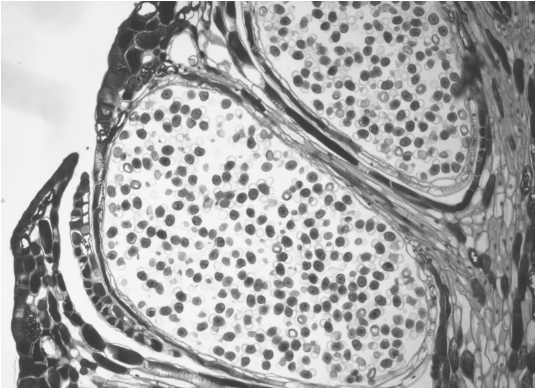
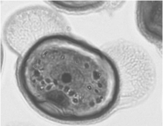
The microgametophyte in gymnosperms is the four-celled, winged pollen grain. Within the pollen grain, you can distinguish the generative cell and the tube cell nucleus. The two prothallial cells are not apparent under the microscope. On either side of the pollen grain, two ear-like structures emerge. Thes air sacs may help orient the pollen grain toward the ovule.
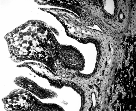
Similarly, the megastrobilus, or seed cone, contains diploid megasporocytes that are produced within a megasporangium. Each megasporocyte undergoes meiosis. However, unlike the microsporocytes, only one of the four cells will survive to develop into a megagametophyte and the other three will die.
The megagametophyte is part of the ovule and contains (usually) two archegonia, each with an egg cell inside. The megagametophyte is retained within the megasporangium, which becomes the nucellus. Surrounding the nucellus is the integument, which is initially continuous with the ovuliferous scale and has a small opening called a micropyle.
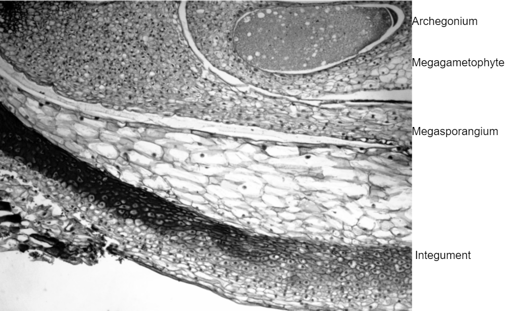
A grain of pollen will be transported on the wind and, if lucky, it will land on a seed cone. The seed cone has a drop of sugary liquid that it secretes, then retracts, pulling the pollen in toward the ovule. This stimulates the tube cell to germinate a pollen tube, while the generative cell divides by mitosis to produce two sperm. These sperm travel down the pollen tube, through the micropyle, and into an archegonium where one will fertilize an egg. When fertilization occurs, the micropyle closes and the integument becomes the seed coat.
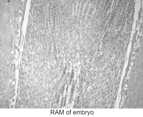
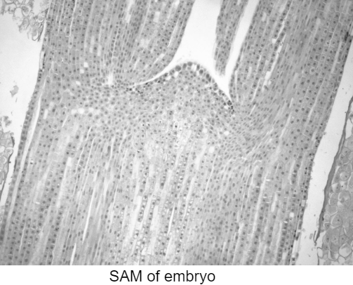
The zygote will grow and develop as an embryo, nourished by the megagametophyte tissue, as well as the nucellus. If you look in a long section of a pine seed, you can see the embryo’s RAM and SAM. The seed will be dispersed by wind or animals and germinate to grow into a diploid pine tree once again.
Pine Life Cycle Diagram
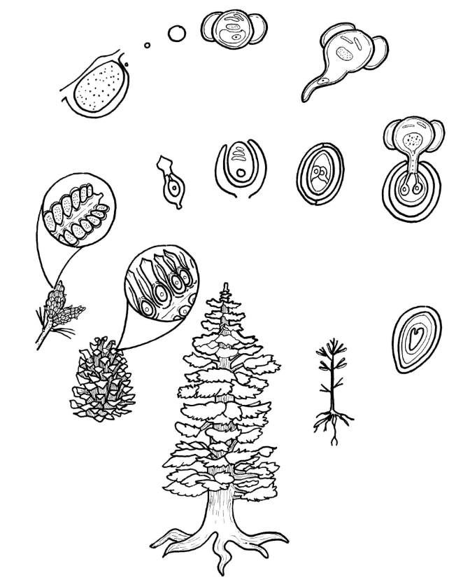
Label the pine life cycle diagram above with the bolded terms from the “Pine Life Cycle” section. Indicate where fertilization and meiosis occur. Draw and label arrows to indicate mitosis. Choose a different color to represent haploid and diploid tissues.
Pine Leaf
In lab Leaf Anatomy, you learned about leaf anatomy and were introduced to the concept of xerophytic leaves. One of the examples in that lab of xerophytic leaves was the pine needle. In the image below, you can see a cross section through a pine needle. Label the following features: xylem, phloem, transfusion tissue, endodermis, mesophyll, hypodermis, epidermis, and cuticle.
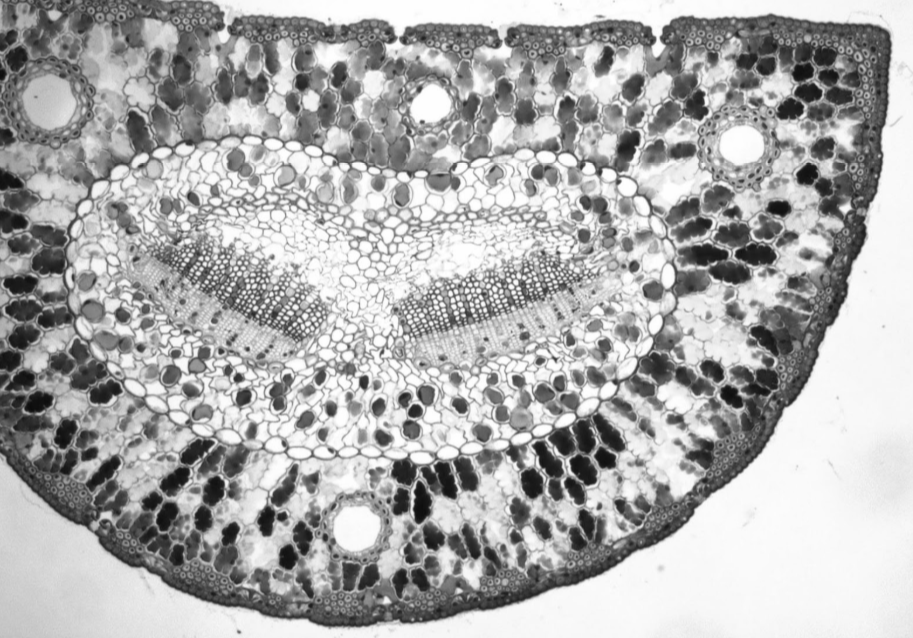
The three large holes you see in the leaf above are resin canals. These conduct a thick, sticky compound called resin that aids in plant defense. What might the resin in the pine needle help pines defend against? Explain your reasoning.
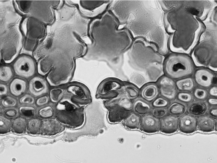
The image above shows a close up of a sunken stoma. Label the mesophyll, guard cells, stoma, hypodermis, epidermis, and cuticle.
How do sunken stomata help leaves reduce water loss?

