Biology LibreTexts

7.4: Conifers

  • Page ID
    35346
  • \( \newcommand{\vecs}[1]{\overset { \scriptstyle \rightharpoonup} {\mathbf{#1}} } \)

    \( \newcommand{\vecd}[1]{\overset{-\!-\!\rightharpoonup}{\vphantom{a}\smash {#1}}} \)

    \( \newcommand{\id}{\mathrm{id}}\) \( \newcommand{\Span}{\mathrm{span}}\)

    ( \newcommand{\kernel}{\mathrm{null}\,}\) \( \newcommand{\range}{\mathrm{range}\,}\)

    \( \newcommand{\RealPart}{\mathrm{Re}}\) \( \newcommand{\ImaginaryPart}{\mathrm{Im}}\)

    \( \newcommand{\Argument}{\mathrm{Arg}}\) \( \newcommand{\norm}[1]{\| #1 \|}\)

    \( \newcommand{\inner}[2]{\langle #1, #2 \rangle}\)

    \( \newcommand{\Span}{\mathrm{span}}\)

    \( \newcommand{\id}{\mathrm{id}}\)

    \( \newcommand{\Span}{\mathrm{span}}\)

    \( \newcommand{\kernel}{\mathrm{null}\,}\)

    \( \newcommand{\range}{\mathrm{range}\,}\)

    \( \newcommand{\RealPart}{\mathrm{Re}}\)

    \( \newcommand{\ImaginaryPart}{\mathrm{Im}}\)

    \( \newcommand{\Argument}{\mathrm{Arg}}\)

    \( \newcommand{\norm}[1]{\| #1 \|}\)

    \( \newcommand{\inner}[2]{\langle #1, #2 \rangle}\)

    \( \newcommand{\Span}{\mathrm{span}}\) \( \newcommand{\AA}{\unicode[.8,0]{x212B}}\)

    \( \newcommand{\vectorA}[1]{\vec{#1}}      % arrow\)

    \( \newcommand{\vectorAt}[1]{\vec{\text{#1}}}      % arrow\)

    \( \newcommand{\vectorB}[1]{\overset { \scriptstyle \rightharpoonup} {\mathbf{#1}} } \)

    \( \newcommand{\vectorC}[1]{\textbf{#1}} \)

    \( \newcommand{\vectorD}[1]{\overrightarrow{#1}} \)

    \( \newcommand{\vectorDt}[1]{\overrightarrow{\text{#1}}} \)

    \( \newcommand{\vectE}[1]{\overset{-\!-\!\rightharpoonup}{\vphantom{a}\smash{\mathbf {#1}}}} \)

    \( \newcommand{\vecs}[1]{\overset { \scriptstyle \rightharpoonup} {\mathbf{#1}} } \)

    \( \newcommand{\vecd}[1]{\overset{-\!-\!\rightharpoonup}{\vphantom{a}\smash {#1}}} \)

    \(\newcommand{\avec}{\mathbf a}\) \(\newcommand{\bvec}{\mathbf b}\) \(\newcommand{\cvec}{\mathbf c}\) \(\newcommand{\dvec}{\mathbf d}\) \(\newcommand{\dtil}{\widetilde{\mathbf d}}\) \(\newcommand{\evec}{\mathbf e}\) \(\newcommand{\fvec}{\mathbf f}\) \(\newcommand{\nvec}{\mathbf n}\) \(\newcommand{\pvec}{\mathbf p}\) \(\newcommand{\qvec}{\mathbf q}\) \(\newcommand{\svec}{\mathbf s}\) \(\newcommand{\tvec}{\mathbf t}\) \(\newcommand{\uvec}{\mathbf u}\) \(\newcommand{\vvec}{\mathbf v}\) \(\newcommand{\wvec}{\mathbf w}\) \(\newcommand{\xvec}{\mathbf x}\) \(\newcommand{\yvec}{\mathbf y}\) \(\newcommand{\zvec}{\mathbf z}\) \(\newcommand{\rvec}{\mathbf r}\) \(\newcommand{\mvec}{\mathbf m}\) \(\newcommand{\zerovec}{\mathbf 0}\) \(\newcommand{\onevec}{\mathbf 1}\) \(\newcommand{\real}{\mathbb R}\) \(\newcommand{\twovec}[2]{\left[\begin{array}{r}#1 \\ #2 \end{array}\right]}\) \(\newcommand{\ctwovec}[2]{\left[\begin{array}{c}#1 \\ #2 \end{array}\right]}\) \(\newcommand{\threevec}[3]{\left[\begin{array}{r}#1 \\ #2 \\ #3 \end{array}\right]}\) \(\newcommand{\cthreevec}[3]{\left[\begin{array}{c}#1 \\ #2 \\ #3 \end{array}\right]}\) \(\newcommand{\fourvec}[4]{\left[\begin{array}{r}#1 \\ #2 \\ #3 \\ #4 \end{array}\right]}\) \(\newcommand{\cfourvec}[4]{\left[\begin{array}{c}#1 \\ #2 \\ #3 \\ #4 \end{array}\right]}\) \(\newcommand{\fivevec}[5]{\left[\begin{array}{r}#1 \\ #2 \\ #3 \\ #4 \\ #5 \\ \end{array}\right]}\) \(\newcommand{\cfivevec}[5]{\left[\begin{array}{c}#1 \\ #2 \\ #3 \\ #4 \\ #5 \\ \end{array}\right]}\) \(\newcommand{\mattwo}[4]{\left[\begin{array}{rr}#1 \amp #2 \\ #3 \amp #4 \\ \end{array}\right]}\) \(\newcommand{\laspan}[1]{\text{Span}\{#1\}}\) \(\newcommand{\bcal}{\cal B}\) \(\newcommand{\ccal}{\cal C}\) \(\newcommand{\scal}{\cal S}\) \(\newcommand{\wcal}{\cal W}\) \(\newcommand{\ecal}{\cal E}\) \(\newcommand{\coords}[2]{\left\{#1\right\}_{#2}}\) \(\newcommand{\gray}[1]{\color{gray}{#1}}\) \(\newcommand{\lgray}[1]{\color{lightgray}{#1}}\) \(\newcommand{\rank}{\operatorname{rank}}\) \(\newcommand{\row}{\text{Row}}\) \(\newcommand{\col}{\text{Col}}\) \(\renewcommand{\row}{\text{Row}}\) \(\newcommand{\nul}{\text{Nul}}\) \(\newcommand{\var}{\text{Var}}\) \(\newcommand{\corr}{\text{corr}}\) \(\newcommand{\len}[1]{\left|#1\right|}\) \(\newcommand{\bbar}{\overline{\bvec}}\) \(\newcommand{\bhat}{\widehat{\bvec}}\) \(\newcommand{\bperp}{\bvec^\perp}\) \(\newcommand{\xhat}{\widehat{\xvec}}\) \(\newcommand{\vhat}{\widehat{\vvec}}\) \(\newcommand{\uhat}{\widehat{\uvec}}\) \(\newcommand{\what}{\widehat{\wvec}}\) \(\newcommand{\Sighat}{\widehat{\Sigma}}\) \(\newcommand{\lt}{<}\) \(\newcommand{\gt}{>}\) \(\newcommand{\amp}{&}\) \(\definecolor{fillinmathshade}{gray}{0.9}\)

    Conifers

    Conifers are the most species-rich lineage of gymnosperms. From the fossil record, we think there were over 20,000 species of conifers. However, their diversity declined with the dinosaurs. Currently, there are around 600 extant species. These amazing plants represent some of the oldest, tallest, and most massive organisms on the planet. Though currently low in diversity, these amazing plants make up 30% of Earth’s forests. Conifers share the following characteristics:

    Note: The Pinaceae is currently the largest family of conifers, so many of our examples for this group of gymnosperms will be from the type genus Pinus (pines).

    Seeds & Pollen

    Seed Cones
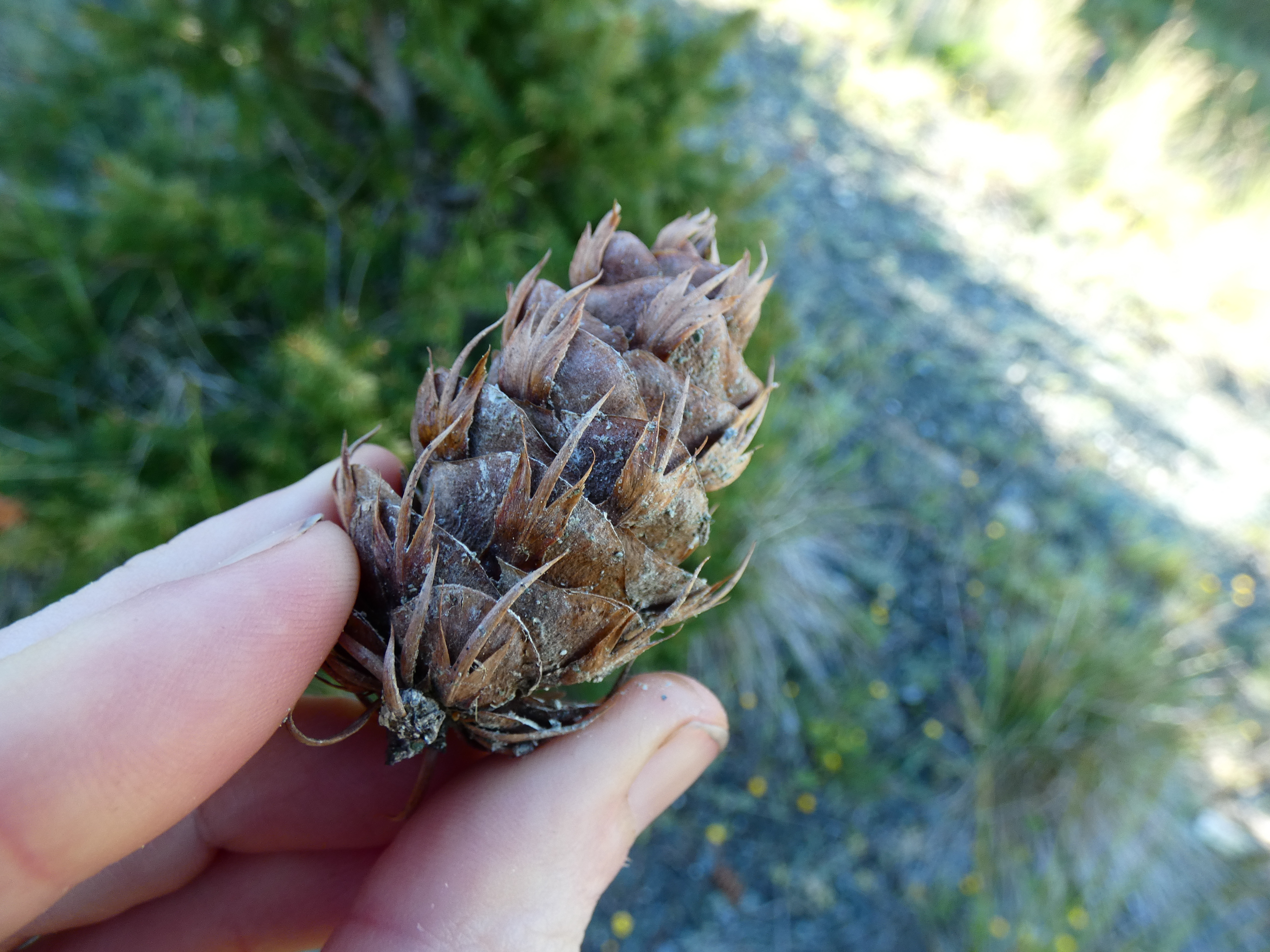
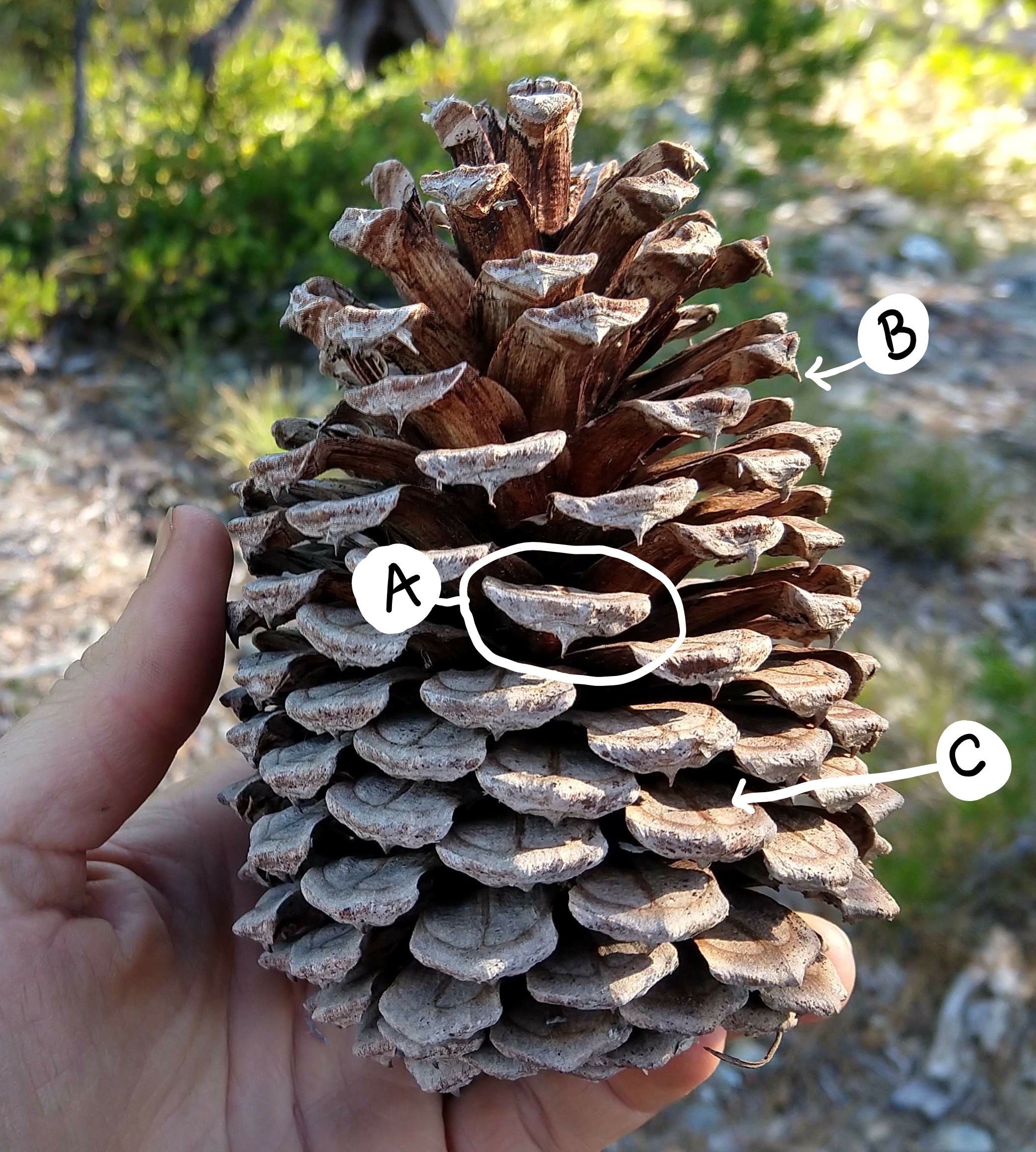
    A Douglas-fir seed cone A jefferey pine seed cone
    Figure \(\PageIndex{1}\): The two images above show seed cones, also called megastrobili. The first shows a Douglas-fir cone shows off its sterile bracts, which are found below the ovuliferous scales and stick out quite far in this species. In the second image, a seed cone from a Jeffrey pine does not have visible bracts but it does showcase a pointy umbo (B) at the tip of the ovuliferous scale (A). Dual markings where two seeds developed on each scale (C). The area that we can see in the image is where the wing of the seed sat. Photos by Maria Morrow, CC BY-SA.

    The megastrobilus, or seed cone, contains diploid megasporocytes that are produced within a megasporangium.
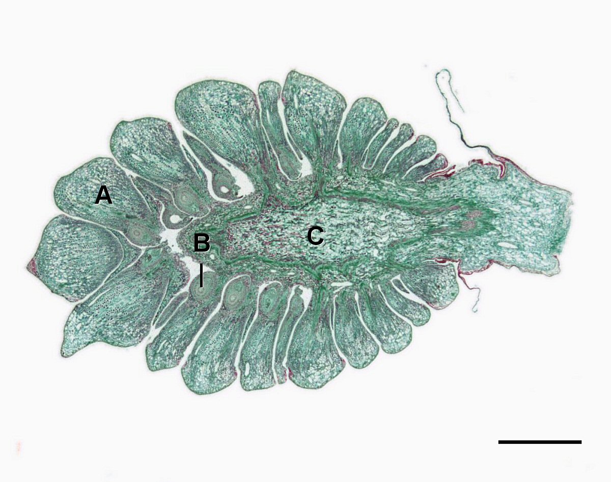
    A long section through a young megastrobilus
    Figure \(\PageIndex{2}\): A micrograph of a longitudinal section through a yearling Pinus megastrobilus, labeled as follows: A=Ovuliferous scale, B=Megasporaphyll, C=Cone axis. Many ovuliferous scales surround the cone axis. Each scale has a megasporangium at its base, where it connects to the axis. Scale=1.0mm. Image by Jon Houseman, CC BY-SA, via Wikimedia Commons.

    Each megasporocyte (also called a megaspore mother cell) undergoes meiosis. Only one of the four cells produced will survive to develop into a megagametophyte and the other three will die.
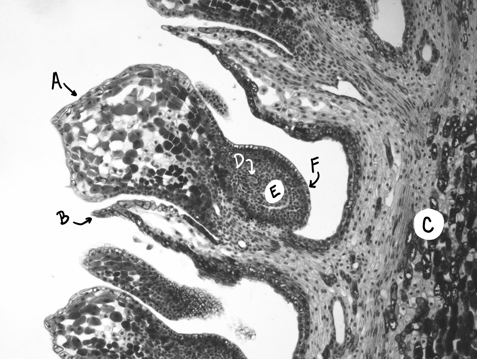
    A long section through a megastrobilus, showing an ovuliferous scale
    Figure \(\PageIndex{3}\): A long section through a megastrobilus allows us to see what is happening on each ovuliferous scale (A). The sterile bract (B) emerges below each scale. On top of the scale, toward the cone axis (C), the megagametophyte (D, this is likely in the megaspore mother cell stage, pre-megagametophyte formation) develops within the megasporangium (E). The megasporangium is surrounded by a protective layer called the integument (F). Photo by Maria Morrow, CC BY-SA.

    The megagametophyte is part of the ovule and contains archegonia, each with an egg cell inside. The megagametophyte is retained within the megasporangium, which becomes the nucellus. Surrounding the nucellus is the integument, which is initially continuous with the ovuliferous scale and has a small opening called a micropyle.
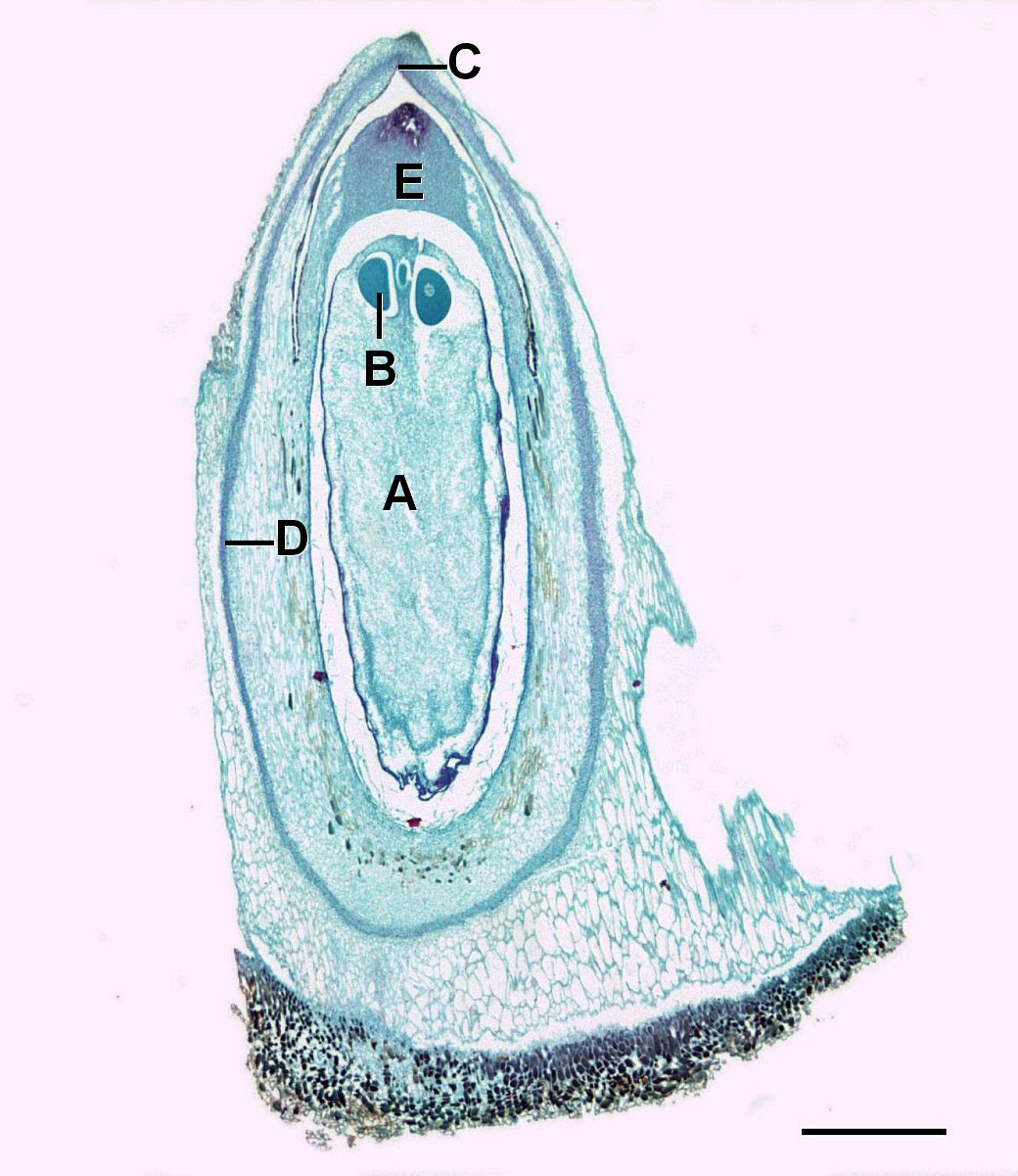
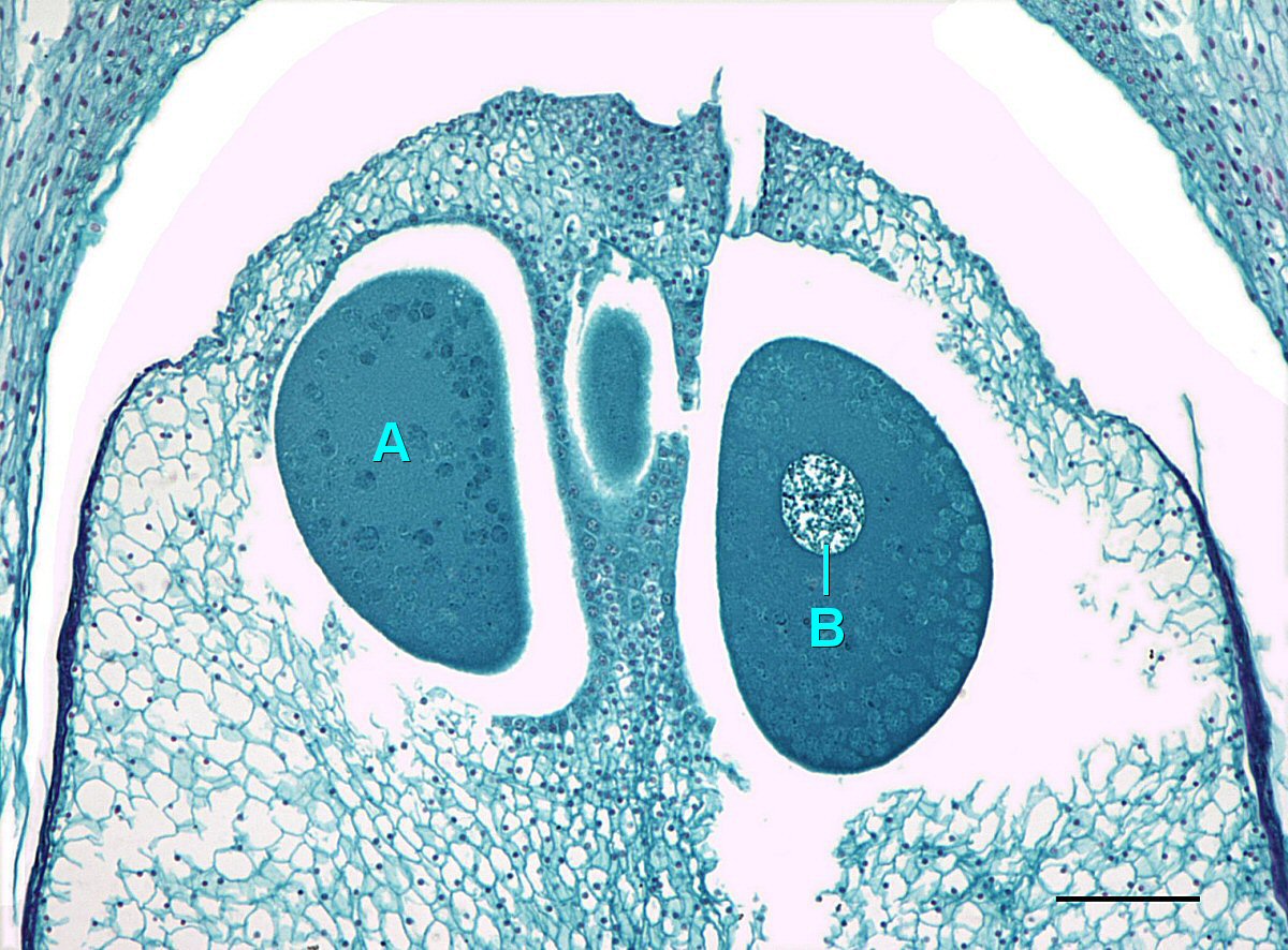
    A longitudinal section through a pine ovule A close up on the pine ovule, showing two egg cells, one of which has a nucleus visible
    Figure \(\PageIndex{4}\): The first image is a micrograph of a Pinus ovule, labeled as follows: A=Gametophyte, B=Egg cell, C=Micropyle, D=Integument, E=Megasporangium. The integument lines the outside with a small hole at the tip (micropyle). Inside the integument is the megasporangium. The megasporangium surrounds the gametophyte, which has two egg cells within it, on the end closest to the micropyle. Scale=0.8mm. The second image is a closer view, showing the eggs within the megagametophyte, labeled as follows: A=Egg cell, B=Egg nucleus. The eggs are large and one has a nucleus visible. Scale=0.31mm. Images by Jon Houseman, CC BY-SA 4.0, via Wikimedia Commons.

    A grain of pollen will be transported on the wind and, if lucky, it will land on a seed cone. The seed cone has a drop of sugary liquid that it secretes, then retracts, pulling the pollen in toward the ovule. This stimulates the tube cell to germinate a pollen tube, while the generative cell divides by mitosis to produce two sperm. These sperm travel down the pollen tube, through the micropyle, and into an archegonium where one will fertilize an egg. When fertilization occurs, the micropyle closes and the integument becomes the seed coat.
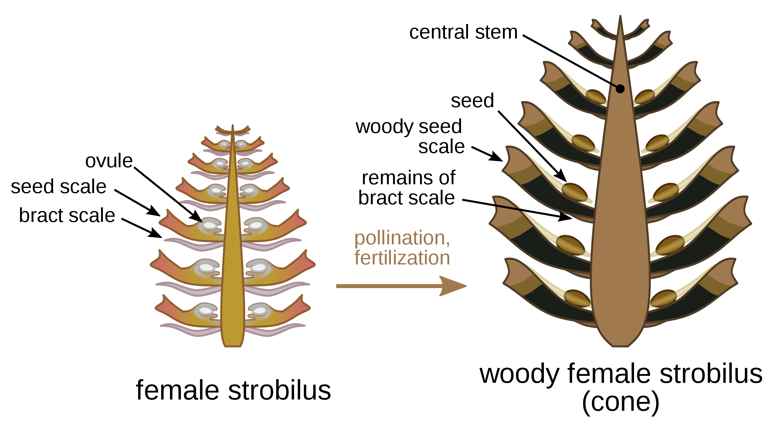
    A long section through a pine megastrobilus before and after fertilization
    Figure \(\PageIndex{5}\): The megastrobilus before (left) and after (right) fertilization. In the unfertilized cone on the left, there is an ovule on top of each ovuliferous scale. In the fertilized cone on the right, the ovule has become a seed, the cone has become woodier, and the sterile bract has been reduced. Image by Nefronus, CC BY-SA, via Wikimedia Commons.
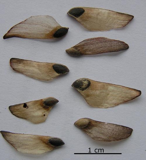
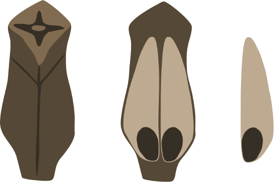
    An ovuliferous scale with two winged seeds Eight winged pine seeds. The wings are tan and thin. The seeds are blackish-brown and ovoid.
    Figure \(\PageIndex{6}\): On the left is a drawing of an individual seed scale from a mature cone of the scots pine (Pinus sylvestris). Two winged seeds are located on each scale. In the image on the right, there are eight pine seeds from the same species. Just like in the drawing, the dark, oval region at the base is the seed and the rest of the tissue (tan) is the wing. First image by Dpaczesniak, CC BY-SA, via Wikimedia Commons. Second image is licensed under the Creative Commons Attribution-Share Alike 3.0 Unported license.

    The zygote will grow and develop as an embryo, nourished by the megagametophyte tissue, as well as the nucellus. If you look in a long section of a pine seed, you can see the embryo’s RAM and SAM. The seed will be dispersed by wind or animals and germinate to grow into a diploid pine tree once again.
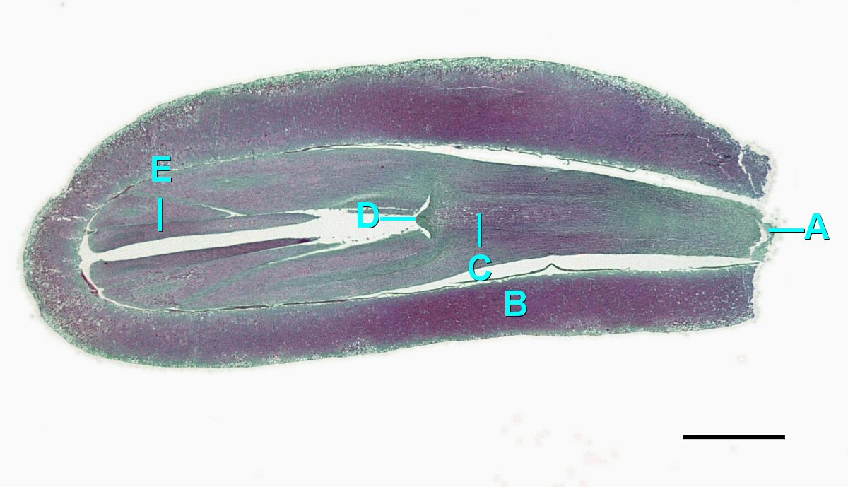
    A longitudinal section of a developing pine embryo within the seed. The shoot apical meristem is located between the two cotyledons.
    Figure \(\PageIndex{7}\): A longitudinal section through a fertilized pine seed. The embryo is developing within the integument, labeled as follows: A=Root cap, B=Integument, C=Hypocotyl-root axis, D=Shoot apical meristem, E=Cotyledons. The integument forms the outer layer, surrounding the embryo. The embryo has a root cap at one end, connected to the shoot apical meristem by the hypocotyl-root axis. Two arm-like cotyledons flank the shoot apical meristem. Scale=1.3mm. Photo by Jon Houseman, CC BY-SA 4.0, via Wikimedia Commons.

    Pollen Cones

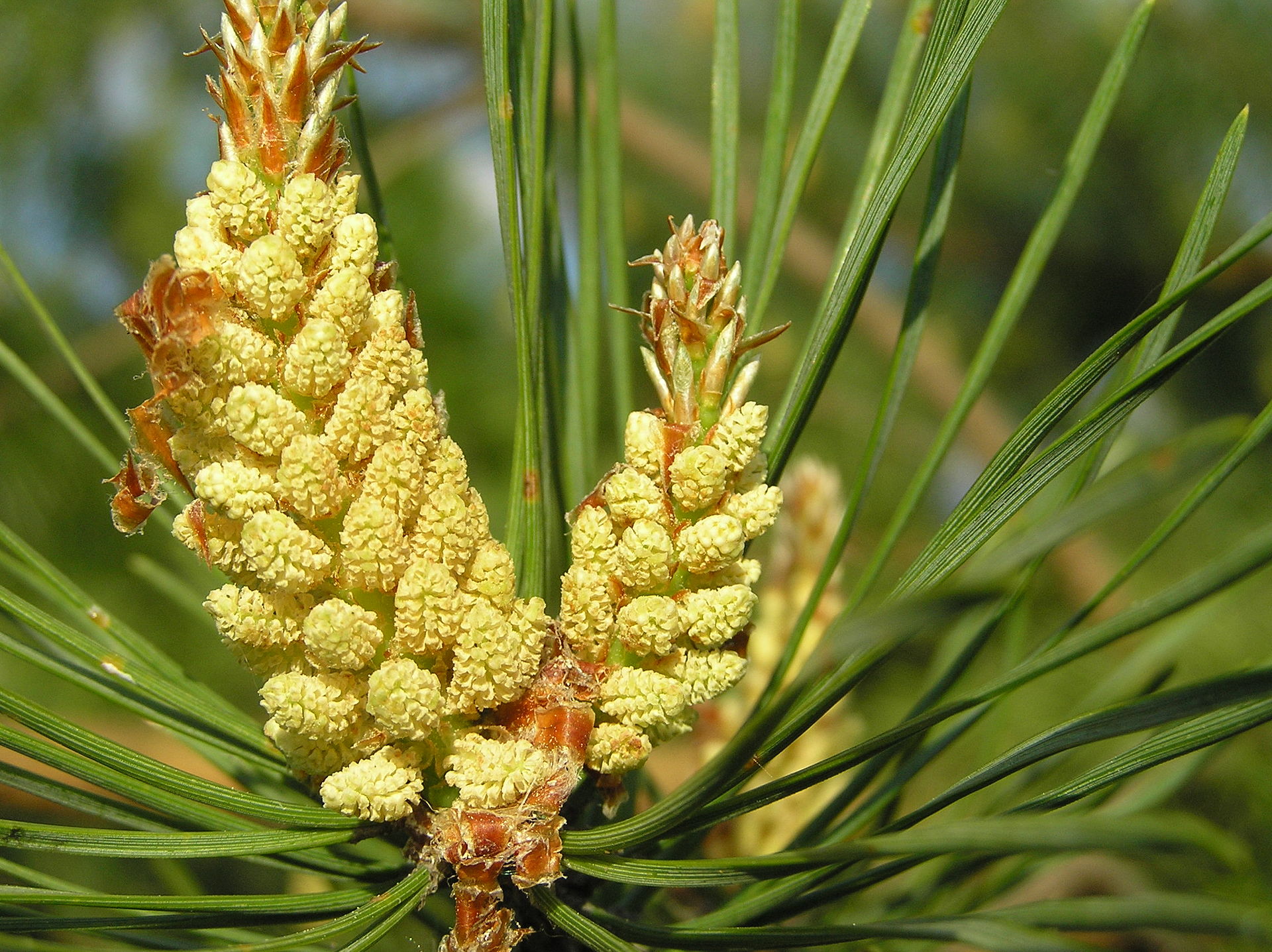
    Two clusters of small pollen cones at the tip of a pine branch
    Figure \(\PageIndex{8}\): Microstrobili on Pinus sylvestris. Unlike the woody seed cones, pollen cones are small, fragile, and ephemeral. Microgametophytes are formed within the microsporangia of the microstrobilus, or pollen cone. These structures are all diploid. Within the microsporangium, there are microsporocytes (also called microspore mother cells), diploid cells that undergo meiosis to produce haploid microspores. Microspores grow by mitosis into microgametophytes, AKA pollen. Author unknown (possibly Beentree), CC BY-SA.

    The microgametophyte in gymnosperms is the four-celled, "winged" pollen grain. Within the pollen grain, you can distinguish the generative cell and the tube cell nucleus. The two prothallial cells are not apparent under the microscope. On either side of the pollen grain, two ear-like structures emerge. These air sacs may help orient the pollen grain toward the ovule.
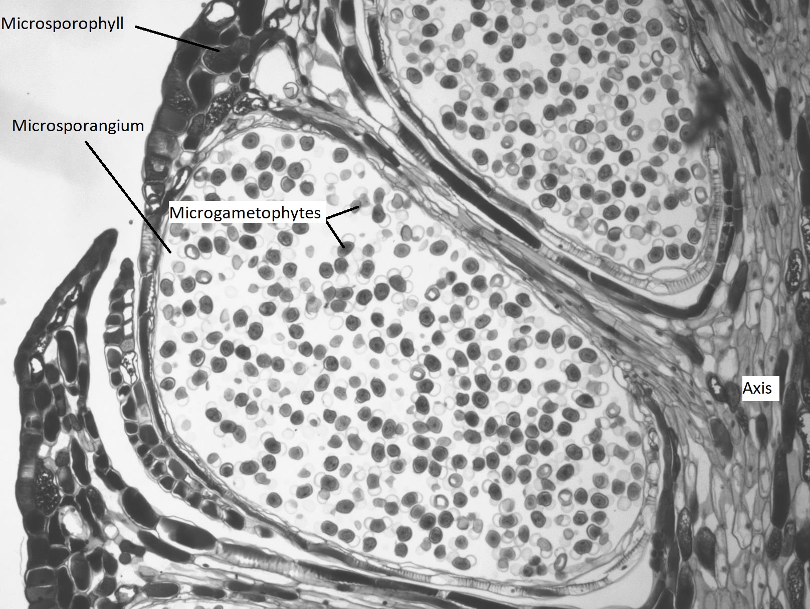
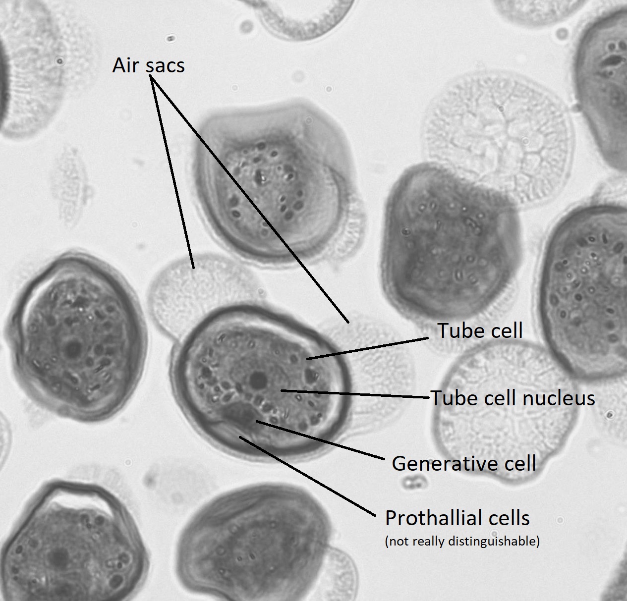
    A pine microstrobilus long section, showing a microsporangium with mature pollen grains Pine pollen with cells and air sacs labeled
    Figure \(\PageIndex{9}\): The first image is a long section through a pine microstrobilus that shows inside a microsporangium. Each microsporangium is produced on a leaf-like structure called a microsporophyll that emerges from the cone axis. Within the microsporangium, many mature pollen grains are visible. You can tell the pollen grains are mature at this magnification by the presence of the air sacs. In the second image, we see a closer view of pollen grains. The two ear-like air sacs flank the four cells enclosed in the center: the tube cell, generative cell, and two prothallial cells. The tube cell takes up most of the internal space and its nucleus is about the same size, if not a little bigger, than the entire generative cell. The two prothallial cells are squished against the edge opposite the air sacs and are not really distinguishable in this image. Photos by Maria Morrow, CC BY-SA.

    Secondary Growth

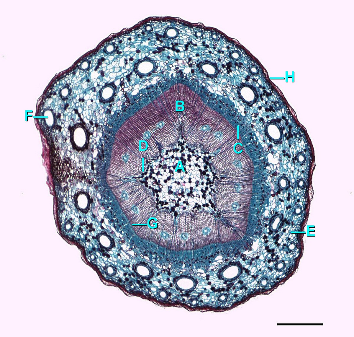
    A cross section through a one year old stem, tissues labeled from innermost to outermost
    Figure \(\PageIndex{10}\): A cross section of a one-year Pinus stem undergoing secondary growth, labeled as follows: in the center is A=Pith, which is surrounded by B=Secondary xylem, C=Secondary phloem forms a ring around the secondary xylem (the vascular cambium would be found between these two layers). Some D=Primary xylem is found near the pith. A layer of E=Cortex surrounds the vascular tissue and pith, dotted with several F=Resin ducts. G=Vascular cambium, H=Epidermis, which is around the outside. Scale=0.625mm. Image by Jon Houseman, CC BY-SA, via Wikimedia Commons.
    A cross section through a mature pine stem section
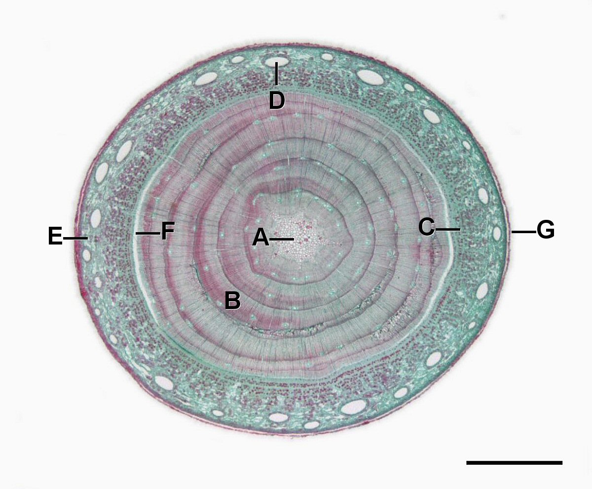
    Figure \(\PageIndex{11}\): A cross section of a mature Pinus stem, labeled as follows: A=Pith, B=Secondary xylem, C=Primary phloem, D=Resin duct, E=Cortex, F=Vascular cambium, G=Epidermis. Scale=0.7mm. Image by Jon Houseman, CC BY-SA, via Wikimedia Commons.
    A cross section through a mature pine stem showing the details of the secondary tissues
    Figure \(\PageIndex{12}\): A closer view (100x) of some of the tissues in a mature Pinus stem cross section, labeled as follows: A and B are part of the secondary xylem, A=Late wood with densely packed, small tracheids, B=Early wood with larger diameter tracheids, C=Resin duct, D=Xylem ray, E=Vascular cambium, F=Secondary phloem, G=Phloem ray, H=Cortex. From the original figure caption: "During the first year of growth the cutinized epidermis is replaced by protective growth of cork rich periderm. The outer periderm consists of layers of cork cells, the phellem, which produces waterproofing suberin. Cork cells are dead at maturity. Deep to the phellem is a living layer of cork cambium or phellogen and beneath that, layers of cork parenchyma or phelloderm. Many cells in the periderm contain dark staining tannins. The cortex is divided into a thin outer hypodermis of lignified sclerenchyma cells and thicker inner cortex of thin walled parenchyma cells containing chloroplasts. Large resin ducts are surrounded by secretory parenchyma that produces resins and turpentines. Some cells enlarge into dark staining tyloses. Both endoderm and pericycle are inconspicuous. The vascular cylinder or stele in young stems consists of a ring of vascular bundles interspaced with medullary rays of parenchyma cells. Seasonal activity of the cambium replaces the isolated vascular bundles with well-defined annual rings of secondary phloem and xylem. Xylem is endarch with protoxylem found towards center of the stem and younger metaxylem towards the periphery of the stem. Protoxylem consists of annular and spiral tracheids with only tracheids found in metaxylem. True xylem vessels are lacking. Because of the greater production of xylem, the vascular cylinder is dominated by radially arranged rays of secondary xylem interspaced with medullary rays of parenchyma cells. Conspicuous resin ducts are present throughout the xylem. Phloem is endarch but annual growth the of stem makes it difficult to distinguish between older protophloem to the periphery and younger metapholem towards center of the stem. Phloem lacks companion cells, consisting entirely of sieve tubes and phloem parenchyma. Medullary rays in the secondary phloem include protein rich albuminous cells. A well-defined pith of parenchyma cells is occasionally interrupted by a few large resin ducts. Visit the BCC Bioscience Image featuring the Microscopic World of Plants. Berkshire Community College Bioscience Image Library, CC0, via Wikimedia Commons with labels added by Maria Morrow.

    Xerophytic Leaves

    Xerophytic leaves are adapted to withstand drought conditions. In conifers, we see a wide range of xerophytic leaves with different morphologies that can be shaped by their local environment. Consider the leaves of the coast redwood and the giant sequoia, shown below. Though these two trees belong to different genera--Sequoia and Sequoiadendron, respectively--they are sister taxa. However, the coast redwood has adapted to life on the coast, where the giant sequoia has evolved in inland, higher elevation forests with much more extreme climatic conditions. How can this be seen in the structure of their leaves?
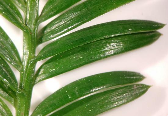
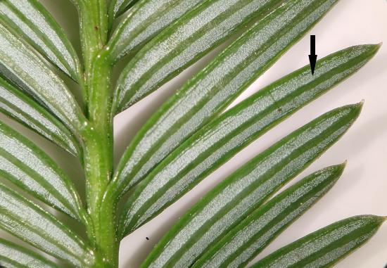
    Coast redwood needles, adaxial side Coast redwood needles, abaxial side
    Figure \(\PageIndex{13}\): The image on the left shows the adaxial (upper) side and the image on the right shows the abaxial (under) side of coast redwood leaves. Note that, particularly in the photo on the left, the leaves look shiny. This is due to the presence of a thick cuticle. The stomata are also concentrated on the underside of the leaf, as indicated by the black arrow in the image on the right. They are clustered in two silvery rows on each leaf, called a stomatal bloom. However, for xerophytic leaves, these are relatively flat, with a fairly standard surface area to volume ratio. Photos by Maria Morrow, CC BY-SA.
    Giant sequoia leaves
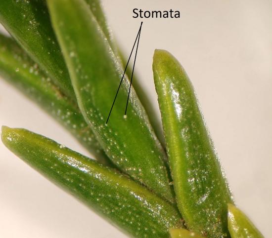
    Figure \(\PageIndex{14}\): These giant sequoia leaves were retrieved from a tree planted in a coastal environment, so the differences here represent evolutionary history, alone. Much like the coast redwood, these shiny leaves have a thick cuticle. The big difference to note is the thickness of these leaves, maintaining a much lower surface area to volume ratio than the coast redwood. The second thing to note is the orientation of the leaves. They are held much more closely together, reducing exposure to the wider environment. Two stomata are indicated in the image, though there are many. They are present on both adaxial and abaxial leaf surfaces. Photo by Maria Morrow, CC BY-SA.
    A cross section through a round pine needle
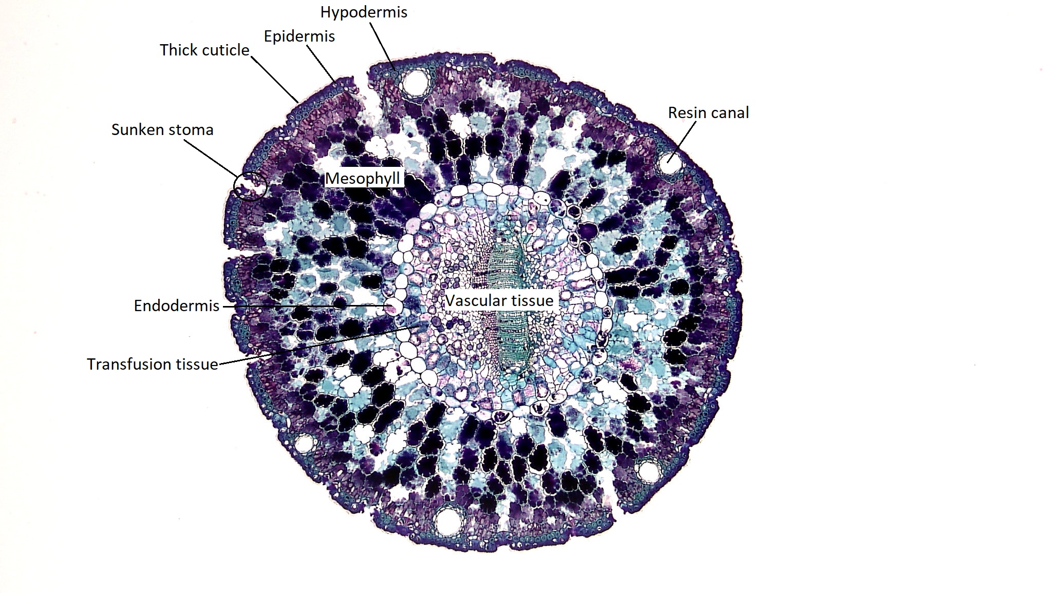
    Figure \(\PageIndex{15}\): Most pine needles you see in botany are flat on one side, however, they also come in round. The tissues of this xerophytic leaf are labeled in the diagram. A thick cuticle surrounds the outside, coating the epidermis. Beneath the epidermis are several layers of tightly packed, small cells: the hypodermis. Stomata are sunken, located within the hypodermis. Under the hypodermis are the highly invaginated mesophyll cells. Further to the inside is a suberized ring of cells called the endodermis, just like in the root, which surrounds the vascular tissue. Transfusion tissue is located between the endodermis and the vascular tissue. There are also resin canals that ring the needle, appearing as holes surrounded by small cells. These secrete resin to protect the plant. Image by Fayette A. Reynolds M.S., Public Domain with labels added by Maria Morrow.
    A close up of the vascular tissue from the cross section of the pine needle
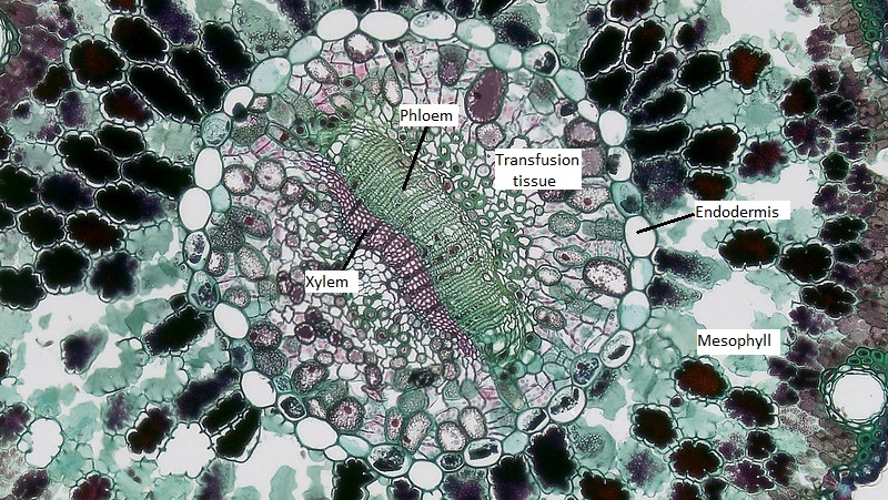
    Figure \(\PageIndex{16}\): A close up of the vascular tissue from the image above. The xylem is composed of ranks of red-stained tracheids and is located to the left of the phloem. Image by Fayette A. Reynolds M.S., Public Domain with labels added by Maria Morrow.

    This page titled 7.4: Conifers is shared under a CC BY 4.0 license and was authored, remixed, and/or curated by Maria Morrow (ASCCC Open Educational Resources Initiative) .

    • Was this article helpful?