
2.3: Organization of the Human Body
- Page ID
- 184822

\( \newcommand{\vecs}[1]{\overset { \scriptstyle \rightharpoonup} {\mathbf{#1}} } \)
\( \newcommand{\vecd}[1]{\overset{-\!-\!\rightharpoonup}{\vphantom{a}\smash {#1}}} \)
\( \newcommand{\dsum}{\displaystyle\sum\limits} \)
\( \newcommand{\dint}{\displaystyle\int\limits} \)
\( \newcommand{\dlim}{\displaystyle\lim\limits} \)
\( \newcommand{\id}{\mathrm{id}}\) \( \newcommand{\Span}{\mathrm{span}}\)
( \newcommand{\kernel}{\mathrm{null}\,}\) \( \newcommand{\range}{\mathrm{range}\,}\)
\( \newcommand{\RealPart}{\mathrm{Re}}\) \( \newcommand{\ImaginaryPart}{\mathrm{Im}}\)
\( \newcommand{\Argument}{\mathrm{Arg}}\) \( \newcommand{\norm}[1]{\| #1 \|}\)
\( \newcommand{\inner}[2]{\langle #1, #2 \rangle}\)
\( \newcommand{\Span}{\mathrm{span}}\)
\( \newcommand{\id}{\mathrm{id}}\)
\( \newcommand{\Span}{\mathrm{span}}\)
\( \newcommand{\kernel}{\mathrm{null}\,}\)
\( \newcommand{\range}{\mathrm{range}\,}\)
\( \newcommand{\RealPart}{\mathrm{Re}}\)
\( \newcommand{\ImaginaryPart}{\mathrm{Im}}\)
\( \newcommand{\Argument}{\mathrm{Arg}}\)
\( \newcommand{\norm}[1]{\| #1 \|}\)
\( \newcommand{\inner}[2]{\langle #1, #2 \rangle}\)
\( \newcommand{\Span}{\mathrm{span}}\) \( \newcommand{\AA}{\unicode[.8,0]{x212B}}\)
\( \newcommand{\vectorA}[1]{\vec{#1}} % arrow\)
\( \newcommand{\vectorAt}[1]{\vec{\text{#1}}} % arrow\)
\( \newcommand{\vectorB}[1]{\overset { \scriptstyle \rightharpoonup} {\mathbf{#1}} } \)
\( \newcommand{\vectorC}[1]{\textbf{#1}} \)
\( \newcommand{\vectorD}[1]{\overrightarrow{#1}} \)
\( \newcommand{\vectorDt}[1]{\overrightarrow{\text{#1}}} \)
\( \newcommand{\vectE}[1]{\overset{-\!-\!\rightharpoonup}{\vphantom{a}\smash{\mathbf {#1}}}} \)
\( \newcommand{\vecs}[1]{\overset { \scriptstyle \rightharpoonup} {\mathbf{#1}} } \)
\(\newcommand{\longvect}{\overrightarrow}\)
\( \newcommand{\vecd}[1]{\overset{-\!-\!\rightharpoonup}{\vphantom{a}\smash {#1}}} \)
\(\newcommand{\avec}{\mathbf a}\) \(\newcommand{\bvec}{\mathbf b}\) \(\newcommand{\cvec}{\mathbf c}\) \(\newcommand{\dvec}{\mathbf d}\) \(\newcommand{\dtil}{\widetilde{\mathbf d}}\) \(\newcommand{\evec}{\mathbf e}\) \(\newcommand{\fvec}{\mathbf f}\) \(\newcommand{\nvec}{\mathbf n}\) \(\newcommand{\pvec}{\mathbf p}\) \(\newcommand{\qvec}{\mathbf q}\) \(\newcommand{\svec}{\mathbf s}\) \(\newcommand{\tvec}{\mathbf t}\) \(\newcommand{\uvec}{\mathbf u}\) \(\newcommand{\vvec}{\mathbf v}\) \(\newcommand{\wvec}{\mathbf w}\) \(\newcommand{\xvec}{\mathbf x}\) \(\newcommand{\yvec}{\mathbf y}\) \(\newcommand{\zvec}{\mathbf z}\) \(\newcommand{\rvec}{\mathbf r}\) \(\newcommand{\mvec}{\mathbf m}\) \(\newcommand{\zerovec}{\mathbf 0}\) \(\newcommand{\onevec}{\mathbf 1}\) \(\newcommand{\real}{\mathbb R}\) \(\newcommand{\twovec}[2]{\left[\begin{array}{r}#1 \\ #2 \end{array}\right]}\) \(\newcommand{\ctwovec}[2]{\left[\begin{array}{c}#1 \\ #2 \end{array}\right]}\) \(\newcommand{\threevec}[3]{\left[\begin{array}{r}#1 \\ #2 \\ #3 \end{array}\right]}\) \(\newcommand{\cthreevec}[3]{\left[\begin{array}{c}#1 \\ #2 \\ #3 \end{array}\right]}\) \(\newcommand{\fourvec}[4]{\left[\begin{array}{r}#1 \\ #2 \\ #3 \\ #4 \end{array}\right]}\) \(\newcommand{\cfourvec}[4]{\left[\begin{array}{c}#1 \\ #2 \\ #3 \\ #4 \end{array}\right]}\) \(\newcommand{\fivevec}[5]{\left[\begin{array}{r}#1 \\ #2 \\ #3 \\ #4 \\ #5 \\ \end{array}\right]}\) \(\newcommand{\cfivevec}[5]{\left[\begin{array}{c}#1 \\ #2 \\ #3 \\ #4 \\ #5 \\ \end{array}\right]}\) \(\newcommand{\mattwo}[4]{\left[\begin{array}{rr}#1 \amp #2 \\ #3 \amp #4 \\ \end{array}\right]}\) \(\newcommand{\laspan}[1]{\text{Span}\{#1\}}\) \(\newcommand{\bcal}{\cal B}\) \(\newcommand{\ccal}{\cal C}\) \(\newcommand{\scal}{\cal S}\) \(\newcommand{\wcal}{\cal W}\) \(\newcommand{\ecal}{\cal E}\) \(\newcommand{\coords}[2]{\left\{#1\right\}_{#2}}\) \(\newcommand{\gray}[1]{\color{gray}{#1}}\) \(\newcommand{\lgray}[1]{\color{lightgray}{#1}}\) \(\newcommand{\rank}{\operatorname{rank}}\) \(\newcommand{\row}{\text{Row}}\) \(\newcommand{\col}{\text{Col}}\) \(\renewcommand{\row}{\text{Row}}\) \(\newcommand{\nul}{\text{Nul}}\) \(\newcommand{\var}{\text{Var}}\) \(\newcommand{\corr}{\text{corr}}\) \(\newcommand{\len}[1]{\left|#1\right|}\) \(\newcommand{\bbar}{\overline{\bvec}}\) \(\newcommand{\bhat}{\widehat{\bvec}}\) \(\newcommand{\bperp}{\bvec^\perp}\) \(\newcommand{\xhat}{\widehat{\xvec}}\) \(\newcommand{\vhat}{\widehat{\vvec}}\) \(\newcommand{\uhat}{\widehat{\uvec}}\) \(\newcommand{\what}{\widehat{\wvec}}\) \(\newcommand{\Sighat}{\widehat{\Sigma}}\) \(\newcommand{\lt}{<}\) \(\newcommand{\gt}{>}\) \(\newcommand{\amp}{&}\) \(\definecolor{fillinmathshade}{gray}{0.9}\)Imagine a machine that has all of the following attributes. It can generate a “wind” of 166 km/hr (100 mi/hr) and relay messages faster than 400 km/hr (249 mi/hr). It contains a pump that moves about a million barrels of fluid over its lifetime, and it has a control center that contains billions of individual components. The machine in question can even repair itself if necessary and not wear out for up to a century or more. It has all these abilities, and yet it consists mainly of water. What is it? It is the human body.
Humans come in all shapes, sizes, and shades (Figure \(\PageIndex{1}\)). We are among the most diverse animal species in appearance, due to different environmental conditions that have shaped various populations, as you read about in the prior section. However, humans are all made of the same components and are all the same species.
In this section of the text, we will explore the very basics of the human body. It is important to note that this is only a starting point for understanding the vast diversity of the human species because every individual is different. For example, individuals vary in their metabolic activity, skin color, reproductive organs, visual acuity, average blood pressure, running speed, white blood cell count, and number of nociceptors, among other traits.

Figure \(\PageIndex{1}\): Humans are one of the most diverse animal species (Omar Sahel CC0).
Organization of the Human Body
The human body is a complex, highly organized structure composed of trillions of parts that work together to perform the functions necessary for sustaining life. The biology of the human body incorporates the body’s structure, the study of which is called anatomy, and the body’s functioning, the study of which is called physiology.
The organization of the human body can be seen as a hierarchy of increasing size and complexity, from atoms and molecules to the entire organism, an individual living thing.
Chemical and Cellular Levels
The simplest level of organization is the chemical level (Figure \(\PageIndex{2}\)). At this level, simple atoms combine to form relatively simple molecules. For example, carbon dioxide (CO2) is made up of one carbon atom and two oxygen atoms, and water (H2O) is made up of two hydrogen atoms and one oxygen atom. Macromolecules (macro: big) are larger and more complex, and include four key types in the human body: carbohydrates (sugars), lipids (fats), proteins, and nucleic acids (DNA).
These four macromolecules form the building blocks of the next level of organization: the cellular level (Figure \(\PageIndex{2}\)). Cells are the smallest and most basic units of structure and function of all living things, including humans. Humans consist of an amazing 37 trillion cells by the time they reach adulthood!
Each cell must perform basic life processes to survive. The four macromolecules interact to perform complex tasks for the cell, such as generating energy (ATP) or producing muscle contractions (through the interactions of two protein complexes: actin and myosin), allowing cells to regulate themselves.
In addition, most human cells are specialized in structure and function to perform specific roles. In fact, the human body may contain as many as 200 different types of cells, each with a specific function and a different structure.

Figure \(\PageIndex{2}\): This diagram shows the chemical and cellular levels of organization of the human body (OpenStax CC-BY 4.0).
Tissue Level
The tissue level of organization consists of groups of connected cells that work together to accomplish one or more specific functions (Figure \(\PageIndex{3}\)). These tissues make up all of the organs of the human body.
There are only four distinct types of tissue in an adult human:
- Muscle tissue is specialized for contraction to generate movement. It is composed of tightly backed cells. Some types of muscle, such as the skeletal muscle shown in Figure \(\PageIndex{4}\), contain striations due to the organization of muscle fibers.
- Nervous tissue is specialized for generating action potentials to rapidly communicate within the body. It is composed of cells with long extensions.
- Epithelial tissue provides a physical barrier to entry into the body and secretes specialized substances via glands. It is mainly composed of tightly packed cells arranged in sheets.
- Connective tissue shows the greatest variability of all of the tissues and forms much of the structure of the body (among many other things). It is composed of cells suspended in a matrix.

Figure \(\PageIndex{3}\): This diagram illustrates the tissue level of organization (OpenStax CC-BY 4.0).

Figure \(\PageIndex{4}\): This diagram illustrates the four tissue types within the human body (NIH CC0).
Organ Level
The organ level of organization is when two or more tissues work together for a specific function (Figure \(\PageIndex{5}\)). For example, the bladder consists of an inner lining of epithelial tissue, bound by various connective tissues to muscle tissue (smooth in this case). Throughout the bladder, there are also neurons that control the muscle tissue, directing it to contract or relax during the urination reflex.

Figure \(\PageIndex{5}\): This diagram illustrates the organ level of organization (OpenStax CC-BY 4.0).
Organ System Level
The organ system level of organization is when two or more organs work together to carry out a complex overall function (Figure \(\PageIndex{6}\)). Each organ of the system does part of the larger job. For example, the bladder, when combined with the kidneys (another organ) and the ureters (“tubes” connecting the kidneys to the bladder), forms the urinary system (or urinary tract). The kidneys filter the blood, and the waste products drain from the kidneys through the ureters into the bladder. The waist is eliminated from the body when we urinate. The urinary system is one of the eleven body systems that can be examined using systemic anatomy.
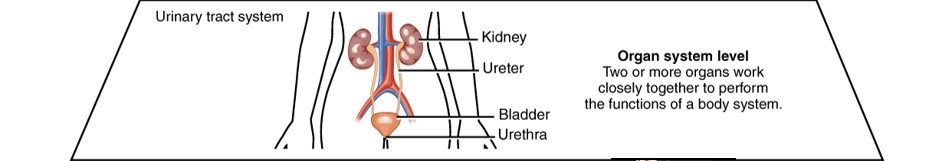
Figure \(\PageIndex{6}\): This diagram illustrates the organ system level of organization (OpenStax CC-BY 4.0).
Organismal Level
The most complex level of organization is the organismal level, where all eleven organ systems function in the human organism, the whole living person (Figure \(\PageIndex{7}\)).
All of the organs and organ systems of the human body normally work together like a well-oiled machine. This is because they are closely regulated by the nervous and endocrine systems. The nervous system controls virtually all body activities, and the endocrine system secretes hormones that help regulate them. Together, the organ systems supply body cells with the substances they need and eliminate their waste products. Through a process called homeostasis, they also keep temperature, pH, and other conditions at just the right levels to support life.

Figure \(\PageIndex{7}\): This diagram illustrates the organismal level of organization (OpenStax CC-BY 4.0).
Rob Knight describes how the human body consists of more than just human components. It also includes a large number of single-celled organisms that live within the body and play a significant, largely unexplored role in our health.
Question after watching: What is your reaction to the many microbes you rely on as a human?
Anatomical Terminology
Use this video to help you understand the terminology used in identifying directions, areas, and region of the body.
What Are Body Cavities?
The human body, like that of many other multicellular organisms, is divided into several body cavities. A body cavity is a fluid-filled space inside the body that holds and protects internal organs. Human body cavities are separated by membranes and other structures. The two largest cavities in the human body are the ventral and dorsal cavities. These two body cavities are subdivided into smaller body cavities (Figure \(\PageIndex{8}\)).

Figure \(\PageIndex{8}\): The ventral cavity includes the thoracic and abdominopelvic cavities and their subdivisions. The abdominopelvic cavity is further divided into the abdominal and pelvic cavities. The dorsal cavity includes the cranial and spinal cavities (NCI CC0).
Ventral Cavity
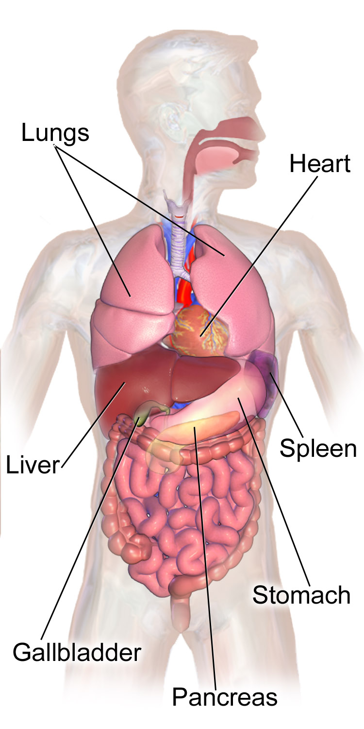
The ventral cavity is at the anterior, or front, of the trunk of the body.
The ventral cavity is subdivided into the thoracic and abdominopelvic cavities (Figure \(\PageIndex{8}\)).
- The thoracic cavity fills the chest and is subdivided into two pleural cavities and the pericardial cavity. The pleural cavities hold the lungs, and the pericardial cavity holds the heart.
- The abdominopelvic cavity fills the lower half of the trunk and is subdivided into the abdominal cavity and the pelvic cavity. The abdominal cavity holds digestive organs and the kidneys, and the pelvic cavity holds reproductive organs and organs of excretion.
Organs contained within this body cavity include the lungs, heart, stomach, intestines, and reproductive organs (some of them are pictured in Figure \(\PageIndex{9}\)).
The ventral cavity allows considerable flexibility in organ size and shape, as each organ performs its functions. For example, organs such as the lungs, stomach, or uterus can expand or contract without distorting other tissues or disrupting the activities of nearby organs.
Dorsal Cavity
The dorsal cavity is at the posterior, or back, of the body, including both the head and the back of the trunk.
The dorsal cavity is subdivided into the cranial and spinal cavities (Figure \(\PageIndex{8}\)).
- The cranial cavity fills most of the upper part of the skull and contains the brain.
- The spinal cavity is a very long, narrow cavity inside the vertebral column. It runs the length of the trunk and contains the spinal cord.
The brain and spinal cord are protected by the bones of the skull and the vertebrae of the spine.
They are further protected by the meninges, a three-layer membrane that encloses the brain and spinal cord. A thin layer of cerebrospinal fluid is maintained between two of the meningeal layers. This clear fluid is produced by the brain, and it provides extra protection and cushioning for the brain and spinal cord.
The meningeal membranes that protect the brain and spinal cord inside their cavities may become inflamed, generally due to a bacterial or viral infection. This condition is called meningitis. Meningitis can lead to serious long-term consequences such as deafness, epilepsy, or cognitive deficits, especially if not treated quickly, and can rapidly become life-threatening.
Learning the symptoms of meningitis is important for getting prompt medical attention. Common symptoms include fever, headache, and neck stiffness. Other symptoms may include confusion or altered consciousness, vomiting, and intolerance to light or loud noises. Young children often exhibit less specific symptoms, such as irritability, drowsiness, or poor feeding.
Meningitis is diagnosed with a lumbar puncture (commonly known as a "spinal tap"), in which a needle is inserted into the spinal canal to collect a sample of cerebrospinal fluid. The fluid is analyzed for pathogens and, if meningitis is diagnosed, treatment consists of antibiotics (for bacterial infections) and sometimes antiviral drugs (for viral infections). IV fluids and corticosteroids may also be used to reduce inflammation and lower the risk of complications such as brain damage.
Some types of meningitis can be prevented with a vaccine, so it is important to speak to healthcare providers about your ability to receive the vaccine. If you have been in contact with someone who has recently been diagnosed with meningitis, see your doctor if you are concerned about your risk.
This 5-minute video examines how meningitis can affect our bodies.