
1.3: Domains of Life
- Page ID
- 88897

\( \newcommand{\vecs}[1]{\overset { \scriptstyle \rightharpoonup} {\mathbf{#1}} } \)
\( \newcommand{\vecd}[1]{\overset{-\!-\!\rightharpoonup}{\vphantom{a}\smash {#1}}} \)
\( \newcommand{\id}{\mathrm{id}}\) \( \newcommand{\Span}{\mathrm{span}}\)
( \newcommand{\kernel}{\mathrm{null}\,}\) \( \newcommand{\range}{\mathrm{range}\,}\)
\( \newcommand{\RealPart}{\mathrm{Re}}\) \( \newcommand{\ImaginaryPart}{\mathrm{Im}}\)
\( \newcommand{\Argument}{\mathrm{Arg}}\) \( \newcommand{\norm}[1]{\| #1 \|}\)
\( \newcommand{\inner}[2]{\langle #1, #2 \rangle}\)
\( \newcommand{\Span}{\mathrm{span}}\)
\( \newcommand{\id}{\mathrm{id}}\)
\( \newcommand{\Span}{\mathrm{span}}\)
\( \newcommand{\kernel}{\mathrm{null}\,}\)
\( \newcommand{\range}{\mathrm{range}\,}\)
\( \newcommand{\RealPart}{\mathrm{Re}}\)
\( \newcommand{\ImaginaryPart}{\mathrm{Im}}\)
\( \newcommand{\Argument}{\mathrm{Arg}}\)
\( \newcommand{\norm}[1]{\| #1 \|}\)
\( \newcommand{\inner}[2]{\langle #1, #2 \rangle}\)
\( \newcommand{\Span}{\mathrm{span}}\) \( \newcommand{\AA}{\unicode[.8,0]{x212B}}\)
\( \newcommand{\vectorA}[1]{\vec{#1}} % arrow\)
\( \newcommand{\vectorAt}[1]{\vec{\text{#1}}} % arrow\)
\( \newcommand{\vectorB}[1]{\overset { \scriptstyle \rightharpoonup} {\mathbf{#1}} } \)
\( \newcommand{\vectorC}[1]{\textbf{#1}} \)
\( \newcommand{\vectorD}[1]{\overrightarrow{#1}} \)
\( \newcommand{\vectorDt}[1]{\overrightarrow{\text{#1}}} \)
\( \newcommand{\vectE}[1]{\overset{-\!-\!\rightharpoonup}{\vphantom{a}\smash{\mathbf {#1}}}} \)
\( \newcommand{\vecs}[1]{\overset { \scriptstyle \rightharpoonup} {\mathbf{#1}} } \)
\( \newcommand{\vecd}[1]{\overset{-\!-\!\rightharpoonup}{\vphantom{a}\smash {#1}}} \)
\(\newcommand{\avec}{\mathbf a}\) \(\newcommand{\bvec}{\mathbf b}\) \(\newcommand{\cvec}{\mathbf c}\) \(\newcommand{\dvec}{\mathbf d}\) \(\newcommand{\dtil}{\widetilde{\mathbf d}}\) \(\newcommand{\evec}{\mathbf e}\) \(\newcommand{\fvec}{\mathbf f}\) \(\newcommand{\nvec}{\mathbf n}\) \(\newcommand{\pvec}{\mathbf p}\) \(\newcommand{\qvec}{\mathbf q}\) \(\newcommand{\svec}{\mathbf s}\) \(\newcommand{\tvec}{\mathbf t}\) \(\newcommand{\uvec}{\mathbf u}\) \(\newcommand{\vvec}{\mathbf v}\) \(\newcommand{\wvec}{\mathbf w}\) \(\newcommand{\xvec}{\mathbf x}\) \(\newcommand{\yvec}{\mathbf y}\) \(\newcommand{\zvec}{\mathbf z}\) \(\newcommand{\rvec}{\mathbf r}\) \(\newcommand{\mvec}{\mathbf m}\) \(\newcommand{\zerovec}{\mathbf 0}\) \(\newcommand{\onevec}{\mathbf 1}\) \(\newcommand{\real}{\mathbb R}\) \(\newcommand{\twovec}[2]{\left[\begin{array}{r}#1 \\ #2 \end{array}\right]}\) \(\newcommand{\ctwovec}[2]{\left[\begin{array}{c}#1 \\ #2 \end{array}\right]}\) \(\newcommand{\threevec}[3]{\left[\begin{array}{r}#1 \\ #2 \\ #3 \end{array}\right]}\) \(\newcommand{\cthreevec}[3]{\left[\begin{array}{c}#1 \\ #2 \\ #3 \end{array}\right]}\) \(\newcommand{\fourvec}[4]{\left[\begin{array}{r}#1 \\ #2 \\ #3 \\ #4 \end{array}\right]}\) \(\newcommand{\cfourvec}[4]{\left[\begin{array}{c}#1 \\ #2 \\ #3 \\ #4 \end{array}\right]}\) \(\newcommand{\fivevec}[5]{\left[\begin{array}{r}#1 \\ #2 \\ #3 \\ #4 \\ #5 \\ \end{array}\right]}\) \(\newcommand{\cfivevec}[5]{\left[\begin{array}{c}#1 \\ #2 \\ #3 \\ #4 \\ #5 \\ \end{array}\right]}\) \(\newcommand{\mattwo}[4]{\left[\begin{array}{rr}#1 \amp #2 \\ #3 \amp #4 \\ \end{array}\right]}\) \(\newcommand{\laspan}[1]{\text{Span}\{#1\}}\) \(\newcommand{\bcal}{\cal B}\) \(\newcommand{\ccal}{\cal C}\) \(\newcommand{\scal}{\cal S}\) \(\newcommand{\wcal}{\cal W}\) \(\newcommand{\ecal}{\cal E}\) \(\newcommand{\coords}[2]{\left\{#1\right\}_{#2}}\) \(\newcommand{\gray}[1]{\color{gray}{#1}}\) \(\newcommand{\lgray}[1]{\color{lightgray}{#1}}\) \(\newcommand{\rank}{\operatorname{rank}}\) \(\newcommand{\row}{\text{Row}}\) \(\newcommand{\col}{\text{Col}}\) \(\renewcommand{\row}{\text{Row}}\) \(\newcommand{\nul}{\text{Nul}}\) \(\newcommand{\var}{\text{Var}}\) \(\newcommand{\corr}{\text{corr}}\) \(\newcommand{\len}[1]{\left|#1\right|}\) \(\newcommand{\bbar}{\overline{\bvec}}\) \(\newcommand{\bhat}{\widehat{\bvec}}\) \(\newcommand{\bperp}{\bvec^\perp}\) \(\newcommand{\xhat}{\widehat{\xvec}}\) \(\newcommand{\vhat}{\widehat{\vvec}}\) \(\newcommand{\uhat}{\widehat{\uvec}}\) \(\newcommand{\what}{\widehat{\wvec}}\) \(\newcommand{\Sighat}{\widehat{\Sigma}}\) \(\newcommand{\lt}{<}\) \(\newcommand{\gt}{>}\) \(\newcommand{\amp}{&}\) \(\definecolor{fillinmathshade}{gray}{0.9}\)We believe with good reason that all life on earth evolved from a common ancestral cell that existed soon after the origins of life on our planet. At one time, all life was divided into two groups: the true bacteria and everything else!
Now we group life into one of three domains:
Prokaryotes are among the first descendants of that common ancestral cell. They lack nuclei (pro meaning before and karyon meaning kernel, or nucleus). They include bacteria and cyanobacteria (blue-green algae).
Eukaryotes include all higher life forms, characterized by cells with true nuclei (Eu, true; karyon, nucleus)
Archaebacteria, (meaning “old” bacteria) include many extremophile bacteria (‘lovers’ of life at extreme high temperatures, salinity, etc.). Originally classified as ancient prokaryotes, Archaebacteria were shown by 1990 to be separate from prokaryotes and eukaryotes, a third domain of life.
The archaea are found in such inhospitable environments as boiling hot springs or arctic ice, though some also live in conditions that are more temperate. Based on comparison of the DNA sequences of genes for ribosomal RNAs in eukaryotes, normal bacteria (eubacteria) and extremophiles, Carl Woese proposed the three-domain phylogeny illustrated in Figure1.1:

Based on sequence similarities and differences, Woese concluded that the archaebacteria (Archaea) are not just a domain separate from the rest of the bacteria and from eukaryotes but are more closely related to eukarya than eubacteria! In fact, the Achaea share genes, proteins, and metabolic pathways found in eukaryotes but not in bacteria, supporting their close evolutionary relationship to eukaryotes. This unique sharing is further testimony to their domain status. Understanding that all living organisms belong to one of three domains has dramatically changed our understanding of evolution. Archea may be prokaryotes, but their immediate ancestors are more closely related to us than to E. coli! For a review, see (Woese, C.2004,A new biology for anew century. Microbiol. Mol. Biol. Rev. 68:173-186) The three domains of life (Archaea, Eubacteria and Eukarya) quickly replaced the older division of living things into Five Kingdoms, the Monera (prokaryotes), Protista, Fungi, Plants, and Animals (all eukaryotes!)
At this point you may be asking, “What about viruses?” Where are they on, or do they even belong in the tree of life? You may already know that viruses require live cellular hosts to reproduce, but that they are not themselves alive. In fact, much about the place of viruses in evolution is an open question that we will consider in a later chapter. For now, let’s look at how we come to know about viruses and some of their peculiarities.
1.3.1. Viruses: Dead or Alive; Big or Small - A History of Surprises
Viruses that infect bacteria are called bacteriophage (phage meaning eaters, hence bacteria eaters). Eukaryotic viruses include DNA and RNA viruses, with DNA and RNA genomes. Smallpox, hepatitis B, herpes, chicken pox/shingles, and adenovirus are caused by DNA viruses. Common colds, influenza, SARS, andCOVID-19 are caused bypositive strand RNA viruses that upon infecting a cell, replicate their RNA genome to make RNA negative strand RNAs encoding all necessary information to make new viruses. HIV AIDS, Ebola, Zika, yellow fever, and some cancers are caused by retroviruses, RNA viruses whose genome is reverse-transcribed into a cDNA intermediate that replicates and is transcribed to generate new viruses. Viruses were not identified as agents of disease until late in the nineteenth century, and we have learned much in the ensuing century. In 1892, Dmitri Ivanofsky, a Russian botanist, was studying plant diseases. One that damaged tobacco (and was therefore of agricultural significance) was themosaic disease (Figure1.2,below).
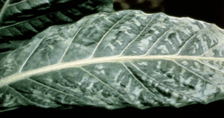
Fig 1.2: Tobacco mosaic virus symptoms (white patches) on a tobacco leaf.
Ivanofsky showed that extracts of infected tobacco leaves were themselves infectious. The assumption was that the extracts would contain infectious bacteria.But his extracts remained infectious even after passing them through a Chamberland-Pasteur filter with a pore size so small that bacteria would not pass into the filtrate. Thus, the infectious agent(s) couldn’t be bacterial. Since the infectious material was not cellular and depended on a host for reproduction with no independent life of its own, they were soon given the name virus, a term that originally just meant toxin, or poison.This marked the start of virology, the study of viruses. The virus that Ivanofsky studied is now called Tobacco Mosaic Virus, or TMV.
Invisible by light microscopy, viruses are sub-microscopic non-cellular bits of life-chemistry that only become reproductive (come alive) when they parasitize a host cell. Since many viruses cause disease in humans, we have learned much about how they are similar and how they differ. In other chapters, we’ll learn how viruses have even become tools for the study of cell and molecular biology. Let’s start with a recent surprise from the study of viruses.
As eventually seen in the electron microscope, viruses (called virions or viral particles) are typically 150 nm or less in diameter. And that is how we have thought of viruses for over a century! But in 2002, a particle inside an amoeba, originally believed to be a bacterium, was shown by electron microscopy to be a giant virus! Since then, several more giant, or Megavirales were discovered.
Megavirales fall into two groups, pandoraviruses and mimiviruses. At 1000nm (\(1 \mu \rm m\)) Megavirus chilensis (a pandoravirus) may be the largest. Compare a few giant viruses to a bacterium (E. coli) and the AIDS virus in Figure 1.3 below.

Consider that a typical virus contains a small genome, encoding an average of 10 genes. In contrast, the M.chilensis genome contains \(2.5 \times 10^6\) base pairs (bp) encoding up to 1,100 proteins. Still, it requires host cell proteins to infect and replicate. More surprising is that 75%of the sequenced \(1.2 \times 10^6\)bp mimivirus genome code putative proteins with no counterparts in other viruses or cells! Equally surprising, some mimiviruses genes encode proteins homologous to those used for translation in prokaryotes and eukaryotes. If all viruses, including the Megavirales, only use host cell enzymes and ribosomal machinery to synthesize proteins, what are these genes doing in a mimivirus genome? Think of the surprises here as questions. The big ones concern where and when Megavirales (giant viruses) evolved:
- What are those genes with no cellular counterparts all about?
- What were the selective advantages to a virus of large size and large genomes?
- Were Megavirales once large cells that invaded other cells, eventually becoming viral parasites and losing most but not all of their genes? Or were they once small viruses that incorporated host cell genes, increasing their genome size and coding capacity?
What information would you need (or what questions would you ask and/or what experiments could you do) to find out what the unique proteins encoded by those uniquely viral genes in e.g., mimivirus are doing for the virus?
Viruses are typically identified because they are harmful. Luc Montagnier, FrançoiseBé-Sinoussi, and Harald Z. Hausen earned the 2008 Nobel Prize in Physiology or Medicine for the discovery of HIV. Asthisis being written, the wealthy nations are emerging from theCOVID-19 pandemic caused by the SARS-CoV-2 RNA virus. Bacteria, also discovered because of the harm they do, are not only beneficial, but necessary symbionts in our many microbiomes.The book is still out on any health benefits of viruses but may someday be written!
Robert Koch discovered bacterial causes of many diseases and won the 1905 Nobel prize in Physiology or Medicine for showing that Mycobacterium tuberculosis caused tuberculosis. What other bacterial diseases did he discover and why are his four postulates still relevant?
Let’s now turn our attention to cells, entities that we define as living, with all of the properties of life..., starting with eubacteria
1.3.2 The Prokaryotes (Eubacteria = Bacteria and Cyanobacteria)
Prokaryotic cells lack nuclei and other eukaryotic organelles, such as mitochondria, chloroplasts, endoplasmic reticulum, and assorted eukaryotic vesicles and internal membranes. Transmission and scanning electron micrographs and an illustration of rod-shaped bacteria are shown below (Figure 1.4).

Bacteria do contain bacterial microcompartments (BMCs), made up entirely of protein and not surrounded by a phospholipid membrane. These function for example, in \(\rm CO_2\) fixation to sequester toxic metabolites toxic in cells. Check out Bacterial Organelles for more information. Bacteria are typically unicellular, although a few (like some cyanobacteria) live colonial lives at least some of the time.
1.3.2.a Bacterial Reproduction
Without the compartments afforded by the internal membrane systems of eukaryotic cells, all intracellular chemistries, (reproduction and gene expression (DNA replication, transcription, translation, and all the metabolic biochemistry of life) happen in the cytoplasm. Bacterial DNA is a circular double helix that duplicates as the cell grows. While not enclosed in a nucleus, bacterial DNA is concentrated in a region of the cell called the nucleoid. When not crowded at high density, bacteria replicate their DNA throughout the life of the cell, dividing by binary fission. The result is the equal partition of duplicated bacterial chromosomes into new cells. The bacterial chromosome is basically naked DNA, unassociated with proteins.
1.3.2.b Cell Motility and the Possibility of a Cytoskeleton
Movement of cells is a response to environment. Some respond to chemicals (chemotaxis), some to light (phototaxis) or even gravity (geotaxis). Bacteria move to or from nutrients, noxious chemicals, light, dark, gravitational force, etc., by one of several mechanisms. Some use a flagellum made up largely of the bacterial protein flagellin. The main proteins of eukaryotic cellflagella and cilia are the tubulins. Together with actin and other proteins, tubulins are also part of the eukaryotic cell cytoskeleton of rods and tubes. Prokaryotes were long thought to lack similar cytoskeletal components. But two bacterial homologues of eukaryotic actin and tubulin genes were recently discovered. MreB is the actin homologue. Like actin, MreB monomers polymerize to form filaments that lie under the cell membrane of bacteria (e.g.,E. coli), helping to maintain their rod-like shape. In fact, E. coli with a mutant MreB gene is spherical..., and normally spherical bacteria lack an MreB gene! MreB was also thought to form an actin-like cortical ring that in dividing eukaryotic cells constricts to pinchoff two new cells. But this function seems to be served by the FtsZ protein that encodes a eukaryotic tubulin homologue. FtsZ polymers form filaments that are seen in a Z ring at the center of a bacterial cell during binary fission. FtsZ mutants divide, but abnormally; thus the role of FtsZ in separating bacterial cells during binary fission is not yet clear. Figure 1.5 shows a micrograph and an illustration of FtsZ in the Z rings of dividing E. coli cells.

It seems that together with flagellin, the MreB and FtsZ proteins may be part of a primitive prokaryotic cytoskeleton involved in cell structure and motility, from which our own evolved!
1.3.2.c Some Bacteria Have Internal Membranes
While bacteria lack organelles (the membrane-bound structures of eukaryotic cells), internal membranes in some bacteria form as inward extensions (invaginations) of plasma membrane. Some of these capture energy from sunlight (photosynthesis) or from inorganic molecules (chemolithotrophy). Photosynthetic vesicles called Carboxysomes (Figure 1.6) are membrane bound photosynthetic vesicles in which \(\rm CO_2\) is fixed (reduced) in cyanobacteria. Photosynthetic bacteria have less elaborate internal membrane systems.

1.3.2.d Bacterial Ribosomes Do the Same Thing as Eukaryotic Ribosomes... and Look Like Them!
Ribosomes are protein-synthesizing machines. Those of prokaryotes are smaller than those of eukaryotes but can translate eukaryotic messenger RNA (mRNA) in vitro. This is because the sequences and structures of ribosomal RNAs are shared by all species, indicating long conserved evolutionary relationships. Recall that it was ribosomal sequence similarities that revealed our closer relationship to archaea than bacteria.
The prokarya (eubacteria) are a diverse group, occupying almost every wet, dry, or hot and cold nook-and-cranny of our planet. Yet, all prokaryotic cells share structural and functional metabolic properties with each other and with archaea and eukaryotes! As we’ve seen with ribosomes, this sharing supports the common ancestry of all life.
Finally, we share not only common ancestry, but living arrangements with bacteria. There are microbiomes in our gut, on our lips, in belly buttons,and in fact all over our skin(see Our Skin Microbiome for more about that!). Gut microbiome bacteria alone number~10X more than our own cells! And microbiomes are invisible but not quiet (The HumanMicrobiome). Interest in our microbiomes even earned them their own The NIH Human Microbiome Project.
Your microbiome is unique and could be another ‘fingerprint’ (Microbiomes are Fingerprints). Why is this so? Suggest circumstances in which microbiome fingerprinting might be unreliable
1.3.3 The Archaebacteria (Archaea)
Allessandro Volta, a physicist who gave his name to the ‘volt’ (electrical potential energy), discovered methane producing bacteria (methanogens) way back in 1776! He found them living in the extreme environment at the bottom of Lago Maggiore, a lake shared by Italy and Switzerland. These unusual bacteria are chemoautotrophs that get energy from \(\rm H_2\) and \(\rm CO_2\) and generate methane gas in the process. It was not until the 1960s that Thomas Brock (at theUniversity of Wisconsin-Madison) discovered thermophilic bacteria living at temperatures approaching \(100^{\circ} C\) in Yellowstone National Park in Wyoming. Organisms living in any extreme environment were soon nicknamed extremophiles. One of the thermophilic bacteria, now called Thermus aquaticus, became the source of Taq polymerase, the heat-stable DNA polymerase that made the polymerase chain reaction (PCR), now a household name in labs around the world! Extremophile and “normal” bacteria are similar in size and shape(s) and lack nuclei.This initially suggested that most extremophiles were prokaryotes. But as Carl Woese demonstrated, it is the archaea and eukarya that share a more recent common ancestry! While some bacteria and eukaryotes can live in extreme environments, the archaea include the most diverse extremophiles. Here are some of them:
- Acidophiles grow at acidic (low) pH.
- Alkaliphiles grow at high pH.
- Halophiles require high [salt], for example, Halobacterium salinarium (Figure 1.7, below left).
- Thermophiles and hyperthermophiles live at high termperatures. Pyrolobus fumarii, a hyperthermophile, lives at \(113^{\circ}C\). Thermus aquaticus (Figure 1.8, below right) normally lives at \(70^{\circ}C\). It is noted for its role in developing the polymerase chain reaction.

Right: Scanning electron micrograph of ‘heat-loving’ Thermus aquaticus bacteria (Figure 1.8).
- Methanogens produce methane.
- Barophiles grow best at high hydrostatic pressure.
- Psychrophiles grow best at temperature 15 °C or lower.
- Xerophiles grow at very low water activity (i.e.,drought or near drought conditions).
- Toxicolerants grow in the presence of high levels of damaging chemicals, for example, pools of benzene, nuclear waste.
Archaea were originally seen as oddities of life, thriving in unfriendly environments. But they include organisms living in less extreme environments, including soils, marshes, and even in the human colon. They are also abundant in the oceans where they are a major part of plankton, participating in the carbon and nitrogen cycles. In the guts of cows, humans, and other mammals, methanogens facilitate digestion, generating methane gas in the process.Infact, cows have even been cited as a major cause of global warming because of their prodigious methane emissions! On the plus side, methanogenic Archaea are being exploited to create biogas and to treat sewage. Other extremophiles are the source of enzymes that function at high temperatures or in organic solvents. As already noted, some of these have become part of the biotechnology toolbox.
1.3.4. The Eukaryotes
The volume of a typical eukaryotic cell is some 1000 times that of a typical bacterial cell. Imagine a bacterium as a 100 square foot room (the size of a small bedroom, or a large walk-in closet!) with one door. Now imagine a room 1000 times as big.That is, imagine a 100,000 square foot ‘room’. You might expect many smaller rooms inside this room for such a large space to be functional. The eukaryotic cell is a lot like that large space, with lots of interior rooms (i.e., organelles) with their own entryways and exits. In fact, eukaryotic life would not even be possible without a division of labor of eukaryotic cells among different organelles (the equivalence to the small rooms in our metaphor).
The smaller prokaryotic “room” has a much larger plasma membrane surface area-to-volume ratio than a typical eukaryotic cell. This enables required environmental chemicals to enter and quickly diffuse throughout the cytoplasm of e.g., an E. coli cell. The communication between chemicals and structures in a small cell is therefore rapid. In contrast, the communication over a larger expanse of cytoplasm inside a eukaryotic cell requires the coordinated (not to mention regulated!) activities of subcellular components and compartments. Such communication can be relatively slow in a large space. In fact, eukaryotic cells have lower rates of metabolism, growth, and reproduction than prokaryotic cells. Thus, the existence of large cells required the evolution of divided labors supported by compartmentalization.
Fungi, more closely related to animal than plant cells, are a curious beast for several reasons! For one thing, the organization of fungi and fungal cells is somewhat less defined than animal cells. Structures between cells called septa separate fungal hyphae, allow passage of cytoplasm and even organelles between cells. Some primitive fungi have few or no septa, in effect creating coenocytes, which are single giant cell with multiple nuclei. Fungal cells are surrounded by a wall,whose principal component is chitin. Chitin is the same material that makes up the exoskeleton of arthropods (which includes insects and lobsters!). Typical animal and plant cells with organelles and other structures are illustrated below, in Figure 1.9 and in Figure 1.10)


We end this look at the domains of life by noting that, while eukaryotes are a tiny minority of all living species, “their collective worldwide biomass is estimated to be equal to that of prokaryotes” (Wikipedia). And we already noted that the bacteria living commensally with us humans represent 10 times as many cells as our own human cells! Clearly, each of us (and probably most animals and even plants) owes our existence to its microbiome as much we do to our own human cells. For now, keeping in mind that plants and animal cells share many internal structures and organelles that perform the same or similar functions, let’s look at them and briefly describe their functions.