4.6: Green Algae
- Page ID
- 35327

\( \newcommand{\vecs}[1]{\overset { \scriptstyle \rightharpoonup} {\mathbf{#1}} } \)
\( \newcommand{\vecd}[1]{\overset{-\!-\!\rightharpoonup}{\vphantom{a}\smash {#1}}} \)
\( \newcommand{\dsum}{\displaystyle\sum\limits} \)
\( \newcommand{\dint}{\displaystyle\int\limits} \)
\( \newcommand{\dlim}{\displaystyle\lim\limits} \)
\( \newcommand{\id}{\mathrm{id}}\) \( \newcommand{\Span}{\mathrm{span}}\)
( \newcommand{\kernel}{\mathrm{null}\,}\) \( \newcommand{\range}{\mathrm{range}\,}\)
\( \newcommand{\RealPart}{\mathrm{Re}}\) \( \newcommand{\ImaginaryPart}{\mathrm{Im}}\)
\( \newcommand{\Argument}{\mathrm{Arg}}\) \( \newcommand{\norm}[1]{\| #1 \|}\)
\( \newcommand{\inner}[2]{\langle #1, #2 \rangle}\)
\( \newcommand{\Span}{\mathrm{span}}\)
\( \newcommand{\id}{\mathrm{id}}\)
\( \newcommand{\Span}{\mathrm{span}}\)
\( \newcommand{\kernel}{\mathrm{null}\,}\)
\( \newcommand{\range}{\mathrm{range}\,}\)
\( \newcommand{\RealPart}{\mathrm{Re}}\)
\( \newcommand{\ImaginaryPart}{\mathrm{Im}}\)
\( \newcommand{\Argument}{\mathrm{Arg}}\)
\( \newcommand{\norm}[1]{\| #1 \|}\)
\( \newcommand{\inner}[2]{\langle #1, #2 \rangle}\)
\( \newcommand{\Span}{\mathrm{span}}\) \( \newcommand{\AA}{\unicode[.8,0]{x212B}}\)
\( \newcommand{\vectorA}[1]{\vec{#1}} % arrow\)
\( \newcommand{\vectorAt}[1]{\vec{\text{#1}}} % arrow\)
\( \newcommand{\vectorB}[1]{\overset { \scriptstyle \rightharpoonup} {\mathbf{#1}} } \)
\( \newcommand{\vectorC}[1]{\textbf{#1}} \)
\( \newcommand{\vectorD}[1]{\overrightarrow{#1}} \)
\( \newcommand{\vectorDt}[1]{\overrightarrow{\text{#1}}} \)
\( \newcommand{\vectE}[1]{\overset{-\!-\!\rightharpoonup}{\vphantom{a}\smash{\mathbf {#1}}}} \)
\( \newcommand{\vecs}[1]{\overset { \scriptstyle \rightharpoonup} {\mathbf{#1}} } \)
\(\newcommand{\longvect}{\overrightarrow}\)
\( \newcommand{\vecd}[1]{\overset{-\!-\!\rightharpoonup}{\vphantom{a}\smash {#1}}} \)
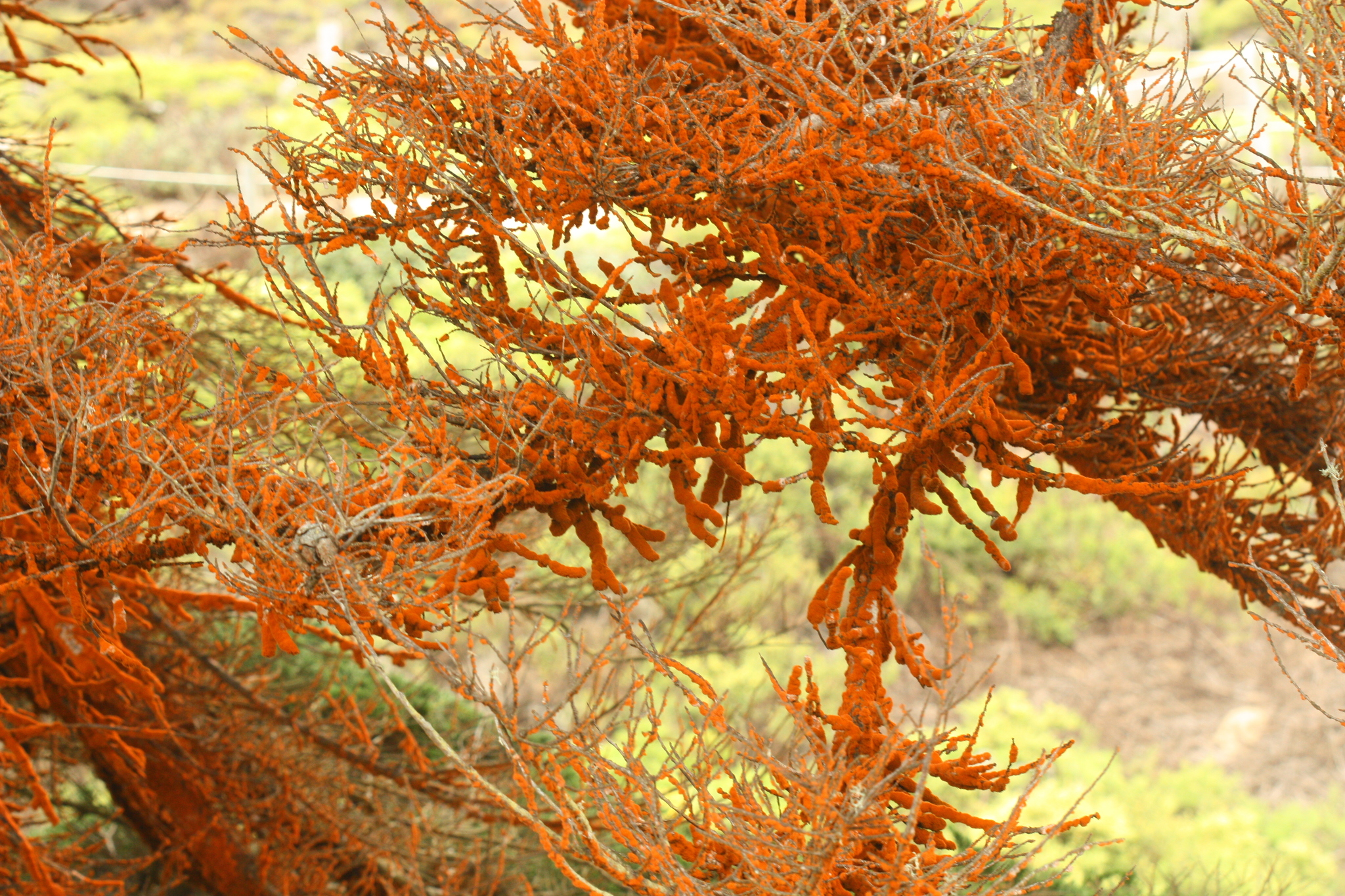
\(\newcommand{\avec}{\mathbf a}\) \(\newcommand{\bvec}{\mathbf b}\) \(\newcommand{\cvec}{\mathbf c}\) \(\newcommand{\dvec}{\mathbf d}\) \(\newcommand{\dtil}{\widetilde{\mathbf d}}\) \(\newcommand{\evec}{\mathbf e}\) \(\newcommand{\fvec}{\mathbf f}\) \(\newcommand{\nvec}{\mathbf n}\) \(\newcommand{\pvec}{\mathbf p}\) \(\newcommand{\qvec}{\mathbf q}\) \(\newcommand{\svec}{\mathbf s}\) \(\newcommand{\tvec}{\mathbf t}\) \(\newcommand{\uvec}{\mathbf u}\) \(\newcommand{\vvec}{\mathbf v}\) \(\newcommand{\wvec}{\mathbf w}\) \(\newcommand{\xvec}{\mathbf x}\) \(\newcommand{\yvec}{\mathbf y}\) \(\newcommand{\zvec}{\mathbf z}\) \(\newcommand{\rvec}{\mathbf r}\) \(\newcommand{\mvec}{\mathbf m}\) \(\newcommand{\zerovec}{\mathbf 0}\) \(\newcommand{\onevec}{\mathbf 1}\) \(\newcommand{\real}{\mathbb R}\) \(\newcommand{\twovec}[2]{\left[\begin{array}{r}#1 \\ #2 \end{array}\right]}\) \(\newcommand{\ctwovec}[2]{\left[\begin{array}{c}#1 \\ #2 \end{array}\right]}\) \(\newcommand{\threevec}[3]{\left[\begin{array}{r}#1 \\ #2 \\ #3 \end{array}\right]}\) \(\newcommand{\cthreevec}[3]{\left[\begin{array}{c}#1 \\ #2 \\ #3 \end{array}\right]}\) \(\newcommand{\fourvec}[4]{\left[\begin{array}{r}#1 \\ #2 \\ #3 \\ #4 \end{array}\right]}\) \(\newcommand{\cfourvec}[4]{\left[\begin{array}{c}#1 \\ #2 \\ #3 \\ #4 \end{array}\right]}\) \(\newcommand{\fivevec}[5]{\left[\begin{array}{r}#1 \\ #2 \\ #3 \\ #4 \\ #5 \\ \end{array}\right]}\) \(\newcommand{\cfivevec}[5]{\left[\begin{array}{c}#1 \\ #2 \\ #3 \\ #4 \\ #5 \\ \end{array}\right]}\) \(\newcommand{\mattwo}[4]{\left[\begin{array}{rr}#1 \amp #2 \\ #3 \amp #4 \\ \end{array}\right]}\) \(\newcommand{\laspan}[1]{\text{Span}\{#1\}}\) \(\newcommand{\bcal}{\cal B}\) \(\newcommand{\ccal}{\cal C}\) \(\newcommand{\scal}{\cal S}\) \(\newcommand{\wcal}{\cal W}\) \(\newcommand{\ecal}{\cal E}\) \(\newcommand{\coords}[2]{\left\{#1\right\}_{#2}}\) \(\newcommand{\gray}[1]{\color{gray}{#1}}\) \(\newcommand{\lgray}[1]{\color{lightgray}{#1}}\) \(\newcommand{\rank}{\operatorname{rank}}\) \(\newcommand{\row}{\text{Row}}\) \(\newcommand{\col}{\text{Col}}\) \(\renewcommand{\row}{\text{Row}}\) \(\newcommand{\nul}{\text{Nul}}\) \(\newcommand{\var}{\text{Var}}\) \(\newcommand{\corr}{\text{corr}}\) \(\newcommand{\len}[1]{\left|#1\right|}\) \(\newcommand{\bbar}{\overline{\bvec}}\) \(\newcommand{\bhat}{\widehat{\bvec}}\) \(\newcommand{\bperp}{\bvec^\perp}\) \(\newcommand{\xhat}{\widehat{\xvec}}\) \(\newcommand{\vhat}{\widehat{\vvec}}\) \(\newcommand{\uhat}{\widehat{\uvec}}\) \(\newcommand{\what}{\widehat{\wvec}}\) \(\newcommand{\Sighat}{\widehat{\Sigma}}\) \(\newcommand{\lt}{<}\) \(\newcommand{\gt}{>}\) \(\newcommand{\amp}{&}\) \(\definecolor{fillinmathshade}{gray}{0.9}\)The nature of the evolutionary relationships between the green algae are still up for debate. As of 2019, genetic data supports splitting the green algae into two major lineages: chlorophytes and streptophytes. The streptophytes include several lineages of green algae and all land plants. Streptophytes and chlorophytes represent a monophyletic group called Viridiplantae (literally “green plants”). Some green algal lineages have adapted to life on land, either inside of lichens or free-living (see Figure \(\PageIndex{1}\)).
General Morphology
Similar to red algae, green algae can be unicellular or multicellular. Many unicellular species form colonies.
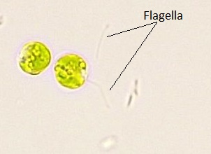
Unicellular
Unicellular species will have two whiplash flagella.
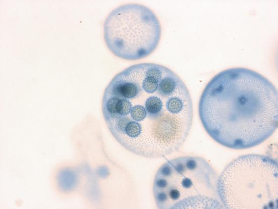
Video \(\PageIndex{1}\): This video shows how sexual reproduction occurs in the colonial green alga Volvox. Sourced from YouTube.
Multicellular
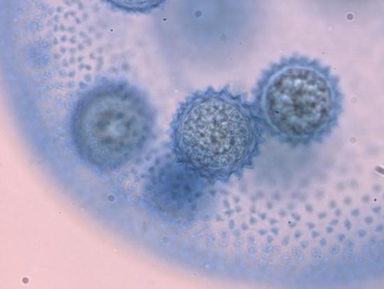
Oedogonium is a genus of filamentous green algae. Some species of Oedogonium are nannandrous. In nannandrous species, the antheridia are small, elongate filaments, usually produced on a different filament than the oogonium. Other species are macrandrous and the antheridia are produced as stacked cells within the same filament as the oogonium.
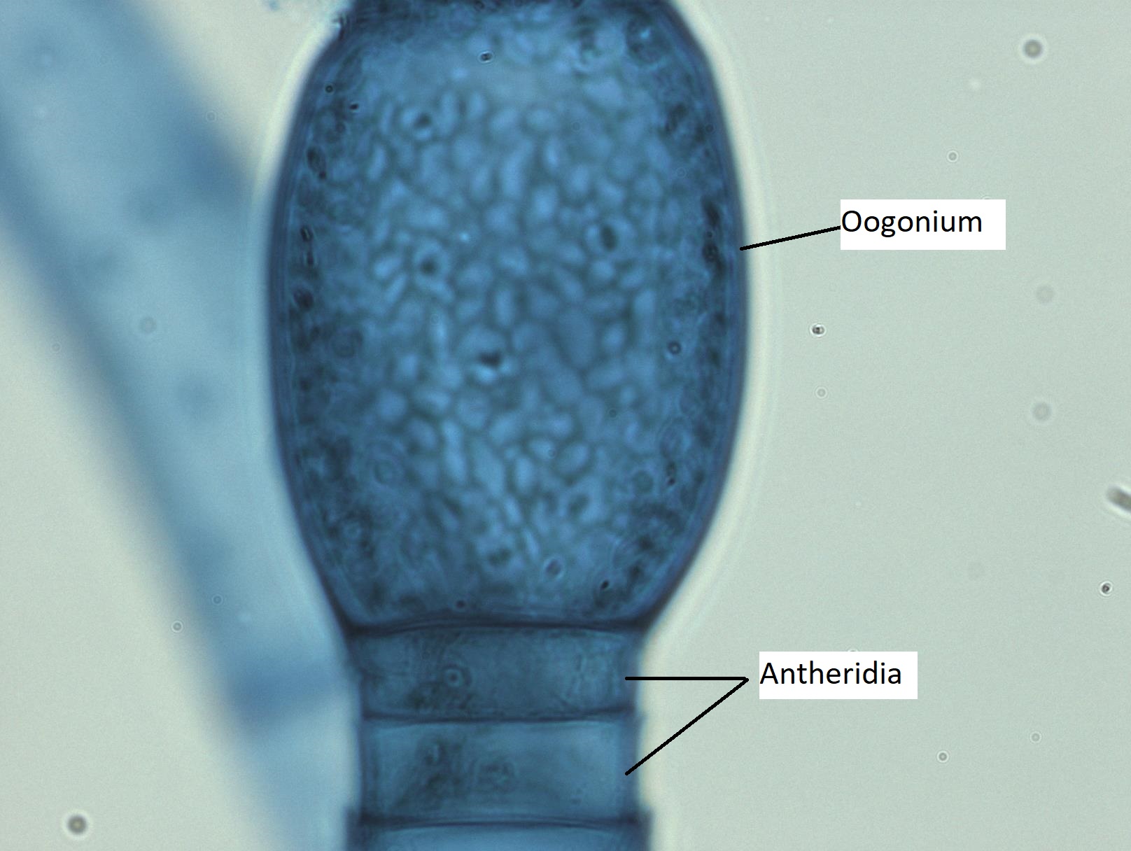
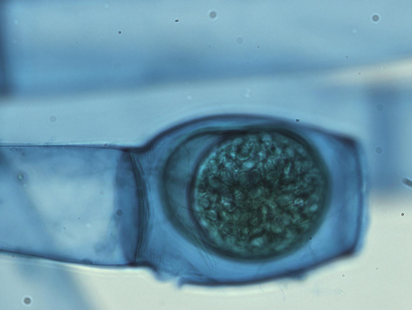
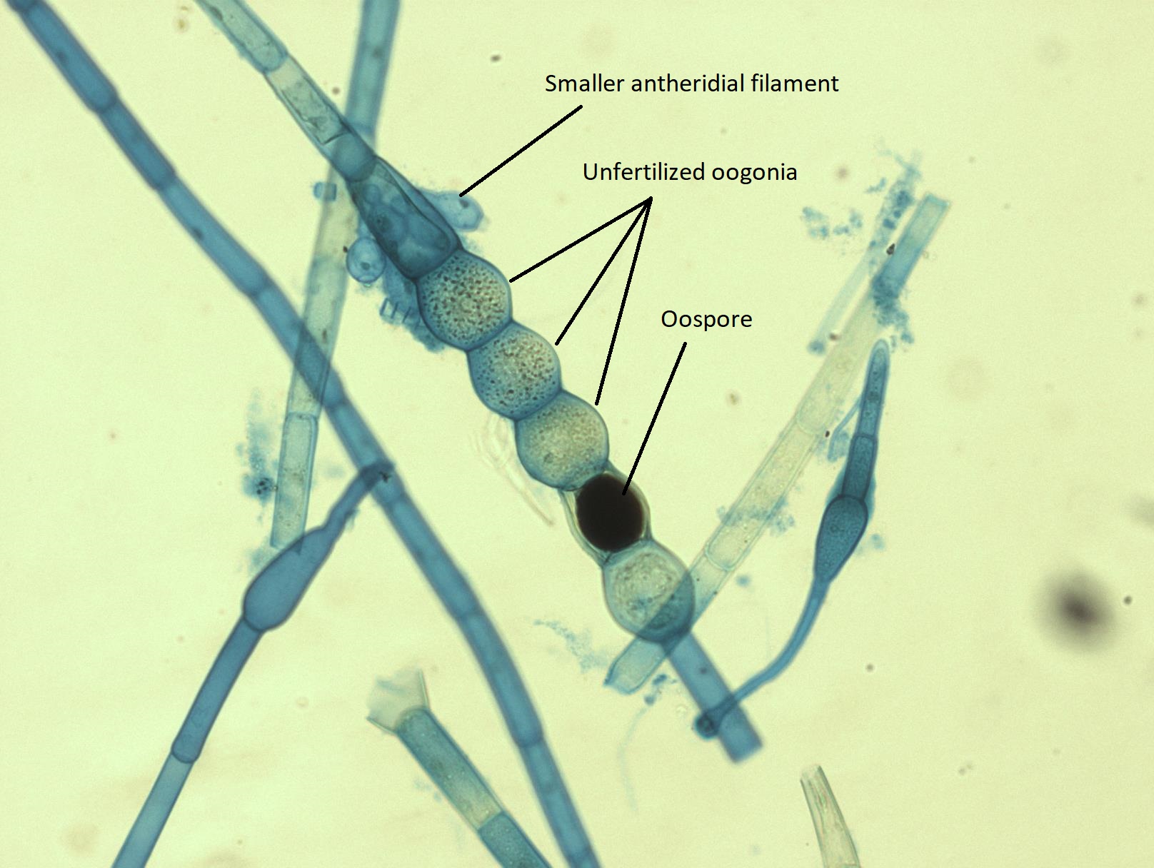
Figure \(\PageIndex{10}\): A close up of the sexual structures of a nannandrous Oedogonium sp. The oogonium is located at the end of the filament and, in this case, is almost lemon-shaped. It is unfertilized, still appearing evenly granular throughout. Many small antheridia are reaching up to try to fuse with the oogonium to fertilize it. These can be seen on the sides of the filament below the oogonium and look like upside down blue bowling pins. Photo by Maria Morrow, CC BY-NC.
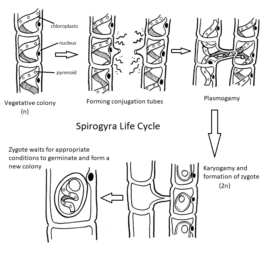
Spirogyra Life Cycle
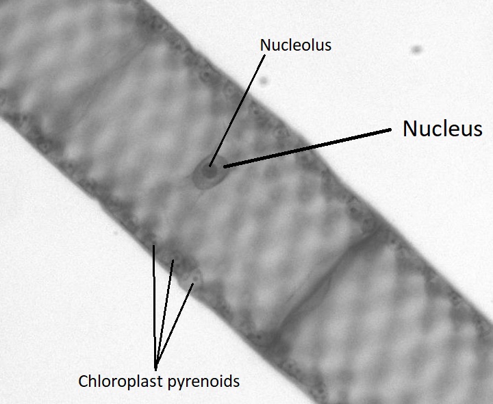
Though green algae display a diversity of life cycles, many have a haplontic life cycle. A model organism for the green algae is Spirogyra. Spirogyra is a unicellular green algae that grows in long, filamentous colonies, making it appear to be a multicellular organism. Even though it is technically unicellular, its colonial nature allows us to classify its life cycle as haplontic. In the haploid vegetative cells of the colony, the chloroplasts are arranged in spirals, containing darkened regions called pyrenoids where carbon fixation happens. Each haploid cell in the filament is an individual, which makes sexual reproduction between colonies an interesting process.
When two colonies of Spirogyra meet that are of a complementary mating type (+/-), sexual reproduction occurs. The two colonies align, each cell across from a complementary cell on the other filament. A conjugation tube extends from each cell in one colony, inducing formation of a tube on the cells in the other colony. The conjugation tubes from each colony fuse together.
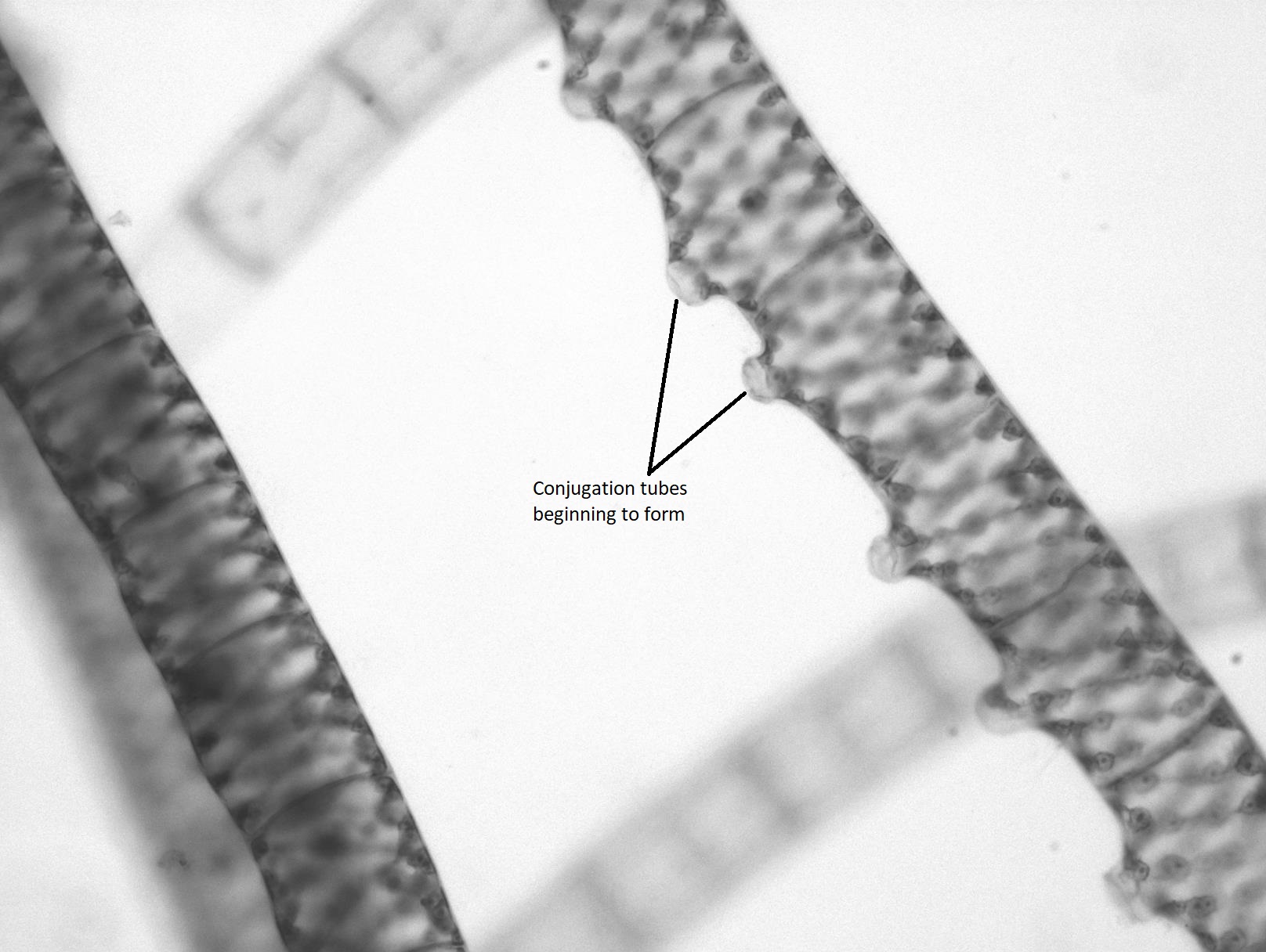
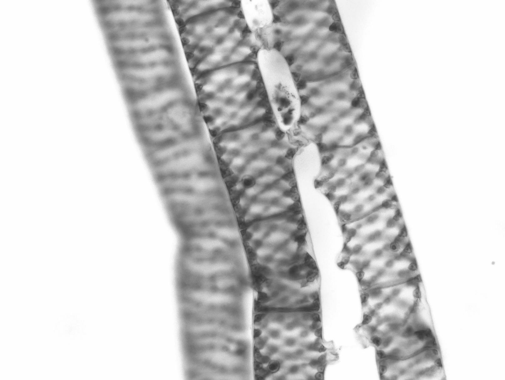
Figure \(\PageIndex{13}\): Spirogyra conjugation tubes meet. These two colonies are both forming conjugation tubes toward each other. Two sets of cells near the top of the image have successfully fused conjugation tubes, forming a connection between the two different organisms. Photo by Maria Morrow, CC BY-NC.
The contents of one cell will move through the conjugation tube and fuse with the contents of the complementary cell, resulting in a diploid zygote.
Figure \(\PageIndex{14}\): Movement of the cytoplasm from one colony to another in Spirogyra. The cytoplasm from one of the cells in the colony on the right has almost completely transferred through the conjugation tube and into the colony on the left. Photo by Maria Morrow, CC BY-NC.
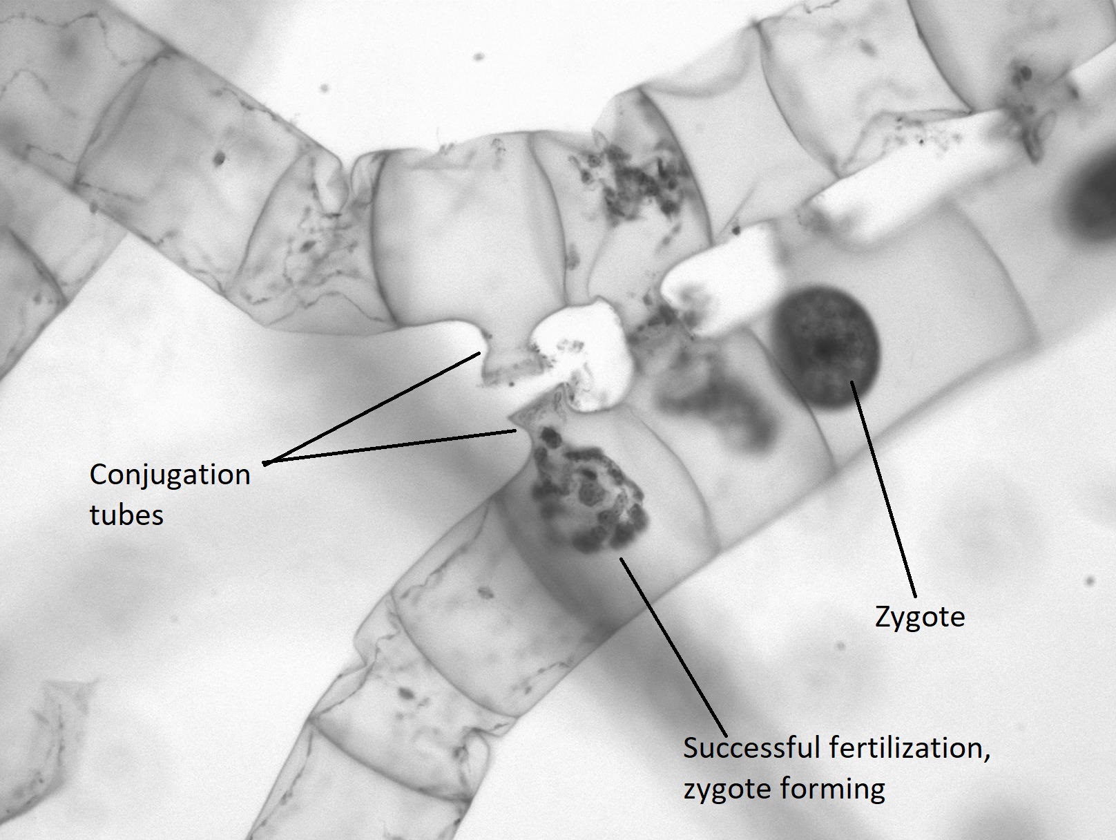
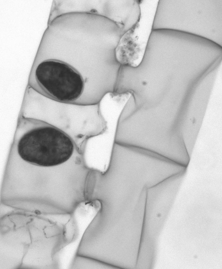
Figure \(\PageIndex{15}\): Cells in various stages of conjugation. Of the cells that have formed conjugation tubes and connected, the one farthest to the left has just recently finished the transfer and fusion of its cytoplasm, but the zygote hasn't fully formed yet. In the cell on the far right, there is a fully formed zygote. It is dark in color and has thick walls. The chloroplasts are not individually distinguishable within it. Photo by Maria Morrow, CC BY-NC.
The zygote appears as a large, egg-like structure contained within the complementary cell. It has a thick wall that provides resistance to desiccation and cold, allowing colonies of Spirogyra to overwinter, when needed. The other colony is now a filament of empty cells that will be broken down by some decomposer.
When conditions are right, the zygote undergoes meiosis to produce another vegetative colony of haploid cells.
Full Life Cycle Diagram