
6.2: Mitosis Teacher's Preparation Notes
- Page ID
- 25120
\( \newcommand{\vecs}[1]{\overset { \scriptstyle \rightharpoonup} {\mathbf{#1}} } \)
\( \newcommand{\vecd}[1]{\overset{-\!-\!\rightharpoonup}{\vphantom{a}\smash {#1}}} \)
\( \newcommand{\dsum}{\displaystyle\sum\limits} \)
\( \newcommand{\dint}{\displaystyle\int\limits} \)
\( \newcommand{\dlim}{\displaystyle\lim\limits} \)
\( \newcommand{\id}{\mathrm{id}}\) \( \newcommand{\Span}{\mathrm{span}}\)
( \newcommand{\kernel}{\mathrm{null}\,}\) \( \newcommand{\range}{\mathrm{range}\,}\)
\( \newcommand{\RealPart}{\mathrm{Re}}\) \( \newcommand{\ImaginaryPart}{\mathrm{Im}}\)
\( \newcommand{\Argument}{\mathrm{Arg}}\) \( \newcommand{\norm}[1]{\| #1 \|}\)
\( \newcommand{\inner}[2]{\langle #1, #2 \rangle}\)
\( \newcommand{\Span}{\mathrm{span}}\)
\( \newcommand{\id}{\mathrm{id}}\)
\( \newcommand{\Span}{\mathrm{span}}\)
\( \newcommand{\kernel}{\mathrm{null}\,}\)
\( \newcommand{\range}{\mathrm{range}\,}\)
\( \newcommand{\RealPart}{\mathrm{Re}}\)
\( \newcommand{\ImaginaryPart}{\mathrm{Im}}\)
\( \newcommand{\Argument}{\mathrm{Arg}}\)
\( \newcommand{\norm}[1]{\| #1 \|}\)
\( \newcommand{\inner}[2]{\langle #1, #2 \rangle}\)
\( \newcommand{\Span}{\mathrm{span}}\) \( \newcommand{\AA}{\unicode[.8,0]{x212B}}\)
\( \newcommand{\vectorA}[1]{\vec{#1}} % arrow\)
\( \newcommand{\vectorAt}[1]{\vec{\text{#1}}} % arrow\)
\( \newcommand{\vectorB}[1]{\overset { \scriptstyle \rightharpoonup} {\mathbf{#1}} } \)
\( \newcommand{\vectorC}[1]{\textbf{#1}} \)
\( \newcommand{\vectorD}[1]{\overrightarrow{#1}} \)
\( \newcommand{\vectorDt}[1]{\overrightarrow{\text{#1}}} \)
\( \newcommand{\vectE}[1]{\overset{-\!-\!\rightharpoonup}{\vphantom{a}\smash{\mathbf {#1}}}} \)
\( \newcommand{\vecs}[1]{\overset { \scriptstyle \rightharpoonup} {\mathbf{#1}} } \)
\(\newcommand{\longvect}{\overrightarrow}\)
\( \newcommand{\vecd}[1]{\overset{-\!-\!\rightharpoonup}{\vphantom{a}\smash {#1}}} \)
\(\newcommand{\avec}{\mathbf a}\) \(\newcommand{\bvec}{\mathbf b}\) \(\newcommand{\cvec}{\mathbf c}\) \(\newcommand{\dvec}{\mathbf d}\) \(\newcommand{\dtil}{\widetilde{\mathbf d}}\) \(\newcommand{\evec}{\mathbf e}\) \(\newcommand{\fvec}{\mathbf f}\) \(\newcommand{\nvec}{\mathbf n}\) \(\newcommand{\pvec}{\mathbf p}\) \(\newcommand{\qvec}{\mathbf q}\) \(\newcommand{\svec}{\mathbf s}\) \(\newcommand{\tvec}{\mathbf t}\) \(\newcommand{\uvec}{\mathbf u}\) \(\newcommand{\vvec}{\mathbf v}\) \(\newcommand{\wvec}{\mathbf w}\) \(\newcommand{\xvec}{\mathbf x}\) \(\newcommand{\yvec}{\mathbf y}\) \(\newcommand{\zvec}{\mathbf z}\) \(\newcommand{\rvec}{\mathbf r}\) \(\newcommand{\mvec}{\mathbf m}\) \(\newcommand{\zerovec}{\mathbf 0}\) \(\newcommand{\onevec}{\mathbf 1}\) \(\newcommand{\real}{\mathbb R}\) \(\newcommand{\twovec}[2]{\left[\begin{array}{r}#1 \\ #2 \end{array}\right]}\) \(\newcommand{\ctwovec}[2]{\left[\begin{array}{c}#1 \\ #2 \end{array}\right]}\) \(\newcommand{\threevec}[3]{\left[\begin{array}{r}#1 \\ #2 \\ #3 \end{array}\right]}\) \(\newcommand{\cthreevec}[3]{\left[\begin{array}{c}#1 \\ #2 \\ #3 \end{array}\right]}\) \(\newcommand{\fourvec}[4]{\left[\begin{array}{r}#1 \\ #2 \\ #3 \\ #4 \end{array}\right]}\) \(\newcommand{\cfourvec}[4]{\left[\begin{array}{c}#1 \\ #2 \\ #3 \\ #4 \end{array}\right]}\) \(\newcommand{\fivevec}[5]{\left[\begin{array}{r}#1 \\ #2 \\ #3 \\ #4 \\ #5 \\ \end{array}\right]}\) \(\newcommand{\cfivevec}[5]{\left[\begin{array}{c}#1 \\ #2 \\ #3 \\ #4 \\ #5 \\ \end{array}\right]}\) \(\newcommand{\mattwo}[4]{\left[\begin{array}{rr}#1 \amp #2 \\ #3 \amp #4 \\ \end{array}\right]}\) \(\newcommand{\laspan}[1]{\text{Span}\{#1\}}\) \(\newcommand{\bcal}{\cal B}\) \(\newcommand{\ccal}{\cal C}\) \(\newcommand{\scal}{\cal S}\) \(\newcommand{\wcal}{\cal W}\) \(\newcommand{\ecal}{\cal E}\) \(\newcommand{\coords}[2]{\left\{#1\right\}_{#2}}\) \(\newcommand{\gray}[1]{\color{gray}{#1}}\) \(\newcommand{\lgray}[1]{\color{lightgray}{#1}}\) \(\newcommand{\rank}{\operatorname{rank}}\) \(\newcommand{\row}{\text{Row}}\) \(\newcommand{\col}{\text{Col}}\) \(\renewcommand{\row}{\text{Row}}\) \(\newcommand{\nul}{\text{Nul}}\) \(\newcommand{\var}{\text{Var}}\) \(\newcommand{\corr}{\text{corr}}\) \(\newcommand{\len}[1]{\left|#1\right|}\) \(\newcommand{\bbar}{\overline{\bvec}}\) \(\newcommand{\bhat}{\widehat{\bvec}}\) \(\newcommand{\bperp}{\bvec^\perp}\) \(\newcommand{\xhat}{\widehat{\xvec}}\) \(\newcommand{\vhat}{\widehat{\vvec}}\) \(\newcommand{\uhat}{\widehat{\uvec}}\) \(\newcommand{\what}{\widehat{\wvec}}\) \(\newcommand{\Sighat}{\widehat{\Sigma}}\) \(\newcommand{\lt}{<}\) \(\newcommand{\gt}{>}\) \(\newcommand{\amp}{&}\) \(\definecolor{fillinmathshade}{gray}{0.9}\)Overview
This minds-on, hands-on activity helps students to understand how mitosis ensures that each new cell gets a complete set of genes. Our instructional philosophy for this activity and our follow-up activity on meiosis and fertilization 2 is that student learning about mitosis and meiosis is most meaningful if students learn how gene - carrying chromosomes move during mitosis, meiosis, and fertilization and understand how these processes result in the transmission of genes from parents to offspring. To provide the background needed for this approach, this mitosis activity begins with an introduction to chromosomes, genes and alleles, and the effects of genes on phenotypic characteristics.
Then, students learn about the basic process of mitosis and use model chromosomes to simulate mitosis. Throughout, students respond to analysis and discussion questions to further develop their understanding of mitosis. We offer two versions of the Student Handout:
- A more complete version (primarily for high school or college non-major students)
- A shorter version (primarily for middle school students).
For this mitosis activity, the more complete version of the Student Handout includes the second pair of homologous chromosomes. For the meiosis and fertilization activity, the more complete version of the Student Handout includes independent assortment, crossing over and a more complete analysis of the sources of genetic differences between siblings. This activity can be used as an introduction to mitosis or to reinforce understanding of mitosis. Depending on your students' background and which version of the Student Handout you use, this activity may require about 1½ 50-minute periods. Some suggested enhancements are presented in "Mitosis, Meiosis, and Fertilization – Major Concepts, Common Misconceptions, and Learning Activities" (available at http://serendip.brynmawr.edu/exchang...MitosisMeiosis). Before beginning this activity, students should know what a cell is and have a basic understanding of the functions of DNA and proteins. For this purpose, we recommend "Understanding the Functions of Proteins and DNA" (available at http://serendip.brynmawr.edu/exchang...ities/proteins). These activities are part of an integrated sequence of learning activities for teaching genetics, presented in "Genetics–Major Concepts and Learning Activities" ( available at http://serendip.brynmawr.edu/exchang...neticsConcepts).
These Teacher Preparation Notes include:
- Learning Goals (pages 2-3)
- Making the Model Chromosomes (pages 3-5)
- Additional Supplies and Requirements for the Modeling Activity (page 5)
- Instructional Suggestions and Background Biology (pages 5-9)
- Background Information on Albinism, Sickle Cell Anemia and Alcohol Sensitivity
- (pages 9-11)
- Follow-up and Related Activities (page11)
- Figures you may want to give your students to help them answer question 16 (page 12)
Learning Goals
In accord with the Next Generation Science Standards (http://www.nextgenscience.org/):
- Students will gain an understanding of several Disciplinary Core Ideas:
- LS1.A: Structure and Function–"All cells contain genetic information in the form of DNA molecules. Genes are regions in the DNA that contain the instructions that code forthe formation of proteins."
- LS1.B: Growth and Development of Organisms–"In multicellular organisms individual cells grow and then divide by a process called mitosis, thereby allowing the organism to grow. The organism begins as a single cell (fertilized egg) that divides successively to produce many cells, with each parent cell passing identical genetic material (two variants of each chromosome pair) to both daughter cells."
- LS3.A: Inheritance of Traits–"Genes are located in the chromosomes of cells, with each chromosome pair containing two variants of each of many distinct genes. Each distinct gene chiefly controls the production of specific proteins, which in turn affects the traits of the individual. Changes (mutations) to genes can result in changes to proteins, which can affect the structures and functions of the organism and thereby change traits." "Each chromosome consists of a single very long DNA molecule, and each gene on the chromosome is a particular segment of that DNA. The instructions for forming species' characteristics are carried in DNA."
- Students will engage in the Scientific Practices, "using models" and "constructing explanations".
- This activity provides the opportunity to discuss the Crosscutting Concepts, "Systems and system models" and "Structure and function".
- This activity helps to prepare students for the Performance Expectations:
- MS-LS3-2, "Develop and use a model to describe why asexual reproduction results in offspring with identical genetic information and sexual reproduction results in offspring with genetic variation."
- HS-LS1-4, "Use a model to illustrate the role of cellular division (mitosis) and differentiation in producing and maintaining complex organisms."
- HS-LS3-1, "Ask questions to clarify relationships about the role of DNA and chromosomes in coding the instructions for characteristic traits passed from parents to offspring."
Specific Learning Goals
- Each cell contains chromosomes and each chromosome contains a long DNA molecule. Each DNA molecule has many genes. A gene provides the instructions for making a protein. Different versions of a gene are called alleles, and different alleles give the instructions for making different versions of a protein. These different versions of a protein can result in different phenotypic characteristics.
- Chromosomes come in pairs of homologous chromosomes. In each pair of homologous chromosomes, both chromosomes have the same genes at the same locations, but a gene may have different alleles in the two chromosomes of a homologous pair.
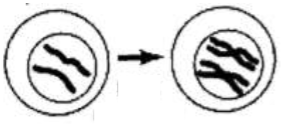
- Before mitosis, the DNA is replicated to form two copies of the original DNA.
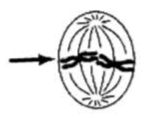
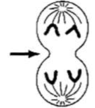
- At the beginning of mitosis, the two copies of the DNA in each chromosome are condensed into compact sister chromatids which are attached at a centromere. During mitosis, spindle fibers line up the chromosomes in the middle of the cell and then separate the sister chromatids of each chromosome, resulting in two complete sets of chromosomes at opposite ends of the cell.
- At the end of each cell division, cytokinesis separates the two halves of the cell to form two daughter cells. Each daughter cell receives a complete set of chromosomes with exactly the same genes as the original cell.
Making the Model Chromosomes
We recommend two students per group for this activity, but student groups can be as large as four students.
For the more complete Student Handout, each student group will need two pairs of homologous model chromosomes. For the follow-up activity "Meiosis and Fertilization–Understanding How Genes Are Inherited" (http://serendip.brynmawr.edu/sci_edu/waldron/#meiosis), you will need the two pairs of homologous model chromosomes to be different colors.
For the shorter Student Handout, you will only need the first pair of homologous model chromosomes, and you should modify the instructions below accordingly. If you plan to use the follow-up activity, "Meiosis and Fertilization–Understanding How Genes Are Inherited", you should be aware that the fertilization part of that activity requires that each student group have a second set of the first pair of homologous model chromosomes, but in a different color. To meet this need, we recommend that you make enough model chromosomes for each pair of students in your largest class, half in one color and half in another color, Then, for the fertilization part of the meiosis and fertilization activity, increase the size of the student groups from two to four.
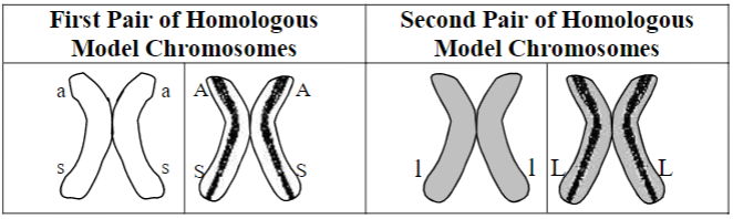
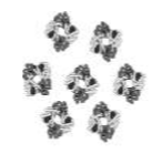
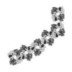
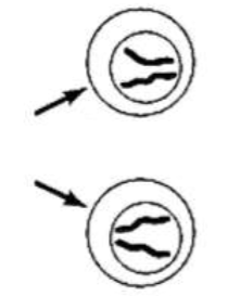
Each model chromosome consists of two sister chromatids which are attached with hook and loop fasteners (Velcro) in the centromere region (approximately the location where the two chromatids touch in the above figures). For each pair of homologous chromosomes, one of the chromosomes has a stripe on each chromatid to represent the multiple differences in alleles between the two chromosomes in a homologous pair.
You can use rolosomes (made from hair roller curlers) or sockosomes (made from socks). The rolosomes provide model chromosomes that are engaging and easy to make. The sockosomes provide three-dimensional models that look like metaphase chromosomes in karyotypes. Sockosomes are much more time-consuming to make, although this may be a good time investment if you will be using the sockosomes year after year. The figures in the chart on the previous page show the approximate shape of sockosome model chromosomes; the shape of rolosomes is shown in the photo below.
Rolosomes
Supplies
For each group of 2-4 students in your largest class:
8 hair roller curlers, 4 in one color and 4 in another color (Hereafter, these hair color rollers will be referred to as rollers. Rollers are readily available online, but you may need to order from two different manufacturers in order to get rollers that have similar diameter but different colors.)
4 pairs of self-stick hook-and-loop dots (Velcro). (The hook and loop dots should have a slightly smaller diameter than the rollers. In our experience, the dots do not stick well if the diameter of the dots is larger than the diameter of the rollers.) You will also need a permanent marker to make the rolosomes. The rolosomes in this photo represent two pairs of homologous chromosomes. Each rolosome has two chromatids attached by Velcro fasteners in the centromere region. The first rolosome has the alleles a and s. The second rolosome is homologous to the first rolosome and has the alleles A and S. The two rolosomes on the right represent a pair of homologous chromosomes with the alleles l and L, respectively. (Both chromatids of the L chromosome have a stripe, although this is not readily visible in this photo.)
Making the Rolosomes
To make the four rolos omes for each group of 2-4 students:
1. For two rollers of the same color, stick a Velcro hook-and-loop dot or piece with hooks on one roller and a matching fuzzy dot or piece with loops on the other roller, so the two rollers can be attached as sister chromatids.
Note
The pair of rollers attached by hook-and-loop dots is a rolosome. In the rolosome, each roller represents a chromatid. After mitosis is completed, each roller represents a chromosome in a daughter cell.
2. Repeat step 1 to make four rolosomes, each with two sister chromatids.
3. Use the chart on page 3 and the figure on page 4 to label the alleles on each pair of homologous model chromosomes. To avoid possible confusion, make the s allele particularly small and the S allele particularly large. Make a long stripe down both chromatids of one of each pair of homologous chromosomes , as shown.
Sockosomes
You do not need these if you make rolosomes. These instructions are provided in case you prefer to make sockosomes.
Supplies
- Small or medium children’s crew socks (no more than half of any one color; even number of pairs of each color sock; four pairs of socks for each group of 2-4 students in your largest class (see chart on page 3); avoid black and dark blue socks typically found in packs of boys socks).
- Fiberfill
- Self-stick squares or circles of hook-and-loop fasteners (Velcro); if you are making more than 36 sockosomes it may be more cost-effective to purchase a roll of self-stick hook-and-loop tape and cut it into 1/2" pieces.
- Needle and thread
- 1" wide masking tape and permanent marker
Making the Sockosomes
- Attach one part of a self-stick hook-and-loop fastener (the fuzzy part) to the heel of one sock, and attach the other part (the part with hooks) to the heel of the other sock; secure with staples or by sewing.
- Fill each sock with fiberfill, and sew the end of each sock closed.
- Stick the socks together at the heels. You now have a chromosome with two chromatids, where each sock represents a chromatid. Note that a sockosome refers to the pair of socks attached by hook-and-loop fasteners, not the individual socks. After mitosis is completed, each individual sock represents a chromosome in a daughter cell.
- Pairs of homologous chromosomes will be represented by two sockosomes of the same color, one with a stripe marked along the length of each sock with a permanent marker. Use the chart on page 3 and the instructions in step 3 on page 4 to guide you in labeling the alleles on each chromatid in your sockosomes. For each allele, add a ring of tape around each sock in each sockosome to mark the allele; the tape stays on best if it goes completely around the sock, overlapping at the ends.
Additional Supplies and Requirements for the Modeling Activity
Students sometimes have difficulty recognizing that the two sets of chromosomes are in two different daughter cells at the end of mitosis. Therefore, we recommend that you provide pieces of string or yarn for students to use as cell membranes. For example, for the modeling activity on page 5 of the Student Handout, each student group will need a piece of string approximately 5 feet long to represent the membrane surrounding the original cell and a pair of scissors to cut the string into two pieces to represent the membranes around the daughter cells.
Students should carry out the modeling activities on a lab table or similar large flat surface, so they can more easily see the processes and outcomes.
Instructional Suggestions and Background Biology
- In the Student Handout, numbers in bold indicate questions for the students to answer.
- If you are using the Word version of the Student Handout, please check the PDF version to make sure that all figures and formatting are displayed properly in the Word version on your computer.
- To maximize student learning, we recommend that you have your students complete groups of related analysis and discussion questions in the Student Handout individually or in pairs and then have a class discussion of these questions. In each discussion, you can probe student thinking and help them to develop a sound understanding of the concepts and information covered before moving on to the next part of the activity.
- If you would like to have a key with the answers to the questions in the Student Handout, please send a message to iwaldron@sas.upenn.edu. The following paragraphs provide additional instructional suggestions and background information.
- For questions which require students to label the s allele in diagrams, you may want to have your students use a lowercase s with a line above it or a cursive s, in order to avoid confusion with the S allele.
- In the Student Handout for this activity, we have introduced multiple technical terms(shown in bold). Students often have difficulty understanding the difference between chromosomes and chromatids, so we have made a special effort to clarify this distinction (e.g. on pages 3-4). We have omitted the technical terms for some of the concepts introduced in the Student Handout in order to allow students to focus on learning the basic concepts without becoming overwhelmed by memorizing vocabulary. If you prefer to introduce additional vocabulary, suggestions about how to include the terms homozygous, heterozygous, dominant and recessive are given on page 7 of these Teacher Preparation Notes and suggestions about how to include the names of the stages of mitosis are given on page 8.
Genes and Chromosomes
Many students have difficulty distinguishing the concepts of DNA, genes, and chromosomes, so you will probably want to reinforce student understanding that a gene is part of a DNA molecule contained in a chromosome. In this activity, we have included both the albinism and sickle cell genes on one pair of model chromosomes in part to counteract the tendency for some students to assume that each chromosome has only a single gene.
 On page 1 of the Student Handout, a gene is defined as a segment of DNA that gives the instructions for making a protein. You should be aware that the definition of a gene has changed as scientific understanding has progressed. Initially, a gene was conceived as a unit of heredity that determines a phenotypic characteristic. A more sophisticated contemporary definition of a gene is a segment of DNA that codes for an RNA molecule, which may be messenger RNA that codes for the sequence of amino acids in one or more proteins, ribosomal RNA, transfer RNA or regulatory RNA. There is no single universally agreed-upon definition of a gene at this time. The changing definition of a gene illustrates the constantly evolving nature of science as scientists develop a progressively more sophisticated understanding of concepts such as the gene. For additional information about the challenges and complexities of defining a gene, see http://www.biologyreference.com/Fo-Gr/Gene.html.
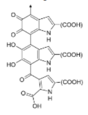
When you discuss question 1, your students will probably recognize that enzymes and hemoglobin are proteins, but they may not know that melanin is not a protein. This figure shows part of the structural formula of the most common type of melanin (eumelanin); the arrow shows where the polymer continues.
We do not introduce the terms homozygous, heterozygous, dominant, or recessive in the Student Handout for this activity. Instead, we introduce them in our Genetics activity (available at http://serendip.brynmawr.edu/sci_edu...dron/#genetics). If you prefer, these terms can easily be introduced when you discuss the information in the table on the bottom of page 1 of the Student Handout. For this purpose, you may want to include the following prose and question after this table in the Student Handout.
- If both copies of a gene have the same allele, the person is homozygous for that gene. If the two copies of a gene have different alleles, the person is heterozygous.
- Often, in a heterozygous individual, a dominant allele determines the observable characteristic and the other recessive allele does not affect the phenotype. Thus, a heterozygous person has the same phenotype as a person who is homozygous for the dominant allele. In our example, the A allele is dominant because it codes for normal, functional enzyme and, even in a heterozygous individual, there is enough of this normal, functional enzyme to produce enough melanin to result in normal skin and hair color. The a allele is recessive because it codes for a non-functional enzyme which does not affect skin or hair color in a heterozygous individual.
- What are two different genotypes for the albinism gene that result in the same phenotype? Explain how two people with different genotypes can have the same phenotype.
In a heterozygous individual, typically each allele is transcribed and both versions of the protein are produced. For many genes, the allele that codes for a functional protein results in the production of enough normal protein to produce a normal phenotype. In these cases, the allele that codes for a functional protein is dominant and the allele that codes for a defective protein is recessive. Two examples are shown on the bottom of page 1 of the Student Handout. The gene for the enzyme that disposes of a harmful molecule produced by alcohol metabolism is an exception where the allele for the defective enzyme is dominant (see page 6 of the more complete Student Handout). A major reason is that the functional enzyme consists of four normal polypeptides bound together, and even one defective polypeptide in this tetramer may inactivate the enzyme.
For more information on each of the genes and associated conditions discussed in the Student Handout, see pages 9-11 of these Teacher Preparation Notes.
At several points in the Student Handouts, there are statements along the lines of "each cell needs to have a complete set of chromosomes". As you no doubt know, there are exceptions to this generalization such as mammalian red blood cells which do not have any chromosomes, and gametes which have only one from each pair of homologous chromosomes. To avoid undue complexities, we have omitted discussion of the special case of red blood cells and we have postponed discussion of gametes to the meiosis and fertilization activity. We have also omitted mention of the important observation that, while all genes are present in each cell, many genes are only active in specific types of cells; e.g. the genes for the hemoglobin polypeptides are expressed in the precursors of red blood cells.
Cell Division—How New Cells Are Made
The discussion on the bottom of page 2 of the Student Handout focuses on cell division (the topic of this activity) and ignores the role of cell differentiation and morphogenesis in the development of embryos. A brief introduction to these topics is available at http://users.rcn.com/jkimball.ma.ult...velopment.html.
The rate of cell replacement by mitosis varies for different circumstances and different types of cells. The rate of cell division and replacement is greater when an injury has occurred, and cells that are routinely exposed to injury (e.g. skin cells or epithelial cells that line the lumen of the stomach and small intestine) are replaced within days or a couple of weeks. In contrast, nerve cells and muscle cells can last a lifetime. Page 3 of the Student Handout shows a simplified description of the cell cycle, including only DNA replication, mitosis, and cytokinesis.
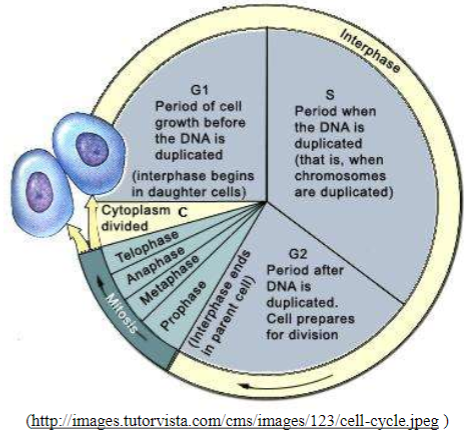
The figure above shown here provides a more complete description of the cell cycle. If you want your students to learn the names of the phases of mitosis, these terms can easily be incorporated on page 4 of the Student Handout. The video available at http://vcell.ndsu.nodak.edu/animatio...ovie-flash.htm provides a helpful explanation of the different phases of mitosis and the cell cycle.
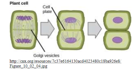
The Student Handout shows cytokinesis in animal cells. Cytokinesis in plant cells is illustrated in this figure above. Mitosis can occur without cytokinesis. For example, this is how multinucleate skeletal muscle fibers are formed.
After completing the introduction to mitosis (pages 3-4 of the Student Handout) and before beginning the mitosis modeling activity, you may want to show an animation of mitosis available at https://www.youtube.com/watch?v=VlN7K1-9QB0. At the end of this mitosis activity, you may want to show the video of mitosis in a lung cell, available at (https://www.youtube.com/watch?v=DD3IQknCEdc).
Modeling Mitosis
To prevent student confusion during the modeling activities:
- It is crucial to circulate among student groups continuously and provide considerable input.
- As the students model mitosis, remind them to check the diagrams (including the figure on page 3 of the Student Handout).
- You will probably want to reinforce student understanding that the modeling activity begins with chromosomes that have replicated DNA in sister chromatids (represented by complete rolosomes) and ends with chromosomes that do not have replicated DNA (represented by a single roller in each daughter cell).
Follow-Up Questions
Question 16 in the more complete Student Handout (question 14 in the shorter Student Handout) engages students in synthesizing and summarizing what they have learned about mitosis in the context of understanding how their bodies have grown. If this question is challenging for your students:
- You may want to rearrange the terms that students are required to include in a more logical sequence, rather than alphabetical order, perhaps: mitosis, genes, chromosomes, DNA replication, sister chromatids, spindle fibers, cytokinesis, daughter cells
- You may want to provide your students with copies of the figures shown on the last page of these Teacher Preparation Notes; students can label and explain these figures as part of their answer to this synthesis/review question.
- Students may benefit from a preliminary small group discussion of this question. However, each student should prepare a written answer in his or her own words. The bottom of page 7 in the more complete Student Handout (page 6 in the shorter Student Handout) introduces students to one example of asexual reproduction, although that term is not introduced in the Student Handout. If you want your students to learn about asexual reproduction, you can introduce this term, emphasize that asexual reproduction results in genetically identical individuals, and give additional examples (see e.g. http://www.mhhe.com/biosci/genbio/tl...Chp18/18_5.pdf, http://education.seattlepi. com/five-examples-organisms-use-asexual-reproduction-5849.html, http://www.saburchill.com/ans02/chapters/chap049.html, and http://www.nature.com/scitable/knowl...antic-13261308).
Background Information on Albinism, Sickle Cell Anemia and Alcohol Sensitivity
Albinism
In the most common form of albinism, the defective enzyme for producing melanin not only results in albino skin and hair color, but also affects the appearance and function of the eyes. The defective enzyme is tyrosinase which is needed for the first two steps in converting tyrosine to melanin. In a heterozygous individual, the normal allele is dominant because it codes for the functioning enzyme and even when there is only one copy of the normal allele there is enough of this functioning enzyme to produce enough melanin to prevent albinism. For additional information about the various forms of albinism see http://www.nlm.nih.gov/medlineplus/e...cle/001479.htm and OMIM = Online Mendelian Inheritance in Man (available at www.ncbi.nlm.nih.gov/omim/; search for 606952 (oculocutaneous albinism)).
Students may ask about the distinction between inherited albinism and vitiligo. Albinism is the inability of the body's cells to produce melanin and affects the whole body. Vitiligo is a patterned loss of melanin pigment resulting from the destruction of melanocytes; the hypopigmented areas appear on the skin of a person with normal pigmentation. (Additional information from the National Vitiligo Foundation is available at www.nvfi.org.)
Sickle Cell Anemia
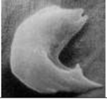
A person who is homozygous for the sickle cell allele and produces only sickle cell hemoglobin has sickle cell anemia. Sickle cell hemoglobin is less soluble in the watery cytosol of the red blood cells than normal hemoglobin, particularly when oxygen concentrations are low. Consequently, sickle cell hemoglobin tends to form long stacks or rods of hemoglobin molecules. These stacks of sickle cell hemoglobin molecules result in the sickled shape of some red blood cells in a person who is homozygous for the sickle cell allele. The sickled red blood cells tend to clog the tiny capillaries, blocking the circulation in different parts of the body. Also, the sickled red blood cells do not survive as long as normal red blood cells, contributing to a tendency to anemia. Resulting symptoms include pain, physical weakness, impaired mental functioning, and damage to organs such as the heart and kidneys.
| Genotype (genes) | ⇒ | Protein | ⇒ | Phenotype (characteristics) |
|
2 copies of the allele that codes for normal hemoglobin (SS) |
⇒ |
Normal hemoglobin dissolves in the cytosol of red blood cells.
|
⇒ |
Disk-shaped red blood cells can squeeze through the small blood vessels ⇒ Normal health
|
|
2 copies of the allele that codes for sickle cell hemoglobin (ss) |
⇒ |
Sickle cell hemoglobin can clump in long rods in red blood cells.
|
⇒ |
When sickle cell hemoglobin clumps in long rods ⇒ Sickle-shaped red blood cells ⇒ Clogged small blood vessels + fragile red blood cells ⇒ Pain, damage to body organs + anemia = sickle cell anemia
|
In a person who is heterozygous for the sickle cell and normal hemoglobin alleles, each red blood cell has both sickle cell and normal hemoglobin. The amount of normal hemoglobin is sufficient to prevent the symptoms of sickle cell anemia in almost all cases. The sickle cell hemoglobin in each red blood cell decreases the severity of malaria in heterozygous individuals because the malaria parasite doesn't grow as well in red blood cells containing sickle cell hemoglobin.
Additional information can be found in "Sickle cell anemia", available at
- www.mayoclinic.com/health/sickle-cell-anemia/DS00324
- OMIM (Online Mendelian Inheritance in Man (http://www.ncbi.nlm.nih.gov/omim/; search for 603903 (sickle cell anemia))
A video, "Sickle cell anemia" is available at http://www.hhmi.org/biointeractive/d...icklecell.html
Alcohol Sensitivity
The enzyme, acetaldehyde dehydrogenase, plays a major role in alcohol metabolism.
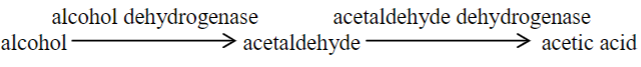
An inactive form of acetaldehyde dehydrogenase results in the accumulation of high levels of acetaldehyde after drinking alcohol. The accumulation of acetaldehyde results in unpleasant symptoms including increased heart rate and stroke volume and associated heart palpitations, increased blood flow to the skin and flushing, and a general "terrible feeling overall". This condition is called alcohol sensitivity or alcohol intolerance.
Heterozygous individuals have a substantial accumulation of acetaldehyde and substantial symptoms, in large part because the functional enzyme is a tetramer and even one abnormal protein in the tetramer may inactivate the enzyme. Although heterozygous individuals are sensitive to alcohol, alcohol sensitivity is more severe in homozygous individuals who experience very unpleasant symptoms whenever they drink alcohol; consequently, homozygous individuals almost never develop alcoholism.
The drug Antabuse (disulfiram), which is given to treat alcohol abuse, works by blocking the enzyme acetaldehyde dehydrogenase. If a person drinks, this results in increased concentrations of acetaldehyde and the resultant highly unpleasant symptoms.
The allele that codes for the relatively inactive version of acetaldehyde dehydrogenase which results in alcohol sensitivity is relatively common in people of East Asian descent, but extremely rare in people of European descent.
Useful general introductions to this topic are available at http://www.mayoclinic.org/diseases-c...n/con-20034907 and http://en.Wikipedia.org/wiki/Alcohol_flush_reaction, and more technical description is available at http://www.omim.org/; search for +100650.
Follow-up and Related Activities
We recommend that you follow this mitosis activity with "Understanding Meiosis and Fertilization–How Genes Are Inherited" (http://serendip.brynmawr.edu/sci_edu/ waldron/#meiosis). These activities are part of an integrated sequence of learning activities for teaching genetics ("Genetics–Major Concepts and Learning Activities"; available at http://serendip.brynmawr.edu/exchang...neticsConcepts). For example, to help students understand the process of DNA replication in preparation for mitosis you may want to use "DNA Structure, Function, and Replication" (http://serendip.brynmawr.edu/exchang...activities/DNA).
"Chromonoodles: Jump into the Gene Pool" by Farrar and Barnhart, The Science Teacher, Summer 2011, 78:34-39 presents an informative series of activities using chromonoodles (made from swim noodles) to demonstrate fertilization, the cell cycle, meiosis, karyotyping, and genetics concepts, including Punnett squares. These activities are whole-class demonstrations, in contrast to the more structured modeling activities for small groups of students presented in the Student Handouts for our activities.
Additional resources that you may find helpful are provided in the podcasts available at http://www.bozemanscience.com/028-ce...s-and-meiosis/.
 |
 |
 |
 |