
8.2.4: Face
- Page ID
- 53620

\( \newcommand{\vecs}[1]{\overset { \scriptstyle \rightharpoonup} {\mathbf{#1}} } \)
\( \newcommand{\vecd}[1]{\overset{-\!-\!\rightharpoonup}{\vphantom{a}\smash {#1}}} \)
\( \newcommand{\dsum}{\displaystyle\sum\limits} \)
\( \newcommand{\dint}{\displaystyle\int\limits} \)
\( \newcommand{\dlim}{\displaystyle\lim\limits} \)
\( \newcommand{\id}{\mathrm{id}}\) \( \newcommand{\Span}{\mathrm{span}}\)
( \newcommand{\kernel}{\mathrm{null}\,}\) \( \newcommand{\range}{\mathrm{range}\,}\)
\( \newcommand{\RealPart}{\mathrm{Re}}\) \( \newcommand{\ImaginaryPart}{\mathrm{Im}}\)
\( \newcommand{\Argument}{\mathrm{Arg}}\) \( \newcommand{\norm}[1]{\| #1 \|}\)
\( \newcommand{\inner}[2]{\langle #1, #2 \rangle}\)
\( \newcommand{\Span}{\mathrm{span}}\)
\( \newcommand{\id}{\mathrm{id}}\)
\( \newcommand{\Span}{\mathrm{span}}\)
\( \newcommand{\kernel}{\mathrm{null}\,}\)
\( \newcommand{\range}{\mathrm{range}\,}\)
\( \newcommand{\RealPart}{\mathrm{Re}}\)
\( \newcommand{\ImaginaryPart}{\mathrm{Im}}\)
\( \newcommand{\Argument}{\mathrm{Arg}}\)
\( \newcommand{\norm}[1]{\| #1 \|}\)
\( \newcommand{\inner}[2]{\langle #1, #2 \rangle}\)
\( \newcommand{\Span}{\mathrm{span}}\) \( \newcommand{\AA}{\unicode[.8,0]{x212B}}\)
\( \newcommand{\vectorA}[1]{\vec{#1}} % arrow\)
\( \newcommand{\vectorAt}[1]{\vec{\text{#1}}} % arrow\)
\( \newcommand{\vectorB}[1]{\overset { \scriptstyle \rightharpoonup} {\mathbf{#1}} } \)
\( \newcommand{\vectorC}[1]{\textbf{#1}} \)
\( \newcommand{\vectorD}[1]{\overrightarrow{#1}} \)
\( \newcommand{\vectorDt}[1]{\overrightarrow{\text{#1}}} \)
\( \newcommand{\vectE}[1]{\overset{-\!-\!\rightharpoonup}{\vphantom{a}\smash{\mathbf {#1}}}} \)
\( \newcommand{\vecs}[1]{\overset { \scriptstyle \rightharpoonup} {\mathbf{#1}} } \)
\(\newcommand{\longvect}{\overrightarrow}\)
\( \newcommand{\vecd}[1]{\overset{-\!-\!\rightharpoonup}{\vphantom{a}\smash {#1}}} \)
\(\newcommand{\avec}{\mathbf a}\) \(\newcommand{\bvec}{\mathbf b}\) \(\newcommand{\cvec}{\mathbf c}\) \(\newcommand{\dvec}{\mathbf d}\) \(\newcommand{\dtil}{\widetilde{\mathbf d}}\) \(\newcommand{\evec}{\mathbf e}\) \(\newcommand{\fvec}{\mathbf f}\) \(\newcommand{\nvec}{\mathbf n}\) \(\newcommand{\pvec}{\mathbf p}\) \(\newcommand{\qvec}{\mathbf q}\) \(\newcommand{\svec}{\mathbf s}\) \(\newcommand{\tvec}{\mathbf t}\) \(\newcommand{\uvec}{\mathbf u}\) \(\newcommand{\vvec}{\mathbf v}\) \(\newcommand{\wvec}{\mathbf w}\) \(\newcommand{\xvec}{\mathbf x}\) \(\newcommand{\yvec}{\mathbf y}\) \(\newcommand{\zvec}{\mathbf z}\) \(\newcommand{\rvec}{\mathbf r}\) \(\newcommand{\mvec}{\mathbf m}\) \(\newcommand{\zerovec}{\mathbf 0}\) \(\newcommand{\onevec}{\mathbf 1}\) \(\newcommand{\real}{\mathbb R}\) \(\newcommand{\twovec}[2]{\left[\begin{array}{r}#1 \\ #2 \end{array}\right]}\) \(\newcommand{\ctwovec}[2]{\left[\begin{array}{c}#1 \\ #2 \end{array}\right]}\) \(\newcommand{\threevec}[3]{\left[\begin{array}{r}#1 \\ #2 \\ #3 \end{array}\right]}\) \(\newcommand{\cthreevec}[3]{\left[\begin{array}{c}#1 \\ #2 \\ #3 \end{array}\right]}\) \(\newcommand{\fourvec}[4]{\left[\begin{array}{r}#1 \\ #2 \\ #3 \\ #4 \end{array}\right]}\) \(\newcommand{\cfourvec}[4]{\left[\begin{array}{c}#1 \\ #2 \\ #3 \\ #4 \end{array}\right]}\) \(\newcommand{\fivevec}[5]{\left[\begin{array}{r}#1 \\ #2 \\ #3 \\ #4 \\ #5 \\ \end{array}\right]}\) \(\newcommand{\cfivevec}[5]{\left[\begin{array}{c}#1 \\ #2 \\ #3 \\ #4 \\ #5 \\ \end{array}\right]}\) \(\newcommand{\mattwo}[4]{\left[\begin{array}{rr}#1 \amp #2 \\ #3 \amp #4 \\ \end{array}\right]}\) \(\newcommand{\laspan}[1]{\text{Span}\{#1\}}\) \(\newcommand{\bcal}{\cal B}\) \(\newcommand{\ccal}{\cal C}\) \(\newcommand{\scal}{\cal S}\) \(\newcommand{\wcal}{\cal W}\) \(\newcommand{\ecal}{\cal E}\) \(\newcommand{\coords}[2]{\left\{#1\right\}_{#2}}\) \(\newcommand{\gray}[1]{\color{gray}{#1}}\) \(\newcommand{\lgray}[1]{\color{lightgray}{#1}}\) \(\newcommand{\rank}{\operatorname{rank}}\) \(\newcommand{\row}{\text{Row}}\) \(\newcommand{\col}{\text{Col}}\) \(\renewcommand{\row}{\text{Row}}\) \(\newcommand{\nul}{\text{Nul}}\) \(\newcommand{\var}{\text{Var}}\) \(\newcommand{\corr}{\text{corr}}\) \(\newcommand{\len}[1]{\left|#1\right|}\) \(\newcommand{\bbar}{\overline{\bvec}}\) \(\newcommand{\bhat}{\widehat{\bvec}}\) \(\newcommand{\bperp}{\bvec^\perp}\) \(\newcommand{\xhat}{\widehat{\xvec}}\) \(\newcommand{\vhat}{\widehat{\vvec}}\) \(\newcommand{\uhat}{\widehat{\uvec}}\) \(\newcommand{\what}{\widehat{\wvec}}\) \(\newcommand{\Sighat}{\widehat{\Sigma}}\) \(\newcommand{\lt}{<}\) \(\newcommand{\gt}{>}\) \(\newcommand{\amp}{&}\) \(\definecolor{fillinmathshade}{gray}{0.9}\)Face
Facial Bones
The facial bones include the inferior nasal conchae (2, one right and one left), the lacrimal bones (2, one right and one left), the maxilla bones (2, one right and one left), the mandible bone (1), the nasal bones (2, one right and one left), the palatine bones (2, one right and one left), vomer bone (1), and zygomatic bones (2, one right and one left). There are therefore a total of 14 bones making up the face, all of which are paired bones (one right and one left) except for the vomer bone and the mandible bone.
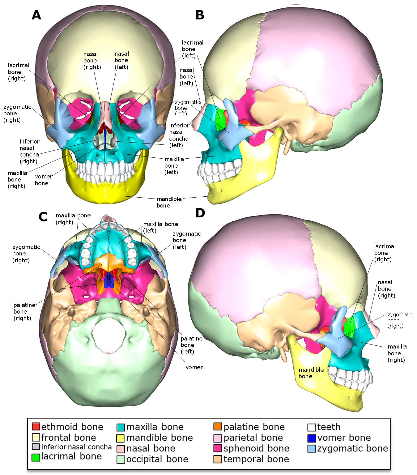
Above: Facial bones with the following views: (A) anterior view, (B) lateral view of the left side of the skull, (C) inferior view with the mandible removed, and (D) lateral view of the right side of the skull.
Clinical Application: Deviated Septum
Deviated septa are common. In more severe cases, a deviated septum can cause difficulty breathing, sinus infections, sleep apnea, snoring, repetitive sneezing, facial pain, nosebleeds, and mild to severe loss of the ability to smell. Although most commonly caused by impact trauma, it can also be a congenital disorder caused by compression of the nose during childbirth. Medications can temporarily relieve symptoms, but a minor surgical procedure, known as septoplasty, is necessary to completely cure the symptoms related to septal deviations.
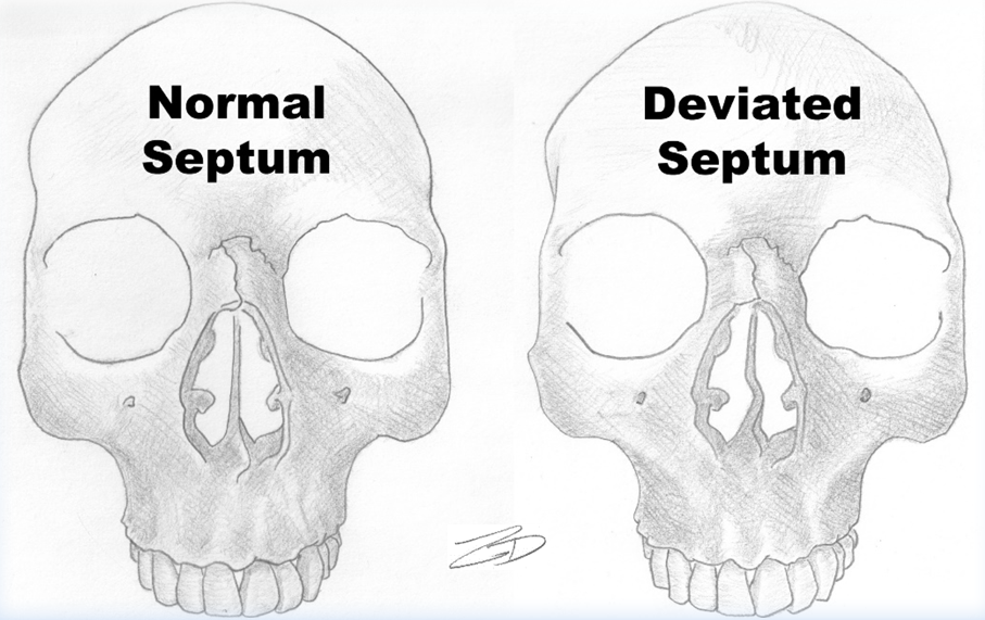
Above: Illustration indicates the difference between a normal septum and a deviated septum. The septum is made up of the perpendicular plate of the ethmoid bone and the vomer bone.
Markings of the Facial Bones
Recall from Chapter 7: Introduction to the Skeletal System, that bones have markings including holes, passageways, basins, and projections.
Before examining the specific markings of the facial bones, review the pertinent types of markings we will see when we examine the face markings:
|
Type of Bone Marking |
Description of Bone Marking Type |
|---|---|
|
angle |
Sharp bony angulations which may serve as bony or soft tissue attachments. |
|
body |
Usually refers to the largest most prominent segment of bone. |
|
foramen (pl. foramina) |
A hole in a bone through which nerves and blood vessels pass. |
|
fossa (pl. fossae) |
A shallow depression in a bone surface. |
|
notch |
A depression in a bone which often, provides stabilization to an adjacent articulating bone. |
|
process (pl. processes) |
Bony projection, allow for muscle attachment. |
|
ramus (pl. rami) |
The curved part of a bone that gives structural support to the rest of the bone. |
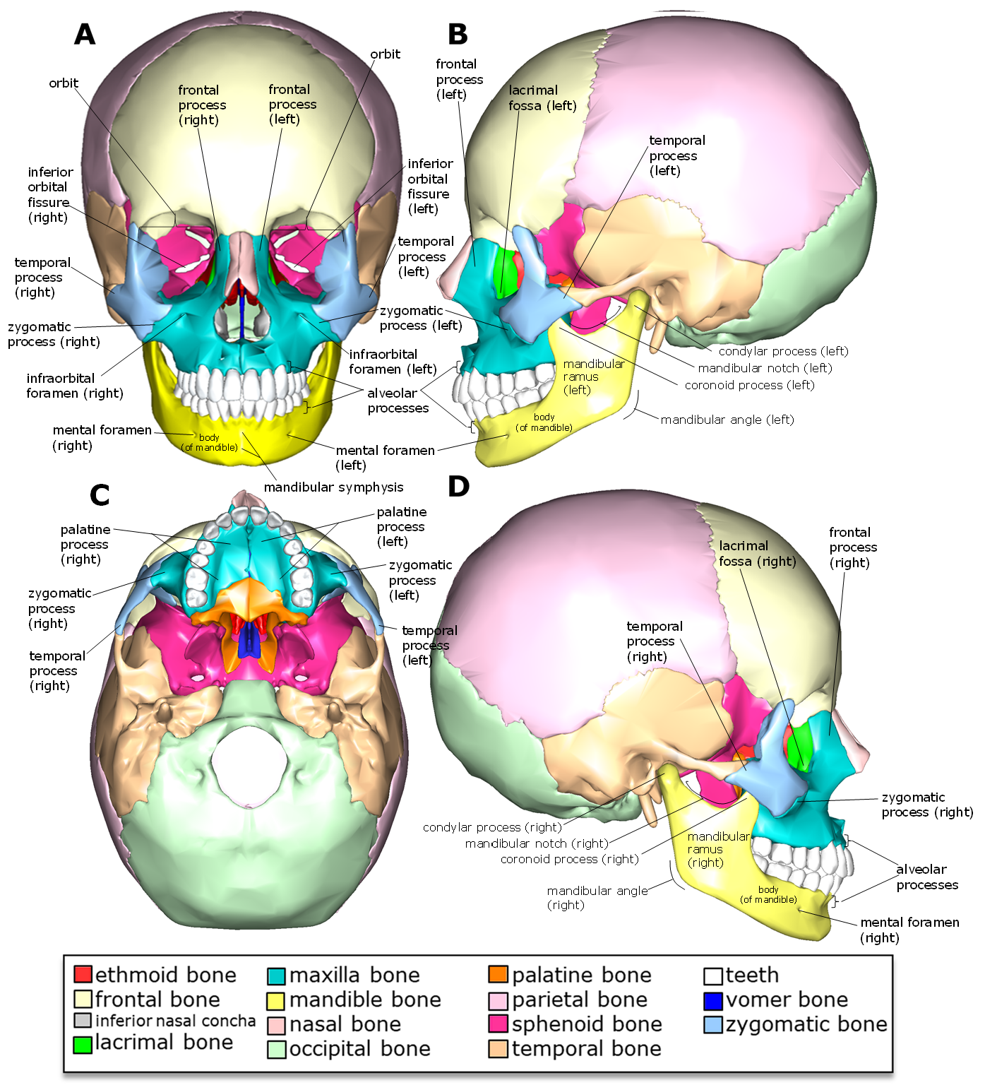
Above: Markings of the facial bones with the following views: (A) anterior view, (B) lateral view of the left side of the skull, (C) inferior view with the mandible removed, and (D) lateral view of the right side of the skull.
|
Marking |
Bone Marking is Part of |
Location |
Function |
|---|---|---|---|
|
alveolar process (3, one on each maxilla bone and one on the mandible) |
maxilla bones & mandible bone |
anterior skull & lateral skull |
entire region of the bone that forms the sockets for the teeth |
|
body (of the mandible) |
mandible bone |
anterior skull & lateral skull |
connects the right and left rami of the mandible; holds the alveolar process; entire horizontal region of the mandible including the chin |
|
condylar process (2, one right & one left) |
mandible bone |
lateral skull |
creates the hinge where the mandible articulates with the temporal bone (within the mandibular fossa) |
|
coronoid process (2, one right & one left) |
mandible bone |
lateral skull |
superior-pointing process anterior to the condylar process; the temporalis muscle attaches here |
|
frontal process (2, one right & one left) |
maxilla bones |
anterior skull & lateral skull |
articulates with the nasal bones, lacrimal bones, and frontal bone forming lateral aspects of the nose |
|
inferior orbital fissure |
formed by maxilla, zygomatic, and sphenoid bones |
anterior skull |
provide passage for nerves and blood vessels |
|
infraorbital foramen (2, one right & one left) |
maxilla bones |
anterior skull |
provide passage for paired (right and left) blood vessels (infraorbital arteries) and nerves (infraorbital nerves) |
|
lacrimal fossa (2, one right & one left) |
lacrimal bones |
lateral skull |
involved in draining tears from the eyes into the nasal cavities |
|
mandibular angle (2, one right & one left) |
mandible bone |
lateral skull |
inferior and posterior aspects of the mandible (right and left) where the body meets the rami |
|
mandibular notch (2, one right & one left) |
mandible bone |
lateral skull |
indentation between the condylar process and the coronoid process |
|
mandibular symphysis |
mandible bone |
anterior skull |
a ridge located medially on the mandible |
|
mental foramen (2, one right & one left) |
mandible bone |
anterior skull & lateral skull |
provides passage for blood vessels and nerves of the chin and lower lip; recall "mental" means "chin" |
|
orbit (2, one right & one left) |
formed by frontal, sphenoid, ethmoid, lacrimal, maxilla, and zygomatic bones |
anterior skull |
eye socket |
|
palatine process (2, one right & one left) |
maxilla bones |
inferior skull |
posterior-projecting processes of the maxilla bones forming the anterior aspect of the roof of the mouth; articulates with the palatine bones |
|
ramus (of the mandible) (2, one right & one left) |
mandible bone |
lateral skull |
regions of the mandible (right and left) connecting the body of the mandible, the condylar process, and the coronoid process |
|
temporal process (2, one right & one left) |
zygomatic bones |
anterior skull & lateral skull |
posterior-pointing process that articulates with the zygomatic process of the temporal bone |
|
zygomatic process (of maxilla bones) (2, one right & one left) |
maxilla bones |
anterior skull & lateral skull |
lateral-pointing processes that articulate with the right and left zygomatic bones |
Attributions (All Skull Sections)
- "Anatomy 204L: Laboratory Manual (Second Edition)" by Ethan Snow, University of North Dakota is licensed under CC BY-NC 4.0
- "Anatomy and Physiology" by J. Gordon Betts et al., OpenStax is licensed under CC BY 4.0
- "Anatomy and Physiology Lab Reference" by Laird C Sheldahl, OpenOregonEducational Resources, Mt. Hood Community College is licensed under CC BY-SA 4.0
- "BIOL 250 Human Anatomy Lab Manual SU 19" by Yancy Aquino, Skyline College is licensed under CC BY-NC-SA 4.0
- "BodyParts3D/Anatomography" by The Database Center for Life Science is licensed under CC BY-SA 2.1
- "Ethmoid.png" by Life Science Databases(LSDB) is licensed under CC BY-SA 2.0
- "Gray151.png" by Henry Vandyke Carter is in the Public Domain
- "Human Skeleton Upper Body Posterior View.jpg" by Andrewmeyerson is licensed under CC BY-SA 3.0
- "Rotation ethmoid.gif" by Life Science Databases(LSDB), animated by was a bee is licensed under CC BY-SA 2.0
- "Sphenoid bone - animation 02.gif" by Database Center for Life Science (DBCLS) is licensed under CC BY-SA 2.1
Updated 2025.