
1.9: Nervous System Anatomy Part 1- Functional Anatomy of the Cerebral Cortex and Spinal Cord
- Page ID
- 58952
\( \newcommand{\vecs}[1]{\overset { \scriptstyle \rightharpoonup} {\mathbf{#1}} } \)
\( \newcommand{\vecd}[1]{\overset{-\!-\!\rightharpoonup}{\vphantom{a}\smash {#1}}} \)
\( \newcommand{\dsum}{\displaystyle\sum\limits} \)
\( \newcommand{\dint}{\displaystyle\int\limits} \)
\( \newcommand{\dlim}{\displaystyle\lim\limits} \)
\( \newcommand{\id}{\mathrm{id}}\) \( \newcommand{\Span}{\mathrm{span}}\)
( \newcommand{\kernel}{\mathrm{null}\,}\) \( \newcommand{\range}{\mathrm{range}\,}\)
\( \newcommand{\RealPart}{\mathrm{Re}}\) \( \newcommand{\ImaginaryPart}{\mathrm{Im}}\)
\( \newcommand{\Argument}{\mathrm{Arg}}\) \( \newcommand{\norm}[1]{\| #1 \|}\)
\( \newcommand{\inner}[2]{\langle #1, #2 \rangle}\)
\( \newcommand{\Span}{\mathrm{span}}\)
\( \newcommand{\id}{\mathrm{id}}\)
\( \newcommand{\Span}{\mathrm{span}}\)
\( \newcommand{\kernel}{\mathrm{null}\,}\)
\( \newcommand{\range}{\mathrm{range}\,}\)
\( \newcommand{\RealPart}{\mathrm{Re}}\)
\( \newcommand{\ImaginaryPart}{\mathrm{Im}}\)
\( \newcommand{\Argument}{\mathrm{Arg}}\)
\( \newcommand{\norm}[1]{\| #1 \|}\)
\( \newcommand{\inner}[2]{\langle #1, #2 \rangle}\)
\( \newcommand{\Span}{\mathrm{span}}\) \( \newcommand{\AA}{\unicode[.8,0]{x212B}}\)
\( \newcommand{\vectorA}[1]{\vec{#1}} % arrow\)
\( \newcommand{\vectorAt}[1]{\vec{\text{#1}}} % arrow\)
\( \newcommand{\vectorB}[1]{\overset { \scriptstyle \rightharpoonup} {\mathbf{#1}} } \)
\( \newcommand{\vectorC}[1]{\textbf{#1}} \)
\( \newcommand{\vectorD}[1]{\overrightarrow{#1}} \)
\( \newcommand{\vectorDt}[1]{\overrightarrow{\text{#1}}} \)
\( \newcommand{\vectE}[1]{\overset{-\!-\!\rightharpoonup}{\vphantom{a}\smash{\mathbf {#1}}}} \)
\( \newcommand{\vecs}[1]{\overset { \scriptstyle \rightharpoonup} {\mathbf{#1}} } \)
\(\newcommand{\longvect}{\overrightarrow}\)
\( \newcommand{\vecd}[1]{\overset{-\!-\!\rightharpoonup}{\vphantom{a}\smash {#1}}} \)
\(\newcommand{\avec}{\mathbf a}\) \(\newcommand{\bvec}{\mathbf b}\) \(\newcommand{\cvec}{\mathbf c}\) \(\newcommand{\dvec}{\mathbf d}\) \(\newcommand{\dtil}{\widetilde{\mathbf d}}\) \(\newcommand{\evec}{\mathbf e}\) \(\newcommand{\fvec}{\mathbf f}\) \(\newcommand{\nvec}{\mathbf n}\) \(\newcommand{\pvec}{\mathbf p}\) \(\newcommand{\qvec}{\mathbf q}\) \(\newcommand{\svec}{\mathbf s}\) \(\newcommand{\tvec}{\mathbf t}\) \(\newcommand{\uvec}{\mathbf u}\) \(\newcommand{\vvec}{\mathbf v}\) \(\newcommand{\wvec}{\mathbf w}\) \(\newcommand{\xvec}{\mathbf x}\) \(\newcommand{\yvec}{\mathbf y}\) \(\newcommand{\zvec}{\mathbf z}\) \(\newcommand{\rvec}{\mathbf r}\) \(\newcommand{\mvec}{\mathbf m}\) \(\newcommand{\zerovec}{\mathbf 0}\) \(\newcommand{\onevec}{\mathbf 1}\) \(\newcommand{\real}{\mathbb R}\) \(\newcommand{\twovec}[2]{\left[\begin{array}{r}#1 \\ #2 \end{array}\right]}\) \(\newcommand{\ctwovec}[2]{\left[\begin{array}{c}#1 \\ #2 \end{array}\right]}\) \(\newcommand{\threevec}[3]{\left[\begin{array}{r}#1 \\ #2 \\ #3 \end{array}\right]}\) \(\newcommand{\cthreevec}[3]{\left[\begin{array}{c}#1 \\ #2 \\ #3 \end{array}\right]}\) \(\newcommand{\fourvec}[4]{\left[\begin{array}{r}#1 \\ #2 \\ #3 \\ #4 \end{array}\right]}\) \(\newcommand{\cfourvec}[4]{\left[\begin{array}{c}#1 \\ #2 \\ #3 \\ #4 \end{array}\right]}\) \(\newcommand{\fivevec}[5]{\left[\begin{array}{r}#1 \\ #2 \\ #3 \\ #4 \\ #5 \\ \end{array}\right]}\) \(\newcommand{\cfivevec}[5]{\left[\begin{array}{c}#1 \\ #2 \\ #3 \\ #4 \\ #5 \\ \end{array}\right]}\) \(\newcommand{\mattwo}[4]{\left[\begin{array}{rr}#1 \amp #2 \\ #3 \amp #4 \\ \end{array}\right]}\) \(\newcommand{\laspan}[1]{\text{Span}\{#1\}}\) \(\newcommand{\bcal}{\cal B}\) \(\newcommand{\ccal}{\cal C}\) \(\newcommand{\scal}{\cal S}\) \(\newcommand{\wcal}{\cal W}\) \(\newcommand{\ecal}{\cal E}\) \(\newcommand{\coords}[2]{\left\{#1\right\}_{#2}}\) \(\newcommand{\gray}[1]{\color{gray}{#1}}\) \(\newcommand{\lgray}[1]{\color{lightgray}{#1}}\) \(\newcommand{\rank}{\operatorname{rank}}\) \(\newcommand{\row}{\text{Row}}\) \(\newcommand{\col}{\text{Col}}\) \(\renewcommand{\row}{\text{Row}}\) \(\newcommand{\nul}{\text{Nul}}\) \(\newcommand{\var}{\text{Var}}\) \(\newcommand{\corr}{\text{corr}}\) \(\newcommand{\len}[1]{\left|#1\right|}\) \(\newcommand{\bbar}{\overline{\bvec}}\) \(\newcommand{\bhat}{\widehat{\bvec}}\) \(\newcommand{\bperp}{\bvec^\perp}\) \(\newcommand{\xhat}{\widehat{\xvec}}\) \(\newcommand{\vhat}{\widehat{\vvec}}\) \(\newcommand{\uhat}{\widehat{\uvec}}\) \(\newcommand{\what}{\widehat{\wvec}}\) \(\newcommand{\Sighat}{\widehat{\Sigma}}\) \(\newcommand{\lt}{<}\) \(\newcommand{\gt}{>}\) \(\newcommand{\amp}{&}\) \(\definecolor{fillinmathshade}{gray}{0.9}\)Objectives:
At the end of this lab, you will be able to…
- Correctly identify the structures of importance within cerebral cortex
- Correctly identify the structures of importance within the spinal cord
- Correctly name the cranial nerves and indicate general functions
- Correctly and independently perform simple dissections
- Use correct anatomical terminology when discussing structures of the nervous system
Pre-Lab Exercise:
After reading through the lab activities prior to lab, complete the following before you start your lab
1. The structures of the Central Nervous System include:
2. The connective tissues that surround the cerebral cortex and spinal cord are known as the and is made of three layers: that connect to the tissues, that connects to the bones and the that contains the cerebral spinal fluid.
3. You will make two incisions during the dissection of the brain (cerebral cortex) either along the fissure to separate the brain into a right and left hemisphere and along the fissure to separate the superior from the inferior.
4. Color the images for use as a reference for identifying the models and dissected specimens.
Materials:
- Brain Models
- Spinal Cord Model
- Sagittal Head Model
- Sheep’s Brain and Dissection Tools
- Prosected Human Brain and Spinal Cord
- Stickers
- Felt Pens
- Colored Pencils
The central nervous system (CNS) is comprised of the neurons and glial cells that form the neural tissue of the cerebral cortex (including the lobes, limbic system, thalamus, hypothalamus, cerebellum, pons, and medulla oblongata), and spinal cord.
Activity 1: Cerebral Cortex:
The cerebral cortex is a gelatinous mass of cells, nothing like the fixed brains that we might think of, that is divided into distinct regions that perform functional tasks for maintaining homeostasis in the body. The neuron mass within the cerebral cortex is highly invaginated (folded) forming structures known as sulcus (fold) and gyrus (cell mass). This folding is generated by the synaptic connections occurring between cells that hold layers of cells together and move the layers based on movement of neurons in and around each other in order to maintain synaptic connections. The folding and invagination is the effect of having limited volume of the cranial vault for the mass of cells that are contained within the cerebral cortex.
Hindbrain
The Hindbrain is comprised on the pons, medulla oblongata and cerebellum. This is the most conserved region of the cerebral cortex. Within its many functions the hindbrain serves as a relay network that connects cerebral cortex to the spinal cord, forms projections that develop into the cranial nerves and the extra-pyramidal tracts of the spinal cord and serves as a region for regulation of autonomic functions.
Midbrain
The midbrain is the next most conserved region of the cerebral cortex. The structures of the midbrain, figure 14a-b and 15 c, are involved with many of the regulatory functions of homeostasis along with establishing neural relays key to cognitive functions and learning. The midbrain establishes connections between forebrain and hindbrain structures along with direct paths to the spinal cord and is comprised of structures that are referred to as the Limbic cortex.
Limbic System
The Limbic structures are the midbrain structures involved with activities of memory integration and automatic/hormonal responses to somatic sensory (homeostatic) information or relays information for coding purposes to higher cortical regions or from higher cortical regions to periphery for response. These structures also include the Hypothalamus, Thalamus, Hippocampus, and various nuclei and ganglion within the central region of the brain.
Forebrain Lobes
The forebrain is comprised of the lobes and structures that are familiarly known and that is associated with controls and regulates complex activities within the body based on the individual lobe. These include the frontal, parietal, occipital and temporal lobes that are named based on the cranial bone that overlays the lobe of the cerebral cortex. The frontal lobe is associated with functions of conscious thought process, memory and emotional responses. The parietal lobe is associated with somatosensation and proprioception and interactions with the frontal lobe to allow for somatic motor control. The occipital lobe is associated with visual processing and spatial recognition. The temporal lobe is associated with auditory processing along with language and symbol recognition.
Cranial Nerves
The cranial nerves are specialized peripheral nerves that originate in the hindbrain and brainstem of the cerebral cortex that provides innervations to the cranium, internal organs of the torso and are involved with autonomic, somatic sensory, visceral sensory, somatic motor and special senses of the body. The naming and identification of the cranial nerves (CN) are done either by classical name, or numerically based on where the nerve exits the cerebral cortex. Where the order of numbering of the cranial nerve is established from the most anterior superior (rostral) being the first and the most posterior-inferior nerve (caudal) being the twelfth. Using the numerical orientation, the nerves are indicated as cranial nerve (CN) and then by Roman Numeral (I-XII) to indicate the order of exiting the cerebral cortex. The site of origin for the cranial nerve (CN) and location of specific nuclei of origination indicates how specific (or diffuse) of a role that nerve has on homeostatic functions for the body. CN I through CN II originate within the proencephalon, while CN III through CN XII originate within the mesencephalon and rhombencephalon from nuclei ganglion near to the Thalamus, Pons and Medulla.
Procedures:
1. Obtain models, stickers, felt pens
2. Write the names of the key anatomical structures of the cerebral cortex on to the stickers
a. Frontal Lobe, Parietal Lobe, Occipital Lobe, Temporal Lobe, Cerebellum, Pons, Medulla Oblongata, Broca’s Area, Wernicke’s Area, Primary Motor Cortex, Primary Somatosensory Cortex
b. Corpus Callosum, Thalamus, Hypothalamus, Fornix, Choroid Plexus, Lateral Ventricle, Fourth Ventricle, Cerebral Aqueduct
3. Select a “team leader” and using the colored images as reference have members of the group take turns labeling the brain model with the key anatomical structures.
4. Using a pen or pencil, take turns within your group identifying the cranial nerves on the model of the cerebral cortex.
5. With your instructor, observe the prosected human brain and spinal cord. Relate the structures that you have identified on the model and in the coloring images to what can be viewed on the human organs.
6. Have your instructor check your work and then move to the next activity.
|
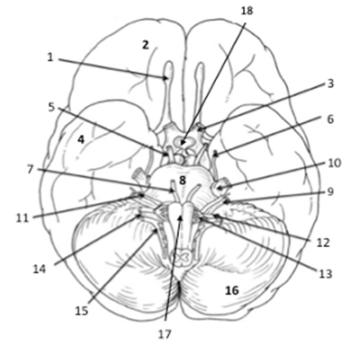
Key Anatomical structures of the cerebral cortex: 1 Cranial Nerve I (Olfactory) 2 Frontal Lobe 3 Cranial Nerve II (Optic) 4 Temporal Lobe 5 Cranial Nerve III (Oculomotor) 6 Cranial Nerve IV (Trochlear) 7 Cranial Nerve VI (Abducens) 8 Pons 9 Cranial Nerve VIII (Cochleovestibular) 10 Cranial Nerve V (Trigeminal) 11 Cranial Nerve VII (Facial) 12 Cranial Nerve IX (Glossopharyngeal) 13 Cranial Nerve X (Vagus) 14 Cranial Nerve XI (Spinal Accessory) 15 Cranial nerve XII (Hypoglossal) 15 Cerebellum 17 Medulla Oblongata 18 Hypothalamus and Pituitary Gland Color each structure a different color for use as a reference when identifying. |
|
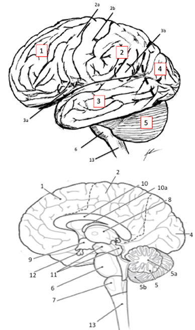
Key Anatomical structures of the cerebral cortex: 1 Frontal Lobe 2 Parietal Lobe 2a Primary Motor Cortex (Pre-Central Gyrus) 2b Primary Somatosensory Cortex (Post-Central Gyrus) 2c Central Sulcus 3 Temporal Lobe 3a Lateral Sulcus 3b Brocca's Area 3c Wernicke’s Area 4 Occipital Lobe 5 Cerebellum 5a Arbor Vitae of Cerebellum 5b Vermis of Cerebellum 6 Pons 7 Medulla Oblongata 8 Thalamus 9 Hypothalamus 10 Corpus Callosum 10a Fornix 11 Tectum & Limbic System 12 Pituitary Glans 13 Spinal Cord Color each structure a different color for use as a reference when identifying. |
Activity 2: Connective Tissue and Ventricles:
The gelatinous mass that is the cerebral cortex is suspended in a viscous solution (cerebral spinal fluid, CSF) within the cranial vault between three fibrous membranes (mater layers) that connect it to the boney structures. The distinct type of mater layer is identified by what the connective tissue is connecting. The pia mater is dense regular connective tissue that connects to the neural tissues of the CNS. The arachnoid is loose areolar and reticular connective tissue that connects the dura mater to the pia mater. The arachnoid allows for the passage of blood and lymph vessels into the cerebral cortex tissues and structures of the CNS. The dura mater is dense regular connective tissue that connects to the bone tissues of the cranium and vertebrae. Within these layers of connective tissues, invaginations and folds develop. These folds are the ventricles of the maters within the cerebral cortex that allows for the movement of the cerebral spinal fluid within and throughout the tissues and connects the movement of CSF to the vascular flow to ensure that the tissues are perfused correctly and materials are flushed from the CNS on a regular basis.
Procedures:
1. Obtain models, stickers, felt pens
2. Write the names of the key anatomical structures of the meninges and ventricles on to the stickers
3. Select a “team leader” and using the colored images as reference have members of the group take turns labeling the sagittal head model with the key anatomical structures.
4. Have your instructor check your work and then move to the next activity.
|
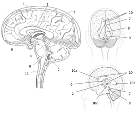
Key Anatomical structures of the cerebral cortex: 1=Pia Mater 2=Choroid Plexus 3=Subdural Space 4=Arachnoid Matter 5=Dura Matter 6=Interventricular Foramen 7=Third Ventricle 8=Cerebral Aqueduct 9=Fourth Ventricle 10=Lateral Ventricle 10a=Anterior Horn of Lateral Ventricle 10b=Posterior Horn of Lateral Ventricle 10c=Lateral Horn of Lateral Ventricle 11=Central Canal Color each structure a different color for use as a reference when identifying. |
Activity 3: Spinal Cord:
The spinal cord is an elongation of axons and interneurons leaving the brain stem and hindbrain of the cerebral cortex and travelling the length the vertebral column. While we may discuss the spinal cord as being a solid bundle of tissues (axons) near the inferior (caudal) aspects the spinal cord becomes slightly less bundled, forming the structure identified as the cauda equinae. This diffusion of axons typically occurs at the level of the second or third lumbar vertebrae. Within the organized axonal bundle region (from the foramen magnum through the initiation of the cauda equinae) there is a rigid structural orientation that the spinal cord possesses.
Within this rigid structure, there exists a general organization to the neural tissues within the spinal cord. The organization is such that we are able to divide the axonal and interneurons into tracts based on a dorsal (posterior) and ventral (anterior) orientation. Where the dorsal (posterior) will primarily have neural networks and tracts that are involved with sensory issues, while the ventral (anterior) will primarily have the neural networks and tracts involved with motor issues. Additionally, the tissue can be indicated based on staining as being “gray” or “white” tissue within the spinal cord. In which the gray is an indication of cell bodies and unmyelinated regions and the white is an indication of axons and myelinated regions.
Procedures:
1. Obtain models, stickers, felt pens
2. Write the names of the key anatomical structures of the spinal cord on to the stickers
a. Dorsal Horn, Dorsal Nerve Root, Dorsal Root Ganglion, Dorsal Sensory Tracts, Commissure, Central Canal, Ventral Horn, Lateral Horn, Ventral Nerve Root, Spinal Nerve
3. Select a “team leader” and using the colored images as reference have members of the group take turns labeling the spinal cord model with the key anatomical structures.
4. Have your instructor check your work and then move to the next activity.
|
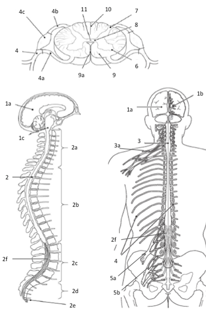
Key Anatomical structures of the cerebral cortex: 1a Cerebral Cortex 1b Thalamic Nuclei 1c Hind Brain 2 Spinal Cord 2a Cervical Nerve Roots 2b Thoracic Nerve Roots 2c Lumbar Nerve Roots 2d Sacral Nerve Roots 2e Coccygeal Nerve Roots 2f Cauda Equinae 3 Cervical Enlargement 3a Brachial Plexus 4 Spinal Nerves 4a Ventral Root 4b Dorsal Root 4c Dorsal Root Ganglion 5a Lumbar Plexus 5b Sacral Plexus 6 Ventral Horn 7 Dorsal Horn 8 Gray Matter 9 White Matter 9a Ventral (Anterior) Median Sulcus 10 Dorsal (Posterior) Median Sulcus 11 Central Canal Color each structure a different color for use as a reference when identifying.
|
Activity 4: Dissection:
1. Observe the external anatomy of the brain. Identify the meningeal layers covering the sheep’s brain. Note the thickening of the meninges that forms between the cerebellum and the occipital lobe.
2. As you observe, turn the brain so that you are able to examine the exterior of the brain superiorly, inferiorly and laterally. In the inferior view, note the various cranial nerves that are visible, if the pituitary is still present, the pons and medulla oblongata
3. Using your dissecting shears, make a small hole near the most anterior part of the frontal lobe in the meninges
4. Take your blunt probe and insert it into the small hole that you just cut in the meninges and carefully lift the meninges to separate it from the brain tissue.
5. Using your dissection shears, scalpel and blunt probe, carefully remove the meninges from rest of the brain. Be careful as you remove meninges from the inferior part of the brain (if necessary, get assistance from your instructor).
6. After removing the meninges, repeat the examination of the external anatomy of the brain.
7. In the posterior examination, gently displace the cerebellum to reveal the Colliculi and Pineal Gland that is now visible between the cerebellum and rest of the brain.
8. After examining the external structure, place the brain on the dissection tray and locate the medulla oblongata.
9. Move the dissection tray to the dissecting scope and adjust the magnification until you are able to clearly see the edge of the medulla oblongata.
a. Make a transverse cut across the medulla to separate the initial segment of the spinal cord from the rest of the brain
b. Lay the spinal cord flat on the dissection tray and move the tray so that the spinal cord is seen in the dissection scope.
c. Examine the spinal cord and note the organization of the horns, white matter, gray matter and commissure. Compare to the model that you have previously examined.
10. In your paired groupings: 1 group will perform the longitudinal dissection and 1 group will perform the superior transverse dissection.
a. Longitudinal Dissection:
i. Remove the dissection tray from the dissection scope and find the longitudinal fissure
ii. Dissect the brain using a sagittal (longitudinal) cut through the longitudinal fissure and the medial structures
iii. Continue the dissection until you have a right and left half of the brain to examine
iv. Examine the anatomical structures of importance that are now visible to the internal medial surface
b. Superior Transverse Dissection:
i. Remove the dissection tray from the dissection scope and find the transverse fissure (“bump” that separates temporal lobe from frontal lobe
ii. Dissect the brain by making a circular incision around the right and left hemispheres to remove the “top” of the brain from the remainder of the sheep’s brain.
iii. The cut should reveal the opening to the lateral ventricles and the thalamus to the center and visible white matter (axonal pathways) through the remainder of the brain
iv. Examine the internal structures of importance that are now visible and then review the inferior aspects of the brain to identify structures of importance.
c. Combine the two groups and take turns within the groups to identify (using pins or probe) structures of importance.